This document summarizes a study examining the role of tropomyosin-related kinase B (TrkB) in metastatic pancreatic cancer. The study found that TrkB was overexpressed in highly metastatic pancreatic cancer cells compared to parental cells. TrkB overexpression correlated with perineural invasion, positive retroperitoneal margins, and shorter time to liver metastasis in patient samples. The metastatic cells also showed increased activation of ERK1/2 and increased expression of IL-8 and VEGF, which are involved in invasion and metastasis. This suggests TrkB overexpression may promote the aggressive growth and metastasis of pancreatic cancer by activating signaling pathways and increasing expression of genes involved in these processes. TrkB may therefore be a novel therapeutic target for pancreatic cancer.

![washed in PBS containing 0.2% Tween 20 and probed with
horseradish peroxidase–coupled secondary goat anti-rabbit or
anti-mouse antibodies (Amersham, Arlington Heights, IL). The
proteins were visualized with Lumi-Light Western blotting
substrate (Roche, Indianapolis, IN) according to the manufac-
turer’s instructions.
Tissue Samples
Tissue samples from 54 patients and normal controls were
obtained from the Pancreatic Tumor Tissue Bank of M.D.
Anderson after institutional review board approval was obtained.
All of the patients had undergone pancreaticoduodenectomy for
ductal adenocarcinoma of the pancreas without prior chemo-
therapy or radiotherapy between 1990 and 2000. All these
pancreatic cancer patients had follow-up CT scans every 3 to 4
mouths for the first two postoperative years and every 6 months
for years 3 to 5. The tissues were fixed in 10% buffered formalin
and paraffin embedded. A representative H&E stained slide was
obtained from each sample and the presence of pancreatic
adenocarcinoma confirmed by M.D. Anderson pathologists.
Mouse tissue samples from orthotopically growing intrapancre-
atic Colo357FG and Colo357L3.6pl tumors and liver metastasis
of Colo357L3.6pl tumors were processed by the same
procedure.
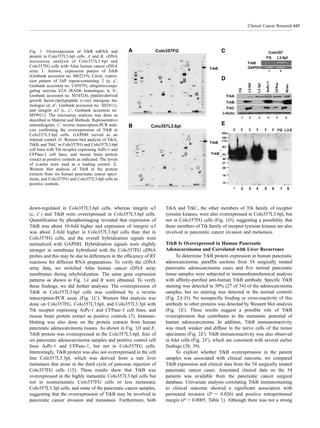
Immunostaining
Paraffin-embedded tissues were used for identification of
TrkB, VEGF, and IL-8. Immunohistochemical staining proce-
dures were done by using polyclonal rabbit antibodies against
TrkB and VEGF and a monoclonal antibody against IL-8 (Santa
Cruz Biotechnology) as previously described (33). Positive
immunoreactivity for TrkB, VEGF, and IL-8 was defined as
brown-colored precipitate in the cell membrane and cytoplasm.
Control samples exposed to secondary antibody alone showed
no specific staining. TrkB expression in the human pancreatic
cancer samples was assessed by a pathologist not aware of the
patients’ clinical status. TrkB, VEGF, and IL-8 expressions
were assessed in the mouse pancreatic tumor tissues and liver
metastases. Quantification of immunostainings was done by
averaging two sets of independently obtained results from five
microscopic fields (34). The area of tumor stained was
determined with a four-tiered scale 0 to 3: ‘‘0’’, no detectable
staining; ‘‘1’’, >0 but <20% of tumor cells stained; ‘‘2’’, 20%
to 50% of tumor cells stained; ‘‘3’’, >50% of tumor cells
stained. The intensity of staining was evaluated with a four-
tiered scale 0 to 3 applying the following scoring criteria: ‘‘0’’,
no detectable staining; ‘‘1’’, weak staining of tumor cells; ‘‘2’’,
moderate staining of tumor cells; ‘‘3’’, strong staining of tumor
cells. Finally, the results of the intensity of staining and area
were calculated for semiquantitating the immunostainings.
Clinicopathologic Correlation
The results from immunohistochemical analysis for TrkB
were correlated with histopathologic and clinical patient
characteristics from a continuously updated database of
surgically treated pancreatic cancer patients at M.D. Anderson
after institutional review board approval was obtained. The
database contains demographic data, surgical details, pathology
reports, and clinical outcome.
Statistical Analysis
All statistical analyses were done with NCSS software
(NCSS, Kaysville, UT). The significance of the data was
determined by using the v2
test and Cox proportional hazard
regression. P < 0.05 was considered significant. To estimate the
degree of association between variables, odds ratio, and hazard
ratio, and corresponding 95% confidence intervals were
computed as appropriate.
Electrophoretic Mobility Shift Assay
An electrophoretic mobility shift assay was done with nuclear
extracts as previously described (35). The 32
P-labeled probes
used were double-stranded oligonucleotides for the Elk-1
(5V-GGATGTCCATATTAGGACATCT-3V), AP-1 (5V-
CGCTTGATGACTCAGCCG GAA-3V), and Oct-1 (5V-
TGTCGAATGCAAATCACTAGAA-3V) consensus sequence
(Sigma, The Woodlands, TX). Equal loading of nuclear extracts
was monitored by assessment of Oct-1 binding.
Kinase Assay
Phosphorylation of TrkB was measured in an in vitro kinase
assay as previously described (36). Briefly, TrkB was immuno-
precipitated and the kinase reaction was carried out in 20 AL
ATP buffer (50 mmol/L HEPES [pH 7.5], 5 mmol/L MgCl2, and
2 mmol/L ATP; ref. 37). After incubation at 37jC for 20
minutes, the reaction products were resolved by SDS-PAGE,
dried, and exposed to X-ray films.
Transfection and Luciferase Assay
One microgram of wild type-AP-1 or mutant AP-1 reporter
plasmid containing the firefly luciferase reporter gene and a
pRL-TK plasmid containing the Renilla luciferase gene under
the control of the herpes simplex virus thymidine kinase
promoter as an internal control were cotransfected into tumor
cells in triplicate by using the lipotransfection method (FuGENE
6; Roche) according to the manufacturer’s recommendation. The
activities of both firefly and Renilla luciferase were determined
48 hours after transfection with the dual luciferase reporter assay
system (Promega, Madison, WI). The firefly luciferase activities
were normalized to the control Renilla luciferase activity.
RNase Protection Assay
RNA was isolated from Colo357FG and Colo357L3.6pl
cells by using TRIZOL Reagent (Life Technologies). A RNase
protection assay was done with the Riboquant multiprobe
protection assay system (PharMingen, San Diego, CA) accord-
ing to the manufacturer’s recommendation. Quantification was
carried out by using phospho-image analysis.
RESULTS
Overexpression of TrkB in Colo357L3.6pl Cells
To search for candidate genes involved in pancreatic tumor
invasion and metastasis, we did cDNA microarray analysis to
identify genes differentially expressed in the highly metastatic
cell line Colo357L3.6pl and its parental cell line Colo357FG. As
shown in Fig. 1A and B, the expression of the genes encoding
IAP repeat-containing 2 (a, aV), ubiquitin-conjugating enzyme
E2A (RAD6 homologue; b, bV), and platelet-derived growth
factor-hpolypeptide (v-sis) oncogene homologue (d, dV) was
TrkB Overexpression in Pancreatic Cancer442](https://image.slidesharecdn.com/93b15509-7430-413b-89dc-a4e098c5c0ca-150818134210-lva1-app6892/85/440-3-320.jpg)


![Ca pancreas [autosaved]](https://cdn.slidesharecdn.com/ss_thumbnails/capancreasautosaved-200627065511-thumbnail.jpg?width=640&height=640&fit=bounds)


