2. drag forces, each of the wells was then placed between the poles of the
magnetic device, which induces an inhomogeneous magnetic field (14).
The forces subjected to one bead were adjusted to 10 dyne/cm2
. Analyses
revealed that this strength was exerted to most of the beads, whereas a
smaller part of the beads in the vicinity of the flat pole was subjected to
lower forces. The forces were applied either with a frequency of 1 Hz if
not otherwise mentioned or permanently during the indicated time. In
general, the following experimental designs were compared: 1) control
cells without any treatment (Ϫ); 2) clustered samples (c), i.e. the cell
monolayer was incubated with anti-integrin antibody-coated beads for
50 min; 3) mechanically stressed samples, i.e. the cell monolayer was
incubated (in a control experiment with anti-CD71) with anti-integrin
antibody-coated beads for 20 min followed by application of forces for 30
min.
Cell Lysis and Immunoblotting—After integrin stimulation as de-
scribed above, adherent cells in each well were washed in phosphate-
buffered saline and lysed in 10 l of SDS sample buffer. Lysates of
several wells were pooled and then boiled. The samples were subjected
to a 7.5% SDS-polyacrylamide gel electrophoresis. For immunoblotting,
the proteins were transferred to polyvinylidene difluoride membranes.
To block nonspecific binding, membranes were incubated with 5% milk
powder in buffer containing 0.01% Tween 20, 100 mM Tris/HCl, pH 9.0,
155 mM NaCl for 30 min at 37 °C. Immunoblotting was performed
with alkaline phosphatase-labeled anti-phosphotyrosine antibody at a
dilution of 1:20,000 and then visualized with chemiluminescence
(CDP star).
Immunoprecipitation—To analyze activation of MAP kinases, cells
were lysed in precipitation buffer containing protease inhibitor, SDS,
and Triton X-100 (0.1%). After centrifugation, the supernatant was
incubated with 100 l of anti-ERK antibody for 1 h at room temperature
followed by adding 50 l of protein A-agarose. After centrifugation, the
pellet was washed in precipitation buffer and analyzed for phosphoty-
rosine as described above.
Preparation of Cytoskeletal Fractions—The cell monolayer was incu-
bated with cell extraction buffer containing 1% Triton X-100, 20 mM
imidazole, 2 mM MgCl2, 80 mM KCl, 2 mM EGTA for 5 min at 4 °C. The
Triton nonsoluble fractions were then collected in SDS sample buffer
and subjected to gel electrophoresis as described above.
Treatment with Cytochalasin and Calcium Chelator—To disrupt the
actin filaments of the cytoskeleton, the cell monolayer was treated with
25 nM cytochalasin D for 20 min at 37 °C. The cells were then washed
and incubated with the microbeads to perform the procedure for me-
chanical loading.
For chelating intracellular calcium, the cells were preincubated with
5 M of 1,2-bis(2-aminophenoxy)ethane-N,N,NЈ,NЈ-tetraacetic acid, ace-
toxymethyl ester (BAPTA-AM) for 15 min. Mechanical strain was then
applied in the presence of 5 M of BAPTA.
RESULTS
The osteosarcoma cell line expressed the 1 as well as the ␣2
integrin subunits on the cell surface (14). Therefore, we exam-
ined the effect of mechanical stress applied to both integrin
subunits on tyrosine phosphorylation of proteins as a mecha-
nism in integrin-mediated signal transduction. First, we were
interested in the time course of tyrosine phosphorylation due to
clustering of the 1 integrin subunit by incubation of the cells
with anti-1-coated microbeads. We observed an increase of
phosphorylation during the time of incubation, which reached
the maximum after 60 min (Fig. 1). Based on this finding,
mechanical stress was applied to integrins for 30 min after an
incubation time of 20 min to bind the beads to the receptors.
Application of forces to the 1 as well as to the ␣2 subunit
induced an increased tyrosine phosphorylation of proteins com-
pared with integrin clustering alone (Fig. 2). Stressing the 1
chain, the effect was more pronounced than with ␣2. To prove
whether the mechanically induced cellular reactions are spe-
cific for integrins, we stressed the transferrin receptor (CD71)
for comparison. Although a slightly increased tyrosine phos-
phorylation was observed compared with untreated cells, the
effect was distinctly lower than after stressing an integrin
receptor (Fig. 3). Next we compared the effect of permanent
mechanical loading with an intermittent stress of 1 Hz on
tyrosine phosphorylation. Application of a stress with a fre-
quency of 1 Hz induced a more profound phosphorylation than
permanent drag forces (Fig. 2). To exclude the possibility that
the influence of different modes of magnetic field application
alone and not the mechanical receptor stress provoked the
differences in tyrosine phosphorylation, we compared controls
in which pure cells were subjected to a permanent and a cyclic
magnetic field. This experiment clearly demonstrated that the
magnetic field alone did not influence tyrosine phosphoryla-
tion, independent of the mode of the magnetic field (Fig. 3).
To evaluate the role of the cytoskeleton in mechanically
induced tyrosine phosphorylation, we examined whether ty-
rosine-phosphorylated proteins are anchored to the cytoskele-
ton. After clustering and mechanically loading of the integrins,
cells were extracted with Triton X-100, and the detergent in-
soluble fraction analyzed for tyrosine-phosphorylated proteins
(Fig. 4). Both clustering and additional stress induced a linkage
of tyrosine-phosphorylated proteins. Again, mechanical load
was more effective than clustering. This observation concerned
the 1 subunit, whereas the linkage of ␣2 to the cytoskeleton
was similar comparing the effect of clustering and additional
mechanical load. Furthermore, cytoskeletally anchored phos-
phorylated proteins were preferably detected in the higher
molecular weight range. The anchorage of phosphorylated pro-
teins to the cytoskeleton was further studied by disruption of
FIG. 1. Time dependence of tyrosine phosphorylation after
clustering of the 1-integrin subunit. Cells were incubated with
anti-1-coated microbeads for the indicated periods in minutes. A total
cell lysate was then electrophoresed and blotted against an anti-phos-
photyrosine antibody. Tyrosine phosphorylation increased up to 60 min
and then decreased.
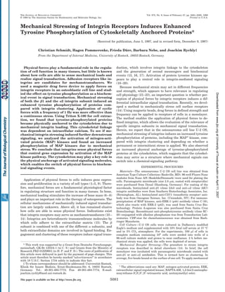
FIG. 2. Tyrosine phosphorylation induced by mechanical
stressing of integrins. Cells in a monolayer were incubated with
microbeads to bind at the 1 or ␣2 integrin subunit followed by appli-
cation of drag forces (s). These samples are compared with clustering of
the integrins by incubation with the microbeads without subsequent
mechanical loading (c), and untreated control cells (Ϫ). Total cell lysates
were electrophoresed and blotted for anti-phosphotyrosine. Most pro-
nounced differences are observed in the 40-kDa region. In all cases,
physical forces induced a significantly enhanced tyrosine phosphoryla-
tion compared with clustering and controls. Mechanical stressing of 1
induced a higher response than loading the ␣2 subunit (compare 1/1
Hz/s versus ␣2/1 Hz/s; or 1/P/s versus ␣2/P/s). A cyclic stress with a
frequency of 1 Hz was more effective than a permanent stress in both
integrin subunits (compare 1/1 Hz/s versus 1/P/s; and ␣2/1 Hz/s
versus ␣2/P/s). The results are representative of four independent
experiments.
Mechanically Induced Tyrosine Phosphorylation5082
atUniversityofTexasatAustinonApril11,2007www.jbc.orgDownloadedfrom
3. the actin filaments by cytochalasin D (Fig. 5). Treatment with
cytochalasin D abolished the cytoskeletal linkage of tyrosine-
phosphorylated proteins indicating the requirement of actin
polymerization.
Because intracellular calcium is induced by integrin stimu-
lation, we determined whether intracellular calcium plays a
role in the association of tyrosine-phosphorylated proteins to
the cytoskeleton. During mechanical loading of integrins, cells
were treated with the intracellular calcium chelator BAPTA-
AM. The following analysis of cytoskeletally anchored proteins,
which are tyrosine-phosphorylated, revealed that BAPTA sig-
nificantly reduced the physical anchorage of these proteins to
the cytoskeleton (Fig. 6). This indicates that calcium regulates
the linkage of activated proteins to the cytoskeleton during
integrin-mediated mechanically induced signal transduction.
Last, we were interested in whether the increased tyrosine
phosphorylation of proteins due to mechanical integrin stress-
ing may lead to downstream signaling, which could be relevant
for gene expression. Therefore, we examined whether among
the tyrosine-phosphorylated proteins the MAP kinases are also
activated. Immunoprecipitation of MAP kinases and analysis
of tyrosine phosphorylation demonstrated that mechanical
loading of both the 1 and the ␣2 integrin subunits induced a
distinctly higher degree of activation of the MAP kinases com-
pared with integrin clustering (Fig. 7). This suggests that reg-
ulation of gene expression by physical forces is controlled by
differential activation of MAP kinases and mediated by
integrins.
DISCUSSION
Tyrosine phosphorylation of several cellular proteins ap-
pears to play an essential role in integrin-mediated signal
transduction because its inhibition blocks gene expression (24).
The mechanisms by which extracellular interactions of inte-
grins regulate tyrosine phosphorylation remains elusive. We
demonstrate that application of physical forces to integrin re-
ceptors enhanced the tyrosine phosphorylation of proteins com-
FIG. 3. Comparison of tyrosine phosphorylation due to me-
chanical stress to integrins, mechanical load to the transferrin
receptor, and application of the magnetic field alone to the
cells. Cells in a monolayer were mechanically stressed at the 1 inte-
grin with 1 Hz as described above (lane 2) or stressed in the same
manner at the transferrin receptor (CD71) (lane 3). Cells in a monolayer
without magnetic beads were subjected to a permanent magnetic field
(lane 4) or a cyclic magnetic field with 1 Hz (lane 5) for 30 min.
Untreated cells were also examined (lane 1). Total cell lysates were
electrophoresed and blotted for anti-phosphotyrosine. Compared with
application of stress to the 1 integrin, mechanical stress to the trans-
ferrin receptor induced a detectable but significantly lower level of
tyrosine phosphorylation. Different modes of the magnetic field applied
to untreated cells had no effect on tyrosine phosphorylation.
FIG. 4. Cytoskeletal anchorage of tyrosine-phosphorylated
proteins due to mechanical stress. After treatment of the cells by
integrin stressing (1 Hz) (s), clustering (c), or without treatment (Ϫ), the
cells were extracted with Triton X-100 to obtain the detergent-insoluble
fraction. This cytoskeletal fraction was then processed for anti-phos-
photyrosine immunoblotting. For ␣2, similar quantities of phosphoryl-
ated proteins were found after mechanical stress and clustering. For 1,
mechanical stress induced a significant enhancement of cytoskeletally
linked tyrosine-phosphorylated proteins compared with clustered inte-
grins. The anchorage of tyrosine-phosphorylated proteins was observed
in the region of 130 kDa but not in the lower molecular weight range.
FIG. 5. Inhibition of the cytoskeletal anchorage of tyrosine-
phosphorylated proteins by cytochalasin D. Cells were treated by
mechanical stressing of the 1 integrin subunit (1 Hz) (s), clustering of
1 (c), or without treatment in the presence of cytochalasin D (ϩ) to
disrupt the actin filaments of the cytoskeleton. For comparison, the
cells were treated in the same way in the absence of cytochalasin D (Ϫ).
The cells were then extracted with Triton X-100 to obtain the insoluble
cytoskeletal fraction. This fraction was then processed for anti-phos-
photyrosine immunoblotting. Cytochalasin D dramatically blocked the
cytoskeletal linkage of tyrosine-phosphorylated proteins. In the absence
of cytochalasin D, mechanical stressing of integrins induced a distinctly
enhanced anchorage of phosphorylated proteins. This represents the
typical results of four independent experiments. (To obtain a back-
ground in the cytochalasin-treated samples, the blots in these experi-
ments were exposed longer to the film.)
FIG. 6. Influence of the calcium chelator BAPTA-AM on the
anchorage of tyrosine-phosphorylated proteins. Cells were
treated by mechanical stressing of the 1 integrin subunit (1 Hz) (s),
clustering of 1 (c), or without treatment (Ϫ) in the presence of
BAPTA-AM (ϩBAPTA) to chelate intracellular calcium. The cells were
then extracted with Triton X-100, and the insoluble fraction was proc-
essed for anti-phosphotyrosine immunoblotting. BAPTA-AM signifi-
cantly inhibited the anchorage of tyrosine-phosphorylated proteins to
the cytoskeleton, which is most obvious in the higher molecular weight
range. For comparison, in the absence of BAPTA-AM (ϪBAPTA), the
most profound tyrosine phosphorylation of cytoskeletally linked pro-
teins was found due to mechanical stressing of the 1 integrin subunit.
The results are representative of four independent experiments.
Mechanically Induced Tyrosine Phosphorylation 5083
atUniversityofTexasatAustinonApril11,2007www.jbc.orgDownloadedfrom
4. pared with integrin clustering alone. These results support the
idea that integrin receptors function as mechanosensors (11,
12, 25). It indicates that physical forces applied to integrin
receptors determine the degree of tyrosine phosphorylation
that may have consequences in gene expression. Furthermore,
tyrosine phosphorylation was also quantitatively influenced by
the mode of receptor stressing. Intermittent forces are more
effective than a permanent stress. Several reports have shown
that application of an intermittent mechanical load on bones is
more effective to stimulate growth than a permanent strain
(26, 27). Therefore, increased tyrosine phosphorylation due to
cyclic integrin stressing possibly provides a cellular-signaling
mechanism for the differential effect of mechanical loads on
bones. One of the initial effects of a mechanical stress to inte-
grins is the induction of their cytoskeletal anchorage. This has
been demonstrated in migrating cells by integrin cross-linking
or mechanical twisting of the integrins (11, 28, 29). It is an
intriguing aspect that the strength of this linkage is variable
and depends on external forces. Using an optical trap to re-
strain integrin bound beads resulted in a proportional
strengthening of the integrin cytoskeletal linkage (30). In the
line of these findings, we suggest that in our experiments
mechanical forces induced an increased strength of the integrin
cytoskeletal linkage. Other experiments have revealed that the
characteristics of the ligand and receptor-ligand interaction
control components of the integrin-mediated signal transduc-
tion resulting in functional consequences (31–33). For example,
integrin clustering induced a restricted accumulation of signal-
ing molecules into the cytoskeletal complex compared with
additional occupancy of the functional receptor epitope, which
led to a model of a hierarchy in signal transduction (34). Fur-
thermore, the physical characteristics of the collagen matrix
was affecting integrin activation and signal transduction,
which inhibited proliferation (32). This clearly suggests that
how integrins interact with a ligand is of importance for the
induced signal transduction. Although the  and ␣ subunits of
integrins form a dimer, we have found a difference in mechan-
ically induced tyrosine phosphorylation between the two inte-
grin subunits. Mechanical load on the  subunit provoked a
more profound tyrosine phosphorylation than stressing the ␣
subunit. We cannot exclude that this finding was due to a
higher expression of 1 compared with ␣2 that we have found
in these cells (14), but it could reflect a different mechanism of
the intracellular interaction of the cytoplasmic domains of both
integrin subunits. The  chain can directly interact with the
cytoskeletal proteins ␣ actinin and talin (35, 36). In addition,
the integrin-linked kinase can associate with the 1 cytoplas-
mic domain and mediate further downstream signaling (37).
Mechanical stress to the  subunit provoked a significant an-
chorage of tyrosine-phosphorylated proteins to the cytoskele-
ton, which was increased compared with integrin clustering.
Tyrosine phosphorylation of cytoskeletally anchored proteins
could be a prerequisite to form the cytoskeletal complex (38),
and a higher degree of phosphorylation may be a prerequisite
for the higher strengthening between receptors and cytoskele-
ton. Regarding the factors that determine the association of
activated signaling molecules to the cytoskeleton, we have
found that intracellular calcium is obviously an important reg-
ulator of the immobilization of proteins to the cytoskeleton.
This concerns not only intracellular-signaling proteins but also
the cytoskeletal anchorage of integrins to the cytoskeleton (28).
The role of calcium for a mechanically induced signal transduc-
tion is also stressed by data that have shown that intracellular
calcium concentrations correlated with increasing force levels
applied to integrins (39). However, our previous experiments
suggest that the differential cytoskeletal anchorage of tyrosine-
phosphorylated proteins and integrin subunits due to stimula-
tion of 1 compared with the ␣ subunit is not controlled by
differences in the magnitude of the calcium response. Incuba-
tion of cells with anti-integrin antibodies prior to mechanical
stimulation of the cells (13), as well as preliminary results
concerning the comparison of the calcium responses due to
mechanical stress applied with magnetic beads to 1 and ␣2,
revealed no quantitative differences in calcium signaling.
Concerning downstream signaling, we argue that the cy-
toskeleton could represent a structure where physical forces
are transformed into a biochemical signal pathway. The differ-
ential anchorage of tyrosine-phosphorylated proteins due to
physically stimulated integrins may regulate downstream in-
tracellular-signaling events.
One of these events is the activation of MAP kinases as a key
mechanism to control the activation of transcription factors,
which therefore mediates gene expression. The involvement of
this pathway in integrin signaling has been established (40,
41). We found that activation of the MAP kinases was signifi-
cantly increased due to physical forces compared with integrin
clustering. Due to the key role of the MAP kinases, our finding
emphasizes that physical forces transduced by integrins differ-
entially regulate cell proliferation and the expression of genes
through the MAP kinase cascade. The fact that activation of
MAP kinase by integrins depends on an intact cytoskeleton (41,
42) and the involvement of cytoskeletally associated signaling
molecules like focal adhesion kinase (43) highlights the signif-
icance of a controlled cytoskeletal anchorage of tyrosine-phos-
phorylated proteins for consequences in cell behavior. Because
the integrin-mediated MAP kinase pathway converges with
growth factor-induced pathways (44), our result suggests a
synergistic effect of mechanical forces and cytokines in the
regulation of cell function.
In conclusion, integrins mediate physical forces and may
regulate physiological consequences in the cell by a well tuned
induction of the degree of tyrosine phosphorylation of proteins.
A significant aspect is the cytoskeletal anchorage of activated
signaling proteins, which depends on the mobilization of intra-
cellular calcium. The functional relevance of these mechanisms
is supported by the result of an enhanced activation of MAP
kinases due to mechanical integrin stimulation.
REFERENCES
1. Wang, D. L., Wung, B. S., Shyy, Y. J., Lin, C. F., Chao, Y. J., Usami, S., and
Chien, S. (1995) Circ. Res. 77, 294–302
2. Diamond, S., Eskin, S., and McIntire, L. (1989) Science 243, 1483–1485
3. Komuro, I., Katoh, Y., Kaida, T., Shibazaki, Y., Kurabayashi, M., Hoh, E.,
Takaku, F., and Yazaki, Y. (1991) J. Biol. Chem. 266, 1265–1268
4. Hasegawa, S., Sato, S., Saito, S., Suzuki, Y., and Brunette, D. M. (1985) Calcif.
Tissue Int. 37, 431–436
5. Wirtz, H. R. W., and Dobbs, L. G. (1990) Science 250, 1266–1269
FIG. 7. Induction of the activation of MAP kinases by mechan-
ical stressing of integrin receptors. Cells were treated by mechan-
ical stressing (1 Hz) of the 1 or the ␣2 integrin subunits (s), clustering
of the subunits (c), or without treatment (Ϫ). After cell lysis, MAP
kinases were immunoprecipitated with anti-ERK antibody. After sep-
aration in the electrophoresis, the samples were probed with anti-
phosphotyrosine antibody. Mechanical stressing of both the 1 and the
␣2 integrin subunit induced an enhanced tyrosine phosphorylation of
MAP kinases p42 (ERK-2) and p44 (ERK-1) compared with controls and
clustering of the integrins. The results are representative of three
independent experiments.
Mechanically Induced Tyrosine Phosphorylation5084
atUniversityofTexasatAustinonApril11,2007www.jbc.orgDownloadedfrom
5. 6. Lanyon, L. E., and Rubin, C. T. (1984) J. Biomech. 17, 897–905
7. Raab-Cullen, D. M., Akhter, M. P., Kimmel, D. B., and Recker, R. R. (1994)
J. Bone Miner. Res. 9, 1143–1152
8. Rubin, C. T., Gross, T. S., McLeod, K. J., and Bain, S. D. (1995) J. Bone Miner.
Res. 10, 488–495
9. Hughes-Fulford, M., and Lewis, M. L. (1996) Exp. Cell Res. 224, 103–109
10. Ingber, D. (1991) Curr. Opin. Cell Biol. 3, 841–848
11. Wang, N., Butler, J. P., and Ingber, D. E. (1993) Science 260, 1124–1127
12. Wang, N., and Ingber, D. E. (1994) Biophys. J. 66, 2181–2189
13. Nebe, B., Rychly, J., Knopp, A., and Bohn, W. (1995) Exp. Cell Res. 218,
479–484
14. Pommerenke, H., Schreiber, E., Du¨rr, F., Nebe, B., Hahnel, C., Mo¨ller, W., and
Rychly, J. (1996) Eur. J. Cell Biol. 70, 157–164
15. Hynes, R. O. (1992) Cell 69, 11–25
16. Juliano, R. L., and Haskill, S. (1993) J. Cell Biol. 120, 577–585
17. Clark, E. A., and Brugge, J. S. (1995) Science 268, 233–239
18. Parson, J. T. (1996) Curr. Opin. Cell Biol. 8, 146–152
19. Kornberg, L., Earp, H. S., Parson, J. T., Schaller, M. D., and Juliano, R. L.
(1992) J. Biol. Chem. 267, 23439–23442
20. Burridge, K., Turner, C. E., and Romer, L. H. (1992) J. Cell Biol. 119, 893–903
21. Sandy, J. R., Meghji, S., Scutt, A. M., Harvey, W., Harris, M., and Meikle, M. C.
(1989) Bone Miner. 5, 155–168
22. Lanyon, L. E. (1984) Calcif. Tissue Int. 36, (suppl.) S56–S61
23. Neidlinger-Wilje, C., Wilke, H, J., and Claes, L. (1994) J. Orthop. Res. 12,
70–78
24. Lin, T. H., Rosales, C., Mondal, K., Bolen, J. B., Haskill, S., and Juliano, R. L.
(1995) J. Biol. Chem. 270, 16189–16197
25. Ingber, D. E. (1991) Curr. Opin. Cell Biol. 3, 841–848
26. Lanyon, L. E., and Rubin, C. T. (1984) J. Biomech. 17, 897–905
27. Hert, J., Liskova, M., and Landa, J. (1971) Folia Morphol. 19, 290–300
28. Nebe, B., Bohn, W., Sanftleben, H., and Rychly, J. (1996) Exp. Cell Res. 229,
100–110
29. Schmidt, C. E., Horwitz, A. F., Lauffenburger, D. A., and Sheetz, M. P. (1993)
J. Cell Biol. 123, 977–991
30. Choquet, D., Felsenfeld, D. P., and Sheetz, P. M. (1997) Cell 88, 39–48
31. Palecek, S. P., Loftus, J. C., Ginsberg, M. H., Lauffenburger, D. A., and
Horwitz, A. F. (1997) Nature 385, 537–540
32. Koyama, H., Raines, E. W., Bornfeldt, K. E., Roberts, J. M., and Ross, R. (1996)
Cell 87, 1069–1078
33. Miyamoto, S., Akiyama, S. K., and Yamada, K. M. (1995) Science 267, 883–885
34. Miyamoto, S., Teramoto, H., Coso, A. A., Gutkind, J. S., Burbelo, P. D.,
Akiyamata, S. K., and Yamada, K. M. (1995) J. Cell Biol. 131, 791–805
35. Otey, C. A., Pavalko, F. M., and Burridge, K. (1990) J. Cell Biol. 111, 721–729
36. Horwitz, A., Duggan, K., Buck, C., Beckerle, M. C., and Burridge, K. (1985)
Nature 320, 531–533
37. Hannigan, G. E., Leung-Hagesteijn, C., Fitz-Gibbon, L., Coppolino, M.,
Radeva, G., Filmus, J., Bell, J., and Dedhar, S. (1996) Nature 379, 91–96
38. Liu, M., Qin, Y., Liu, J., Tanswell, A. K., and Post, M. (1996) J. Biol. Chem.
271, 7066–7071
39. Glogauer, M., Ferrier, J., and McCulloch, C. A. G. (1995) Am. J. Physiol. 269,
C1093–C1104
40. Clark, E. A., and Hynes, R. O. (1996) J. Biol. Chem. 271, 14814–14818
41. Morino, N., Mimura, T., Hamasaki, K., Tobe, K., Ueki, K., Kikuchi, K.,
Takehara, K., Kadowaki, T., Yazaki, Y., and Nojima, Y. (1995) J. Biol.
Chem. 270, 269–273
42. Chen, Q., Kinch, M. S., Lin, T. H., Burridge, K., and Juliano, R. (1994) J. Biol.
Chem. 269, 26602–26605
43. Schlaepfer, D. D., and Hunter, T. (1997) J. Biol. Chem. 272, 13189–13195
44. Sundberg, C., and Rubin, K. (1996) J. Cell Biol. 132, 741–752
Mechanically Induced Tyrosine Phosphorylation 5085
atUniversityofTexasatAustinonApril11,2007www.jbc.orgDownloadedfrom