Call Girl Bangalore Nandini 7001305949 Independent Escort Service Bangalore
Tradus de onisim
1. ORIGINAL ARTICLE
The a-receptor for platelet-derived growth factor as a target
for antibody-mediated inhibition of skeletal metastases
from prostate cancer cells
MR Russell1,4
, WL Jamieson1,4
, NG Dolloff 1,3
and A Fatatis1,2
1
Department of Pharmacology and Physiology, Drexel University College of Medicine, Philadelphia, PA, USA and 2
Department of
Pathology and Laboratory Medicine, Drexel University College of Medicine, Philadelphia, PA, USA
Bone resorption by osteoclasts is thought to promote the
proliferation of prostate cancer cells disseminated to
the skeleton (Mundy, 2002). Using a mouse model of
experimental metastasis, we found that although late-
stage metastatic tumors were indeed surrounded by
osteoclasts, these cells were spatially unrelated to the
small foci of cancer cells in early-stage metastases. This is
the first evidence that survival and growth of disseminated
prostate cancer cells immediately after their extravasation
may not depend on osteoclast involvement. Interestingly,
prostate cancer cells expressing the a-receptor for
platelet-derived growth factor (PDGFRa) progress during
early-stages of skeletal dissemination, whereas cells
expressing lower levels or lacking this receptor fail to
survive after extravasation in the bone marrow. However,
non-metastatic cells acquire bone-metastatic potential
upon ectopic overexpression of PDGFRa. Finally, func-
tional blockade of human PDGFRa on prostate cancer
cells utilizing a novel humanized monoclonal antibody—
soon to undergo phase-II clinical trials—significantly
impairs the establishment of early skeletal metastases.
In conclusion, our results strongly implicate PDGFRa in
prostate cancer bone tropism through its promotion of
survival and progression of early-metastatic foci, provid-
ing ground for therapeutic strategies aimed at preventing
or containing the initial progression of skeletal metastases
in patients affected by prostate adenocarcinoma.
Oncogene (2009) 28, 412–421; doi:10.1038/onc.2008.390;
published online 13 October 2008
Keywords: skeletal metastasis; prostate adenocarcinoma;
PDGFRa
Introduction
Solid tumors frequently colonize distant organs in a
selective manner, a tissue tropism epitomized by the
propensity of prostate adenocarcinoma to metastasize
to the skeleton (Paget, 1889; Chambers et al., 2002).
Patients with advanced prostate cancer frequently
develop skeletal metastases and eventually succumb to
them. Thus, the identification of factors responsible for
promoting the colonization of the skeleton by malignant
prostate phenotypes is paramount in establishing
effective therapies to counteract metastatic complica-
tions of prostate adenocarcinoma. The survival and
growth of cancer cells disseminated to the skeleton is
considered dependent on trophic factors present in the
bone marrow (Mundy, 2002; Fidler, 2003). It has been
shown that bone-metastatic phenotypes produce mole-
cules, such as parathyroid hormone-related peptide,
which stimulate osteoblasts to release a receptor
activator of nuclear factor-kB (RANKL), which in turn
attracts and activates osteoclasts, thereby promoting
osteolysis (Roodman, 2004). The degradation of bone
matrix releases trophic factors, sustaining cancer cell
proliferation and facilitating increased recruitment and
activation of osteoclasts in a self-sustained vicious cycle,
which may promote the expansion of the metastatic
tumor mass within the bone tissue (Kingsley et al.,
2007). Inhibition of osteoclast activity by administration
of either bisphosphonates (Lee et al., 2002) or osteo-
protegerin (Canon et al., 2008) limits the growth of
cancer cells in the skeleton of animal models. These
studies were conducted using bioluminescence and/or
radiographic detection of bone lesions induced by
intravascular injection or, more frequently, direct
intraosseous implantation of cancer cells. However, at
least 2 Â 104
cells are needed in the bone to produce
a detectable bioluminescent signal, which is visible
not earlier than 2–3 weeks, following initial homing
(Wetterwald et al., 2002; van der Pluijm et al., 2005).
Analogously, radiographic analysis detects bone lesions
produced by large numbers of cancer cells but is unable
to identify single cancer cells or small foci (Fritz et al.,
2007). Therefore, investigations of the interactions
established by prostate cancer cells with the surrounding
microenvironment soon after their extravasation into
the bone marrow have been lacking.
Received 23 June 2008; revised 11 August 2008; accepted 8 September
2008; published online 13 October 2008
Correspondence: Dr A Fatatis, Department of Pharmacology and
Physiology, Drexel University College of Medicine, 245 N. 15th Street,
New College Building MS488, Philadelphia, PA 19102, USA.
E-mail: afatatis@drexelmed.edu
3
Current address: Department of Medicine, University of
Pennsylvania, Philadelphia, PA 19104, USA.
4
MR Russell and WL Jamieson contributed equally to this study.
Oncogene (2009) 28, 412–421
& 2009 Macmillan Publishers Limited All rights reserved 0950-9232/09 $32.00
www.nature.com/onc
2. It could be hypothesized that osteoclast recruitment in
early metastatic stages may not occur due to the low
number of cancer cells and the consequent paucity of
secreted chemo-attractant and activating molecules.
Thus, it is not until the tumor reaches a critical mass
that such influence on the microenvironment is exerted.
If this is the case, disseminated single cancer cells and
small foci might rely on trophic molecules readily
available in the bone marrow immediately after their
arrival to ensure a successful lodging in the skeleton—
before mutual interactions with osteoclasts can be
established. The expression of receptors compatible
with these trophic molecules would provide selected
malignant phenotypes with a determinant survival
advantage (Fidler, 2003).
In this study, we used an animal model of hemato-
genous skeletal metastases combined with a fluores-
cence-based histological analysis to monitor the
establishment and progression of bone metastases from
the arrival of individual cells to full-blown macroscopic
tumors. Using this approach, we determined the spatial
relationships between osteoclasts and micro-metastases
and investigated the relevance of platelet-derived growth
factor receptor-a (PDGFRa) expression for the survival
and growth of prostate cancer cells in the bone marrow.
Finally, we evaluated antibody-mediated targeting of
PDGFRa as a strategy to counteract skeletal metastases
from prostate cancer.
Results and discussion
In the first set of experiments, we investigated the
involvement of osteoclasts in early skeletal micro-
metastases. Male SCID mice were inoculated in the left
cardiac ventricle with PC3-ML human prostate cancer
cells, which consistently produce macroscopic metas-
tases at the craniofacial region, tibia and femur of
X80% of animals within 4 weeks. The PC3-ML cells
were previously selected from the widely used PC3
parental line for their invasive ability in vitro and
metastatic potential in vivo (Wang and Stearns, 1991).
We engineered these cells to stably express enhanced
green fluorescent protein and, by the combined use of
fluorescence stereomicroscopy, histological analysis and
digital imaging of serial cryosections, identified small
metastatic foci in the skeleton of mice at 1, 2 and 3
weeks following cell inoculation. A manual caliper was
used to measure the volume of 4-week macro-metastases
(Figure 1a). We found that metastases with a cross-
section area of 28 Â 103
mm2
or larger—which we usually
observe 2 weeks after cell inoculation—were surrounded
by an almost uninterrupted layer of osteoclasts,
identified by TRAcP staining (Figure 1b). However,
the smaller foci identified in the first 2 weeks after
inoculation were found to be spatially unrelated to
osteoclasts (Figure 1c and Supplementary Figure 1).
Thus, our results indicate a later involvement of
osteoclasts in metastatic progression and suggest the
existence of mechanisms alternative to bone degradation
to support the initial survival of prostate cancer cells in
early bone micro-metastases. As osteoclast involvement
appears to be a delayed event in metastatic progression,
single cells and small foci would initially have to rely on
the trophic support exerted by factors immediately
available in the bone marrow stroma, through the
stimulation of compatible cellular receptors. We have
previously reported that PC3-ML cells express high
levels of PDGFRa, a tyrosine-kinase receptor respon-
sible for a strong in vitro activation of the PI3K/Akt
survival pathway in response to human bone marrow
(Dolloff et al., 2007). The PC3-N prostate cancer cells—
a non-invasive counterpart of PC3-ML cells (Wang and
Stearns, 1991)—express low levels of PDGFRa, whereas
brain metastasis-derived DU-145 prostate cells lack this
receptor (Dolloff et al., 2005). When exposed in vitro to
human bone marrow, these cells show a modest or
negligible activation of the PI3-K/Akt pathway, respec-
tively (Dolloff et al., 2007). Inoculation of fluorescent
DU-145 or PC3-N cells in SCID mice failed to produce
macroscopic skeletal metastases after 4 weeks, in
agreement with other studies (Wang and Stearns, 1991;
Nemeth et al., 1999). However, we found that both
PC3-N and DU-145 cells arrived at the skeleton with
efficiency only slightly lower than PC3-ML cells
(Table 1). All three cell types targeted the distal femur
and proximal tibia, locating predominantly at the
metaphysis and in close proximity of the growth plate
(Figures 2a and b). The examination of tissue samples
from the craniofacial region showed a preferential
distribution of all three prostate cancer cell types to
the teeth, indicating access to the mandible primarily
through the dental pulp rather than bone marrow
(Figure 4a). To assess the extravasation of cancer cells
into the bone marrow stroma of femur and tibia,
endothelial cells lining the marrow sinusoids were
fluorescently labeled (Gupta et al., 2007). Twenty-four
hours after their inoculation, the majority of cells were
spatially located outside the sinusoids and in the bone
marrow stroma (Figures 2c–e), in which they were still
detected at 72 h post-inoculation (Table 1). Thus,
despite their differing abilities in inducing macroscopic
metastases, all three prostate phenotypes are able to
arrive to the skeleton and extravasate, features we
previously found might be related to the expression of
the CX3CR1 chemokine receptor (Shulby et al., 2004;
Jamieson et al., 2008). On the other hand, the inability
of PC3-N and DU-145 cells to emulate the metastatic
progression of PC3-ML cells could be correlated to their
different patterns of PDGFRa expression, possibly
leading to impaired survival following their extravasa-
tion into the bone marrow. To test this idea, mice were
inoculated with fluorescent DU-145 or PC3-N cells and
killed 1 week later. Neither cancer foci nor single cells
could be detected in the femora or tibiae of mice
inoculated with DU-145 cells, suggesting that these cells
fail to survive between 72 h and 7 days after their arrival
to the skeleton. Interestingly, although PC3-N cells did
produce tumor foci, they were significantly smaller than
those induced by PC3-ML cells after the same 1-week
interval (6.5±1.5 Â 102
versus 19±5 Â 102
mm2
). The
differences in PDGFRa expression observed among
a-PDGFR and early metastases from prostate adenocarcinoma
MR Russell et al
413
Oncogene
3. the prostate cell lines tested in our study could mirror a
comparable scenario in vivo. In fact, human arrays of
malignant prostate tissue showed uniform and strong
staining for PDGFRa combined with more hetero-
geneous expression patterns, often coexisting in the
same gland (Figures 3a and b). We also consistently
detected PDGFRa in human tissues of skeletal metas-
tases from prostate adenocarcinoma (Figure 3c), which
further implies a role for this receptor in the bone
tropism of malignant prostate cells. Thus, the next series
of experiments investigated whether an increase in
PDGFRa expression could promote skeletal survival
and growth of prostate cells originally lacking bone-
metastatic potential. When PC3-N cells were stably
transduced with the cDNA for human PDGFRa,
they showed a significant increase in downstream Akt
phosphorylation in response to PDGF-AA as compared
to their wild-type counterparts (Figure 3d). We
have previously described that human bone marrow
aspirates are able to activate Akt in PC3-ML cells by
recruiting PDGFRa (Dolloff et al., 2007). When
PC3-N(PDGFRa) cells were tested upon similar condi-
tions, we observed a phosphorylation of Akt compar-
able to that detected in PC3-ML cells (Figure 3e) as well
as an evident PDGFRa phosphorylation (Figure 3f).
As the newly obtained PC3-N(PDGFRa) cells showed
signaling responses similar to the bone-metastatic
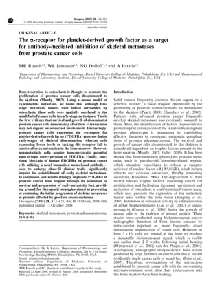
3 weeks2 weeks 4 weeks1 week
Figure 1 Progression of skeletal metastases from prostate cancer cells and spatial relationship with osteoclasts. (a) Enhanced green
fluorescent protein-expressing human PC3-ML cells, inoculated in the left cardiac ventricle of SCID mice, locate to the upper tibia
(shown) and lower femur in X80% of animals and progressively increase in size, measured by the calibrated digital analysis of
fluorescence microscopy images (1 week: 1.9±0.5 Â 103
mm2
; 2 weeks: 35±6 Â 103
mm2
; 3 weeks: 264±60 Â 103
mm2
) or by a manual
caliper (4 weeks: 14±4 mm3
). By the analysis of serial cryosections, the largest cross-section for each metastasis was identified, its
relative length and width measured and the total area calculated using the ellipse formula: l  w  3.14. Macroscopic metastatic tumors
were measured by a caliper and their volume calculated by assimilating them to an ellipsoid using the formula: l  w2
 0.52
(Vantyghem et al., 2005). Bone tissue samples were fixed, decalcified and frozen. Serial cryosections were examined using a fluorescence
stereomicroscope. Between 5 and 8 mice were used for the 1–3 week time points and 17 mice for the 4-week time points. The presence of
active osteoclasts in the bone marrow regions colonized by cancer cells was histologically established by TRAcP staining. (b)
Metastases with cross-section area larger than 28 Â 103
mm2
—indicated by the green fluorescent signal—were surrounded by an evident
layer of active osteoclasts, as shown in the magnified panel; (c) smaller metastases were instead spatially unrelated to osteoclasts, which
appear sparsely distributed (black arrows). Original magnifications:  200 for (a) and  400 for (b and c). Measurement bar is 100 mm.
Table 1 PC3-ML, PC3-N and DU-145 prostate cancer cells detected
in the craniofacial region or femora and tibiae of mice inoculated
24 or 72 h earlier
24 h 72 h
Mandible Legs Mandible Legs
PC3-ML 13±4 5±2 29±3 6±1
PC3-N 12±4 5±1 10±3 9±2
DU-145 6±2 5±0.2 5±2 2±1
Five animals per group were used. A fluorescent stereomicroscope was
used to examine all the serial cryosections obtained from each collected
bone. The values indicate the average number of cells detected in either
mandible or femora and tibiae combined.
a-PDGFR and early metastases from prostate adenocarcinoma
MR Russell et al
414
Oncogene
4. PC3-ML cells, the next experiments aimed to determine
their bone-metastatic potential. Thus, SCID mice
were inoculated with either PC3-N (wt) or
PC3-N(PDGFRa) cells, killed 3 weeks later and
examined for fluorescent metastases in tibiae and
femora. In contrast to PC3-N (wt) cells, which produced
small metastases in only 2 of the 10 inoculated mice, the
PC3-N(PDGFRa) cell induced evident bone tumors in 5
out of 7 mice (Figure 3g). Notably, PC3-N(PDGFRa)
cells produced bone tumors comparable in size to those
induced by PC3-ML cells after a similar 3-week interval
(Figures 1 and 3h). Taken together, these results
strongly support a determinant role for PDGFRa in
the progression of prostate skeletal metastases in the
skeleton. This effect would likely involve the down-
stream stimulation of the PI3K/Akt pathway, which
plays a central role in cellular survival and could
support the trophism and proliferation of cancer cells
(Carnero et al., 2008) before other tissue factors—such
as those released from the bone matrix by osteoclasts
at later stages of metastatic progression—become
accessible. Notably, we have previously shown that
human bone marrow can activate PDGFRa in a ligand-
independent manner (Dolloff et al., 2007), termed
transactivation, suggesting that the expression of this
receptor might be determinant for the survival of cancer
cells even in the absence of relevant levels of PDGF
ligands in the marrow micro-environment. On the basis
of this model, targeting PDGFRa could therefore
impair the bone-metastatic potential of prostate cancer
cells. To examine this possibility, we used the humanized
monoclonal antibody IMC-3G3, which will soon begin
phase-II trials. It has been previously shown (Loizos
et al., 2005)—and further confirmed by our data
(Supplementary Figure 2)—that IMC-3G3 is specific
for human PDGFRa and does not affect the mouse
form of the receptor. After cell inoculation, mice were
randomly assigned to control (saline treated) or
Figure 2 Early arrival of prostate cancer cells to the skeleton and extravasation in the surrounding stroma. (a and b) Fluorescent cells
were detected in the femur and tibia of SCID mice at 24 or 72 h following inoculation in the left cardiac ventricle. Cancer cells (arrows)
consistently located at the metaphysis and in the vicinity of the growth plate (yellow dashed line); the labeling of bone marrow
sinusoids by a fluorescent lectin was used to establish the fraction of cancer cells that were (c) still in the vascular lumen as compared to
cells (d) in the process of extravasating or (e) already migrated into the marrow’s stroma. The white dotted line indicates the sectioned
limits of three different bone marrow sinusoids. The ratios of cancer cells outside or inside the vasculature—24 h after inoculation—
were 19 to 1 for PC3-ML cells (five positive sections from three mice), 12 to 0 for PC3-N cells (six positive sections from four mice) and
13 to 3 for DU-145 cells (12 positive sections from four mice).
a-PDGFR and early metastases from prostate adenocarcinoma
MR Russell et al
415
Oncogene
5. TM
BM
MET
PC3-ML PC3-N
(PDGFRα)
0 15 30 60 0 15 30 60Bone Marrow
Akt
BM - +
PDGFRα
P -PDGFRα
P -Akt
Akt
P -Akt
0
5
10
15
Mice with bone metastases
Mice inoculated
PC3-N (wt)
PC3-N
(PDGFRα)
Numberof
Animals
PC3-N PC3-N
(PDGFRα)
PDGF
0 5 15 30 60 0 5 15 30 60
PDGF
0
100
200
300
400 PC3-N
(PDGFRα)
PC3-ML
µm2
x103
Figure 3 Human tissue arrays staining for platelet-derived growth factor receptor-a (PDGFRa) and induction of ectopic expression
of PDGFRa in prostate cancer cells. (a) Dishomogeneous detection of PDGFRa in prostate adenocarcinoma, emphasized by positive
(black arrow) and negative (white arrow) staining of two different gland acini; (b) homogeneous detection of PDGFRa. The expression
of this receptor on the surface of prostate epithelial cells is confirmed by the peripheral staining shown in the magnified panel; (c)
skeletal metastases from prostate adenocarcinoma also stain positive for PDGFRa, whereas bone marrow (BM) stains negative for the
receptor. Staining intensity was scored based on a scale in which negative specimens were graded (0), tissue prevalently negative for
PDGFRa showing some areas of positive staining were scored as (0–1) and tissue showing uniform staining for PDGFRa were scored
as either (1–2) or (2–3) based on the intensity of the signal observed. Of the 105 cores of malignant human prostate tissue examined, 37
were scored as (0), 40 as (0–1), 22 as (1–2) and 6 as (2–3); (d) the overexpression of PDGFRa in PC3-N cells causes a dramatic increase
of Akt phosphorylation in response to PDGF-AA; (e) PC3-N(PDGFRa) and PC3-ML cells—exposed to human bone marrow after
being serum-starved for 4 h—phosphorylate Akt to a comparable degree in a time-dependent manner; (f) phosphorylation of PDGFRa
in PC3-N(PDGFRa) cells exposed for 30 min to human bone marrow; (g) dramatic increase in the number of bone metastases
observed after 3 weeks in tibiae and femora of mice inoculated with PC3-N(PDGFRa) cells (5 of 7 mice) as compared to PC3-N(wild-
type) cells (2 of 10 mice); (h) comparable size of skeletal tumors induced by PC3-N(PDGFRa) and PC3-ML cells 3 weeks after
intracardiac inoculation in mice (150±70 Â 103
and 263±60 Â 103
mm2
, respectively (t-test; P ¼ 0.27).
a-PDGFR and early metastases from prostate adenocarcinoma
MR Russell et al
416
Oncogene
6. IMC-3G3-treated groups and killed 4 weeks later. The
analysis of femora and tibiae showed 72% reduction in
the number of macroscopic metastases observed in mice
treated with IMC-3G3 (0.23±0.1 tumor per animal)
as compared to control groups (0.82±0.2 tumor per
animal). Most importantly, a much larger fraction of
IMC-3G3-treated mice were free of metastases in either
femur or tibia as compared to control animals (10 of 13
for IMC-3G3 versus 5 of 17 for controls) (Figure 5a). To
ascertain whether IMC-3G3 exerts its effect in early
stages of metastatic progression, subsequent experi-
ments were conducted with inoculated animals ran-
domly assigned to control or IMC-3G3 groups and
killed after only 2 weeks. Similarly to what was observed
after 4 weeks of IMC-3G3 treatment, the blockade of
PDGFRa reduced the number of skeletal micro-
metastases in femora and tibiae by 70% (5.2±1.2 per
animal and 1.6±0.7 per animal for saline and IMC-
3G3, respectively) (Figure 5b). The larger number of
cancer foci detected at 2 weeks compared to that of
macroscopic metastases observed at 4 weeks—in both
control and IMC-3G3 treated mice—is most likely the
result of multiple foci coalescing into a single macro-
metastasis. The few metastases that progressed despite
IMC-3G3 treatment appeared to elude an early effect, as
they did not differ in size when compared to lesions
observed in saline-treated animals (354±60 Â 102
versus
323±119 Â 102
mm2
, respectively). Taken together, these
results indicate that the blockade of PDGFRa
dramatically inhibits the occurrence of skeletal metas-
tasis and its effect is predominantly exerted during the
very initial stages of bone colonization by prostate
cancer cells.
As mentioned above, prostate cancer cells locate to
the teeth of inoculated mice before invading the bone
tissue of the mandible (Figure 4a). Interestingly,
IMC-3G3 did not affect the number or size of metastases
in the craniofacial region after either 2 or 4 weeks of
treatment (Figures 4b and c). Differences between bone
marrow and dental pulp, such as the presence of unique
trophic factors sustaining the continuous dental eruption
in rodents, may explain a more prevailing role of
PDGFRa in the survival of prostate cancer cells in long
bones, which are by far more common sites of metastasis
than the craniofacial region in prostate adenocarcinoma
patients (Schneider et al., 2005).
INCISORS MOLARS
0.00
0.25
0.50
0.75
Control
IMC-3G3
Skeletal
metastases/mouse
0
1
2
Control
IMC-3G3
Skeletal
metastases/mouse
Figure 4 Arrival of prostate cancer cells to the mandible and effect of the immuno-mediated targeting of platelet-derived growth
factor receptor-a (PDGFRa) in a mouse model. Prostate cancer cells show initial homing to the teeth before gaining access to the
craniofacial region. (a) Transverse section of the mandible of a mouse inoculated in the left cardiac ventricle with fluorescent PC3-ML
prostate cancer cells. The magnified panel shows a single cancer cell located in the dental pulp of a molar tooth. The administration of
the humanized monoclonal antibody IMC-3G3 against PDGFRa did not affect the number of metastatic tumors in the mandible at (b)
2 or (c) 4 weeks following cancer cell inoculation. Original magnification: Â 70. Measurement bar is 600 mm.
a-PDGFR and early metastases from prostate adenocarcinoma
MR Russell et al
417
Oncogene
7. In summary, our study differentiates the role of
osteoclasts in early- and late-stage metastases and
provides the first compelling evidence for a central role
of PDGFRa in defining a bone-metastatic prostate
cancer phenotype. The translation of these observations
to the clinic is reflected by the ability of the humanized
monoclonal antibody IMC-3G3 to significantly
reduce—and in most cases impede—the establishment
and progression of skeletal micro-metastases in our
animal model.
Recent clinical trials using imatinib mesylate (STI571,
Gleevec), a small-molecule inhibitor of PDGFRs—
either alone (Mathew et al., 2004; Rao et al., 2005; Lin
et al., 2006; Bajaj et al., 2007) or in combination with
docetaxel (Mathew et al., 2007)—have been disappoint-
ing, showing significant adverse toxic effects combined
with lack of efficacy in counteracting bone metastatic
progression and/or improving overall survival. How-
ever, it should be considered that, although in the adult
organism both PDGFRa and PDGFRb cooperate in
modulating largely overlapping physiological process—
including angiogenesis, wound healing and tissue
homeostasis (Heldin and Westermark, 1999; Betsholtz,
2004)—PDGFRb plays an overall predominant role
(Andrae, Gallini and Betsholtz, 2008). Also, experi-
ments in mice showed that the intracellular domain of
PDGFRb could fully substitute for the PDGFRa,
whereas replacement of PDGFRb cytoplasmic domain
0
5
10
15
20
Mice with tibia/femur metastases
Mice inoculated
CONTROL IMC-3G3
Numberof
Animals
0.00
0.25
0.50
0.75
1.00
1.25
Control
IMC-3G3
∗
Skeletal
metastases/mouse
0.0
2.5
5.0
7.5
Control
IMC-3G3
∗
Skeletal
metastases/mouse
Figure 5 Immuno-mediated targeting of platelet-derived growth factor receptor-a (PDGFRa) inhibits skeletal metastases at femora
and tibiae in a mouse model. (a) Targeting PDGFRa dramatically reduces the number of skeletal macro-metastases in the leg by 76%,
as shown by IMC-3G3 administered to SCID mice inoculated in the left cardiac ventricle with PC3-ML prostate cancer cells and killed
4 weeks later (t-test; P ¼ 0.015). The inset shows the number of control and IMC-3G3-treated mice inoculated with PC3-ML cells as
well as the significantly higher number of animals free from femur and tibia macro-metastases observed in the IMC-3G3-treated group
(5 of 17 for control group and 10 of 13 for IMC-3G3-treated group). The correct execution of cell inoculation in mice free of metastases
in femur and tibia was confirmed by the presence of metastases in the craniofacial region and/or adrenal glands; (b) administration of
IMC-3G3 reduced femur and tibia micro-metastases by 70% as compared to control groups in mice killed 2 weeks following cancer cell
inoculation. (t-test; P ¼ 0.038).
a-PDGFR and early metastases from prostate adenocarcinoma
MR Russell et al
418
Oncogene
8. with that of the a-receptor caused abnormalities in
vascular smooth muscle cell development and function,
among others (Yarden et al., 1986). Thus, the indis-
criminate blockade of both PDGFRa and PDGFRb by
imatinib might be responsible for the systemic adverse
effects observed in clinical trails. On the other hand, the
selective inactivation of PDGFRa—using a monoclonal
antibody rather than a broad-range inhibitor—could
limit the survival of malignant cells that depend
on this receptor while causing limited toxicity, due to
the largely duplicate physiological roles exerted by
PDGFRb.
Furthermore, the preclinical animal studies conducted
to investigate the effects of imatinib were based on bone
tumors mostly produced by implanting large numbers of
prostate cancer cells directly in the tibia. Although
significantly shortening the time required for each
experiment, this approach inherently bypasses the initial
stages of establishment and progression while focusing
on macroscopic bone lesions with characteristics of late
metastases. Thus, the different histopathological fea-
tures of intra-osseous inoculation of cancer cells in the
animal as compared to human skeletal metastases—
combined with the late timing of therapeutic interven-
tion—might also explain the disappointing effects of
imatinib in clinical trials.
Our results indicate that the inhibition of PDGFRa is
very effective during the initial stages of metastatic
progression. Only 15% of men currently present with
macroscopic metastases when first diagnosed with
primitive prostate cancer (Ross et al., 2006). Thus, one
can envision a prophylactic strategy targeting PDGFRa
in prostate cancer patients positive for this receptor that
are metastasis-free at diagnosis or after initial therapy as
well as patients with advanced disease harboring occult
metastases.
Materials and methods
Cell lines and cell culture
All cell lines were cultured at 37 1C and 5% CO2 in Dulbecco’s
modified Eagle’s medium supplemented with 10% fetal
bovine serum (Hyclone, Logan, UT, USA) and 0.1%
gentamicin (Invitrogen, Carlsbad, CA, USA). Cells were
engineered to stably express enhanced green fluorescent
protein by transduction with a proprietary lentiviral vector
(America Pharma Source, Bethesda, MD, USA) in complete
culture medium for 24 h using a multiplicity of infection of 50
i.f.u. per cell.
Cell inoculation
Five-week-old male immunocompromised SCID mice (CB17-
SCRF, Taconic, Germantown, NY, USA) were housed in a
germ-free barrier. At 6 weeks of age, animals were anesthetized
with ketamine (80 mg/kg) and xylazine (10 mg/kg) and
inoculated in the left cardiac ventricle with cancer cells
(5 Â 104
in 100 ml of serum-free Dulbecco’s modified Eagle’s
medium/F12 using a 30-gauge needle). All experiments were
conducted in accordance with NIH guidelines for the humane
use of animals. All protocols involving the use of animals were
approved by the Drexel University College of Medicine
Committee for the Use and Care of Animals.
Ectopic PDGFRa expression
Human PDGFRa (NM_006206) was expressed in the same
lentiviral vector (America Pharma Source) used to obtain
stably fluorescent cells. The PDGFRa cDNA was originally
expressed in a PCDNA3.1 vector (Invitrogen) and had been
cloned in using a newly introduced 50
-BamH1 site and
an existing 30
BamH1 site. To clone it into the lentiviral
vector, we introduced a 50
BamH1 and a 30
Xho1 site by PCR
using the following primers: BamH1: 50
-GGATCCCAGAG
CTATGGGGA-30
, XhoI: 50
-CTCGAGGTGGCCCCAGAAG
T-30
. Cells were transduced using a multiplicity of infection of
50 i.f.u. per cell. As the lentiviral vector contains an enhanced
green fluorescent protein-IRES site, the cells expressing
PDGFRa were isolated and purified by flow cytometry and
sorting based on their fluorescence intensity.
Human bone marrow acquisition and processing
The bone marrow samples from normal male donors (ages
18–45) were supplied by Lonza Biosciences (Poietics Donor
Program, Walkersville, MD, USA). Samples were shipped and
maintained at 4 1C throughout processing and were subse-
quently stored at À80 1C. Briefly, samples were centrifuged at
1500 r.p.m. to separate the soluble and cellular phases.
Supernatant was removed and filtered using 0.8 and 0.22 mm
filters in succession.
In vitro experimental protocol
Cells were starved from serum for 4 h before being exposed to
bone marrow or PDGF-AA (20 ng/ml). Fifty micro-liters of
processed bone marrow were administered to cells in 1 ml of
experimental medium for a final 1:20 dilution.
SDS–polyacrylamide gel electrophoresis and western blotting
Cell lysates were obtained and SDS–polyacrylamide gel electro-
phoresis and western blot analysis were performed as previously
described (Shulby et al., 2004), with few modifications. Mem-
branes were blotted with antibodies targeting phospho-Akt
(Ser-473, Cell Signaling Technology, Danvers, MA, USA),
PDGFRa (R&D Systems, Minneapolis, MN, USA) and total
Akt (Cell Signaling, Danvers, MA). Primary antibody binding
was detected using an horseradish peroxidase-conjugated second-
ary antibody (Pierce, Rockford, IL, USA). Chemiluminescent
signals were obtained using SuperSignal West Femto reagents
(Pierce) and detected with the Fluorochem 8900 imaging system
and relative software (Alpha Innotech, San Leandro, CA, USA).
Densitometry analysis was performed using the UN-SCAN IT
software (Silk Scientific). Samples were run on the same gels for
effective comparison of intensity levels. Each experiment was
repeated at least three times and provided similar results.
Detection of PDGFRa phosphorylation
Cells were washed twice with ice-cold phosphate-buffered saline
and lysed with immunoprecipitation buffer (50 mM Tris, 150 mM
NaCl, 10 mM NaF, 10 mM sodium pyrophosphate, 1% NP40)
supplemented with protease and phosphatase inhibitors (Pro-
tease Inhibitor Cocktail Set III, Phosphatase Inhibitor Cocktail
Set II, Calbiochem, Gibbstown, NJ, USA). Cell lysates (750 mg)
were incubated with agarose-conjugated anti-PDGFRa primary
antibody (Santa Cruz, Santa Cruz, CA, USA) overnight at 4 1C.
Immunoprecipitation was carried out according to the manu-
facturer’s protocol, with immunoprecipitated protein run on a
4–12% polyacrylamide gel (Lonza Biosciences, Walkersville,
MD, USA). Western blotting was carried out as previously
described using an antibody directed against phosphotyrosine
(Cell Signaling). Even loading was confirmed by stripping the
membrane and blotting for total PDGFRa.
a-PDGFR and early metastases from prostate adenocarcinoma
MR Russell et al
419
Oncogene
9. Tissue preparation
Bones and soft-tissue organs were collected and fixed in 4%
formaldehyde solution. Bones were decalcified in 0.5 M EDTA
for 7 days followed by incubation in 30% sucrose. Tissues were
frozen in O.C.T. medium (Electron Microscopy Sciences,
Hatfield, MA, USA) by placement over dry-ice chilled 2-
methylbutane (Fisher, Pittsburgh, PA, USA). Serial sections of
80 mm in thickness were obtained using a Microm HM550
cryostat (Mikron, San Marcos, CA, USA).
Histology
TRAcP staining Slides were incubated at 37 1C for 5 min in a
solution containing naphthol AS-BI phosphate and ethylene
glycol monoethyl ether (Sigma, St Louis, MO, USA). Slides
were then transferred to a solution containing sodium nitrite
and pararosaniline chloride (Sigma) for approximately 3 min.
Evaluation of cell extravasation Tissues were fixed in ice-cold
acetone and then treated with 2 mg/ml biotinylated lycopersi-
con esculentum lectin (Vector Laboratories, Burlingame, CA,
USA) for 1 h at room temperature to label the endothelial cells
of the bone marrow sinusoids. After washing in Tris-buffered
saline, tissues were incubated at room temperature for 1 h in
1.8 mg/ml of CY3-conjugated streptavidin (Jackson Immuno-
research, West Grove, PA, USA). An average of 20 cryosec-
tions were obtained from the mandible and legs of animals
killed at either 24 or 72 h post-inoculation and examined for
fluorescent cancer cells. Digital images were captured, pro-
cessed for the removal of background fluorescence, and
the spatial relationships of green-fluorescent cancer cells with
the red-fluorescent bone marrow sinusoids were determined.
The total numbers of cells located within the red-fluorescent
rim of the sinusoids (intra-vascular), overlapping with it (in the
process of extravasating) or located outside the red-fluorescent
rim (fully extravasated) were counted.
PDGFRa immunostaining Human tissue microarrays PR951,
PR208, PR803 and GL802 were obtained from US Biomax
Inc. (Rockville, MD, USA) Tissue samples were deparaffinized
with Histosolve (Dako Cytomation, Carpinteria, CA, USA)
and re-hydrated with decreasing concentrations of ethanol.
Endogenous peroxidase was quenched using methanol and
hydrogen peroxide. Antigen retrieval was performed using
Dako Cytomation Target Retrieval Solution in a 95 1C water
bath for 30 min. Arrays were blocked with 10% normal
donkey serum in Tris-buffered saline for 60 min, then
incubated in horseradish peroxidase-conjugated IMC-3G3
(10 mg/ml) in blocker overnight at 4 1C. Antibody binding
was visualized with a chromogenic DAB kit (Vector,
Burlingame, CA, USA). Slides were dehydrated with increas-
ing concentrations of ethanol, cleared in Histosolve and
mounted using Permount. The IMC-3G3 antibody was
conjugated to horseradish peroxidase using EZ-Link Plus
Activated Peroxidase kit (Pierce), according to the manufac-
turer’s instructions. The specificity of the IMC-3G3 antibody
for the PDGFRa was validated using human tissue arrays
containing tissue cores of 35 cases of glioblastoma (highly
expressing the receptor; Thorarinsdottir et al., 2008) and 5
cases of normal brain tissue (weakly positive for the receptor).
Microscopy and measurements
Bright-field and fluorescence images of single cells, small foci
and macroscopic metastases were acquired using an SZX12
Olympus stereomicroscope coupled to an Olympus DT70
CCD color camera. Digital images were analysed with ImageJ
software (http://rsb.info.nih.gov/ij/) and calibrated by obtain-
ing a pixel to millimeter ratio.
IMC-3G3 administration
Animals received a loading dose of 214 mg/kg immediately
after inoculation with PC3-ML cells, followed by a main-
tenance dose of 60 mg/kg every 72 h thereafter. Doses and
administration schedule were selected based on tumor-growth
inhibitory studies and pharmacokinetic analyses conducted by
ImClone Systems Inc. Control animals received comparable
volumes of sterile phosphate-buffered saline solution.
Statistics
We analysed number and size of skeletal metastases in control
and IMC-3G3-treated groups using a two-tailed Student’s t-test.
A value of Pp0.05 was considered statistically significant.
Acknowledgements
The cDNA for human PDGFRa was a kind gift of Dr Carl-
Henrik Heldin (Ludwig Institute for Cancer Research,
Uppsala, Sweden). We thank Dr Olimpia Meucci (Department
of Pharmacology and Physiology) for critically reading the
manuscript and helpful discussion, Dr Mark Stearns (Depart-
ment of Pathology and Laboratory Medicine) for helpful
discussion, Dr Gregg Johannes (Department of Pathology and
Laboratory Medicine) for help with the PDGFRa-expressing
vector, Mr Michael Amatangelo for his contribution to the
immuno-detection of PDGFRa in human tissues and Dr Nick
Loizos (ImClone Systems Inc., New York, NY, USA) for
kindly providing the IMC-3G3 antibody. This study was
supported in part by the NIH Grant GM067892 to AF.
References
Andrae J, Gallini R, Betsholtz C. (2008). Role of platelet-derived
growth factors in physiology and medicine. Genes Dev 22:
1276–1312.
Bajaj GK, Zhang Z, Garrett-Mayer E, Drew R, Sinibaldi V, Pili R et al.
(2007). Phase II study of imatinib mesylate in patients with prostate
cancer with evidence of biochemical relapse after definitive radical
retropubic prostatectomy or radiotherapy. Urology 69: 526–531.
Betsholtz C. (2004). Insight into the physiological functions of PDGF
through genetic studies in mice. Cytokine Growth Factor Rev 15:
215–228.
Canon JR, Roudier M, Bryant R, Morony S, Stolina M, Kostenuik PJ
et al. (2008). Inhibition of RANKL blocks skeletal tumor
progression and improves survival in a mouse model of breast
cancer bone metastasis. Clin Exp Metastasis 25: 119–129.
Carnero A, Blanco-Aparicio C, Renner O, Link W, Leal JF. (2008).
The PTEN/PI3K/AKT signalling pathway in cancer, therapeutic
implications. Curr Cancer Drug Targets 8: 187–198.
Chambers AF, Groom AC, MacDonald IC. (2002). Dissemination
and growth of cancer cells in metastatic sites. Nat Rev Cancer 2:
563–572.
Dolloff NG, Russell MR, Loizos N, Fatatis A. (2007). Human bone
marrow activates the Akt pathway in metastatic prostate cells
through transactivation of the alpha-platelet-derived growth factor
receptor. Cancer Res 67: 555–562.
Dolloff NG, Shulby SS, Nelson AV, Stearns ME, Johannes GJ,
Thomas JD et al. (2005). Bone-metastatic potential of human
prostate cancer cells correlates with Akt/PKB activation by alpha
platelet-derived growth factor receptor. Oncogene 24: 6848–6854.
a-PDGFR and early metastases from prostate adenocarcinoma
MR Russell et al
420
Oncogene
10. Fidler IJ. (2003). The pathogenesis of cancer metastasis: the ‘seed and
soil’ hypothesis revisited. Nat Rev Cancer 3: 453–458.
Fritz V, Louis-Plence P, Apparailly F, Noe¨ l D, Voide R, Pillon A et al.
(2007). Micro-CT combined with bioluminescence imaging: a
dynamic approach to detect early tumor-bone interaction in a
tumor osteolysis murine model. Bone 40: 1032–1040.
Gupta GP, Ngyuen DX, Chiang AC, Bos PD, Kim JY, Nadal C et al.
(2007). Mediators of vascular remodeling co-opted for sequential
steps in lung metastasis. Nature 446: 765–770.
Heldin C-H, Westermark B. (1999). Mechanism of action and in vivo
role of platelet-derived growth factor. Physiol Rev 79: 1283–1316.
Jamieson WL, Shimizu S, D’Ambrosio JA, Meucci O, Fatatis A.
(2008). CX3CR1 is expressed by prostate epithelial cells and
androgens regulate the levels of CX3CL1/fractalkine in the bone
marrow: potential role in prostate cancer bone tropism. Cancer Res
68: 1715–1722.
Kingsley LA, Fournier PG, Chirgwin JM, Guise TA. (2007).
Molecular biology of bone metastasis. Mol Cancer Ther 10:
2609–2617.
Lee YP, Schwarz EM, Davies M, Jo M, Gates J, Shang X et al. (2002).
Use of zoledronate to treat osteoblastic versus osteolytic lesions in a
severe-combined-immunodeficient mouse model. Cancer Res 62:
5564–5570.
Lin AM, Rini BI, Weinberg V, Fong K, Ryan CJ, Rosenberg JE et al.
(2006). A phase II trial of imatinib mesylate in patients with
biochemical relapse of prostate cancer after definitive local therapy.
BJU Int 98: 763–769.
Loizos N, Xu Y, Huber J, Liu M, Lu D, Finnerty B et al. (2005).
Targeting the platelet-derived growth factor receptor a with a
neutralizing human monoclonal antibody that inhibits the growth of
tumor xenografts: implications as a potential therapeutic target.
Mol Cancer Ther 4: 369–379.
Mathew P, Thall PF, Bucana CD, Oh WK, Morris MJ, Jones DM
et al. (2007). Platelet-derived growth factor receptor inhibition and
chemotherapy for castration-resistant prostate cancer with bone
metastases. Clin Cancer Res 13: 5816–5824.
Mathew P, Thall PF, Jones D, Perez C, Bucana C, Troncoso P et al.
(2004). Platelet-derived growth factor receptor inhibitor imatinib
mesylate and docetaxel: a modular phase I trial in androgen-
independent prostate cancer. J Clin Oncol 22: 3323–3329.
Mundy GR. (2002). Metastasis to bone: causes, consequences and
therapeutic opportunities. Nat Rev Cancer 2: 584–593.
Nemeth JA, Harb JF, Barroso Jr U, He Z, Grignon DJ, Cher ML.
(1999). Severe combined immunodeficient-hu model of human
prostate cancer metastasis to human bone. Cancer Res 59:
1987–1993.
Paget S. (1889). The distribution of secondary growths in cancer of the
breast. Lancet 1: 571–573.
Rao K, Goodin S, Levitt MJ, Dave N, Shih WJ, Lin Y et al. (2005).
A phase II trial of imatinib mesylate in patients with prostate
specific antigen progression after local therapy for prostate cancer.
Prostate 62: 115–122.
Roodman GD. (2004). Mechanisms of bone metastasis. N Engl J Med
350: 1655–1664.
Ross RW, Oh WK, Hurwitz M, D’Amico AV, Richie JP, Kantoff PW.
(2006). Neoplasm of the prostate. In: Holland-Frei (ed). Cancer
Medicine. BC Decker Inc. publisher, pp 1431–1461.
Schneider A, Kalikin LM, Mattos AC, Keller ET, Allen MJ, Pienta KJ
et al. (2005). Bone turnover mediates preferential localization of
prostate cancer in the skeleton. Endocrinology 146: 1727–1736.
Shulby SA, Dolloff NG, Stearns ME, Meucci O, Fatatis A. (2004).
CX3CR1-fractalkine expression regulates cellular mechanisms in-
volved in adhesion, migration, and survival of human prostate
cancer cells. Cancer Res 64: 4693–4698.
Thorarinsdottir HK, Santi M, McCarter R, Rushing EJ, Cornelison R,
Jales A et al. (2008). Protein expression of platelet-derived growth
factor receptor correlates with malignant histology and PTEN with
survival in childhood gliomas. Clin Cancer Res 14: 3386–3394.
van der Pluijm G, Que I, Sijmons B, Buijs JT, Lowik CW, Wetterwald
A et al. (2005). Interference with the microenvironmental support
impairs the de novo formation of bone metastases in vivo. Cancer Res
65: 7682–7690.
Vantyghem SA, Allan AL, Postenka CO, Al-Katib W, Keeney M,
Tuck AB et al. (2005). A new model for lymphatic metastasis:
development of a variant of the MDA-MB-468 human breast cancer
cell line that aggressively metastasizes to lymph nodes. Clin Exp
Metastasis 22: 351–361.
Wang M, Stearns ME. (1991). Isolation and characterization of PC-3
human-prostatic tumor sublines which preferentially metastasize to
select organs in SCID mice. Differentiation 48: 115–125.
Wetterwald A, van der Pluijm G, Que I, Sijmons B, Buijs J, Karperien
M et al. (2002). Optical imaging of cancer metastasis to bone
marrow: a mouse model of minimal residual disease. Am J Pathol
160: 1143–1153.
Yarden Y, Escobedo JA, Kuang WJ, Yang-Feng TL, Daniel TO,
Tremble PM et al. (1986). Structure of the receptor for platelet-
derived growth factor helps define a family of closely related growth
factor receptors. Nature 323: 226–232.
Supplementary Information accompanies the paper on the Oncogene website (http://www.nature.com/onc)
a-PDGFR and early metastases from prostate adenocarcinoma
MR Russell et al
421
Oncogene