1. Inhibition of constitutive NF-jB activity by IjBaM suppresses
tumorigenesis
Shuichi Fujioka1
, Guido M Sclabas1
, Christian Schmidt1
, Jiangong Niu2
, Wayne A Frederick1
,
Qiang G Dong1
, James L Abbruzzese3
, Douglas B Evans1
, Cheryl Baker4
and Paul J Chiao*,1,2,5
1
Department of Surgical Oncology, The University of Texas, Houston, TX 77030, USA; 2
The Graduate School of Biomedical
Sciences, The University of Texas, Houston, TX 77030, USA; 3
Department of Gastrointestinal Medical Oncology, The University of
Texas, Houston, TX 77030, USA; 4
Department of Cancer Biology, The University of Texas, Houston, TX 77030, USA; 5
Department
of Molecular and Cellular Oncology, The University of Texas MD Anderson Cancer Center, The University of Texas, Houston, TX
77030, USA
We have demonstrated that nuclear factor-jB (NF-jB) is
constitutively activated in human pancreatic adenocarci-
noma and human pancreatic cancer cell lines but not in
normal pancreatic tissues or in immortalized, nontumori-
genic pancreatic epithelial cells, suggesting that NF-jB
plays a critical role in the development of pancreatic
adenocarcinoma. To elucidate the role of constitutive NF-
jB activity in human pancreatic cancer cells, we generated
pancreatic tumor cell lines that express a phosphorylation
defective IjBa (S32, 36A) (IjBaM) that blocks NF-jB
activity. In this study, we showed that inhibiting
constitutive NF-jB activity by expressing IjBaM sup-
pressed the tumorigenicity of a nonmetastatic human
pancreatic cancer cell line, PANC-1, in an orthotopic
nude mouse model. Immunohistochemical analysis showed
that PANC-1-derived tumors expressed vascular endothe-
lial growth factor (VEGF) and induced angiogenesis.
Inhibiting NF-jB signaling by expressing IjBaM sig-
nificantly reduced expression of Bcl-xL and Bcl-2. The
cytokine-induced expression of VEGF and Interleukin-8
in PANC-1 cells is also decreased. Taken together, these
results suggest that the inhibition of NF-jB signaling can
suppress tumorigenesis of pancreatic cancer cells and that
the NF-jB signaling pathway is a potential target for
anticancer agents.
Oncogene (2003) 22, 1365–1370. doi:10.1038/sj.onc.1206323
Keywords: IkBa; NF-kB; tumorigenesis; angiogenesis;
pancreatic cancer
Nuclear factor-kB (NF-kB) is a family of pleiotropic
transcription factors that regulate the transcription of a
large number of genes that play key roles in embryonic
development, lymphoid differentiation, apoptosis, and
immune and inflammatory responses (Verma et al.,
1995; Baldwin, 1996; Gilmore et al., 1996). Several
reports suggest that the members of the NF-kB family
and their inhibitor, IkB family, are involved in the
development of cancer (Sylla and Temin, 1986; Moore
and Bose, 1988; Gilmore et al., 1996). For instance, the
genes encoding c-Rel, Bcl-3, p105 (p50), and p100 (p52)
are located at sites of recurrent genomic rearrangements
in cancer (Ohno et al., 1990; Lu et al., 1991; Neri et al.,
1991; Kitajima et al., 1992; Dejardin et al., 1995). We
first reported that RelA, the p65 subunit of NF-kB, is
constitutively activated in human pancreatic adenocar-
cinoma and human pancreatic cancer cell lines but not
in normal pancreatic tissues or in immortalized, non-
tumorigenic pancreatic epithelial cells (Wang et al.,
1999b). We previously found that NF-kB induces
overexpression of urokinase plasminogen activator and
that Bcl-xL is induced by NF-kB in pancreatic cancer cell
lines (Wang et al., 1999a; Dong et al., 2002). Other
studies showed that IkBaM-mediated inhibition of NF-
kB activity reduced angiogenesis and metastasis in an
ovarian cancer cell line and a prostate cancer cell line
and completely inhibited liver metastasis of a pancreatic
cancer cell line in mice (Huang et al., 2000, 2001;
Fujioka et al., 2002). The purpose of this study was to
determine whether the inhibition of constitutive NF-kB
activity would suppress tumorigenic phenotypes in
pancreatic cancer cells.
To determine the role of constitutive NF-kB activity
in tumorigenesis, we constructed a retroviral vector
encoding a Flag-tagged phosphorylation defective mu-
tant of IkBa (S32, 36A) (IkBaM). The nonmetastatic
human pancreatic tumor cell line PANC-1 was infected
with retrovirus encoding Flag-tagged IkBaM or a
control retrovirus and then selected for puromycin
resistance (800 ng/ml). The puromycin-resistant clones
of PANC-1 cells from each infection were pooled to
establish PANC-1/IkBaM and PANC-1/Puro cell lines.
Both cytoplasmic and nuclear extracts from pooled
puromycin-resistant PANC-1 (PANC-1/puro) and
PANC-1 cells expressing IkBaM (PANC-1/IkBaM)
were prepared for subsequent analysis. Western blot
analysis showed the expression of Flag-tagged IkBaM
(Figure 1a). Constitutive NF-kB activity was found in
Received 7 October 2002; revised 10 December 2002; accepted 11
December 2002
*Correspondence: PJ Chiao, Departments of Molecular and Cellular
Oncology and Surgical Oncology, Unit 107, The University of Texas
MD Anderson Cancer Center, 1515 Holcombe Boulevard, Houston,
TX 77030, USA; E-mail: pjchiao@notes.mdacc.tmc.edu
Oncogene (2003) 22, 1365–1370
& 2003 Nature Publishing Group All rights reserved 0950-9232/03 $25.00
www.nature.com/onc
2. the nuclear extracts from PANC-1/puro cells using
electrophoretic mobility shift assay (EMSA) (Figure 1b),
consistent with our previous report that NF-kB is
constitutively activated in human pancreatic adenocar-
cinoma cells (Wang et al., 1999b). NF-kB DNA binding
activity is completely inhibited in PANC-1/IkBaM cells
(Figure 1b). Competition and supershift assays showed
the presence of RelA and p50 in the NF-kB binding
activity in PANC-1/puro cells (Figure 1c). Expression of
antiapoptotic NF-kB downstream target genes, bcl-2
and bcl-xL, are inhibited in PANC-1/IkBaM cells
(Figure 1d, e), which is consistent with our previous
reports (Dong et al., 2002; Fujioka et al., 2002). These
results suggest that the expression of these and other
NF-kB downstream target genes is decreased through
the IkBaM-mediated inhibition of NF-kB activity. The
expression of the proapoptotic gene bcl-xS, the alter-
native spliced form of bcl-xL, does not decrease
(Figure 1f). It is possible that the half-life of bcl-xS
mRNA or protein is much longer as a number of reports
suggest that bcl-xL and bcl-xS are differentially regulated
at post-transcriptional and post-translational stages
(Chang et al., 1997; Klingler et al., 1997; Tsai et al.,
2000; Parborell et al., 2002).
To determine whether the inhibition of NF-kB
suppresses the tumorigenic phenotype of PANC-1/puro
cells, we performed an in vivo experiment using an
orthotopic nude mouse model. Six weeks after injection
of PANC-1/puro and PANC-1/IkBaM into the pan-
creas of nude mice, all the mice injected with PANC-1
and PANC-1/puro cells became sick. Pathologic exam-
ination was performed to determine the extent of tumor
formation and metastasis. As shown in Figure 2a, 100%
(10/10) of animals injected with PANC-1/puro cells
Figure 1 Effect of IkBaM expression on constitutive and TNF-a-induced NF-kB activity. (a) Flag-IkBaM/puror
and pRetro/puror
control retroviruses were generated and infections were performed as previously described (Naviaux et al., 1996). Pooled puromycin-
resistant control cells and the cells that expressed Flag-tagged IkBaM were used for subsequent analyses. Western blot analysis using
whole cell protein extracts with a rabbit antibody against IkBa (Santa Cruz Biotechnology, Inc., Santa Cruz, CA, USA) was performed
as previously described (Grau et al., 1997). (b) Electrophoretic mobility shift assay (EMSA) was performed to determine NF-kB DNA-
binding activity in PANC-1/puro and PANC-1/IkBaM cells as previously described (Chiao et al., 1994). A probe containing the Oct-1
motif was used as a control for quality and quantity of cell extracts, (c) Specificity of the NF-kB DNA-binding activity, as determined
by competition and supershift experiments in PANC-1/puro cells. Arrows indicate the migration of the induced NF-kB DNA-binding
complexes. Migration of the free probe is not shown. SS, supershifted band; NS, nonspecific band. Western blot analysis of the
expression of Bcl-xL (d), Bcl-2 (e), and Bcl-xs (f) in PANC-1/puro and PANC-1/IkBaM cells. Equal loading of protein extracts was
determined by probing the same membrane filter with anti-b-actin antibody
Suppression of tumorigenesis by inhibition of NF-jB activation
S Fujioka et al
1366
Oncogene
3. presented pancreatic tumors with no metastasis in the
liver or other organs, whereas none of 10 mice injected
with PANC-1/IkBaM cells showed pancreatic tumor
formation, implying that constitutive RelA/NF-kB
activity induces tumorigenicity. In addition, 100% (5/
5) mice injected with PANC-1 cells developed pancreatic
tumor as those observed in the mice injected with
PANC-1/puro (data not shown).
It is now well established that angiogenesis is essential
for the growth of both primary tumors and metastatic
lesions both of which require an adequate blood supply
(Folkman et al., 1989; Folkman, 1990; Folkman, 1992).
Tumor angiogenesis is, in part, regulated by angiogenic
factors that are produced and secreted by tumor cells.
Vascular endothelial growth factor (VEGF) and inter-
leukin-8 (IL-8) are two positive regulators of endothelial
cells that have been identified (Yoshimura et al., 1987;
Liotta et al., 1991; Ferrara, 1995). VEGF binds to the
specific transmembrane tyrosine kinase receptors KDR/
flk-1 and flt-1, which are selectively expressed on
vascular endothelial cells, thus stimulating the growth
of endothelial cells (Millauer et al., 1993). Subsequent
studies have revealed that IL-8 triggers angiogenesis in
vivo via mechanisms that are mediated by direct
stimulation of endothelial cell growth or by indirect
leukocyte-dependent effects (Koch et al., 1992; Hu et al.,
1993). IL-8 can induce proliferation and migration of
human umbilical vein endothelial cells and can stimulate
vascularization in a rat cornea assay (Strieter et al.,
1992). Recently, it was demonstrated that NF-kB
activation is obligatory for retinal angiogenesis (Yoshi-
da et al., 1999). To determine whether PANC-1/puro
cell-derived tumors express NF-kB target genes, such as
VEGF, we performed immunohistochemical staining to
analyse the expression of VEGF and the extent of
angiogenesis in the pancreatic tissues obtained from the
mice injected with PANC-1/puro cells. As shown in
Figure 3, much higher levels of VEGF expression and a
significantly higher level of intratumoral microvessel
formation were found in the tumors derived from
PANC-1 cells than in the adjacent pancreatic tissues.
Because cytokines including TNF-a have been suggested
to play an important role in tumor angiogenesis and
cancer progression (Leibovich et al., 1987; Passaniti
et al., 1992; Leek et al., 1996; Shono et al., 1996;
Yancopoulos et al., 2000; Xu et al., 2001), we
investigated the role of NF-kB activity in regulating
TNF-a-mediated VEGF and IL-8 expression. As shown
Figure 2 IkBaM-mediated suppression of tumorigenicity in an
orthotopic nude mouse model. (a) One million viable PANC-1 and
PANC-1/IkBaM cells suspended in 50 ml phosphate-buffered saline
(PBS) were injected into the pancreatic parenchyma of female
athymic BALB/c nude mice at 8 weeks of age (Charles River
Laboratories, Inc. Wilmington, MA, USA). Six weeks after
injection, all mice were killed, and the degrees of tumorigenesis
and metastasis were investigated. The incidence of tumorigenesis,
ascitic formation, jaundice, liver, or peritoneal metastasis is shown
in a graph. (b) Summary of the weights of tumors resected from
mice injected with PANC-1 cells
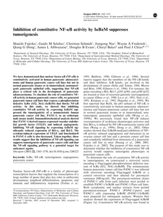
Figure 3 Immunohistochemical staining of pancreatic tumors
derived from orthotopically injected pancreatic cancer cells. (a)
Detection of VEGF expression by a polyclonal anti-VEGF
antibody (Santa Cruz Biotechnology, Inc., Santa Cruz, CA,
USA). (b) Detection of CD31 by a monoclonal anti-CD31
antibody (Pharmingen, San Diego, CA, USA). Paraffin-embedded
sections (for VEGF staining) and frozen sections (for CD-31
staining) were obtained from marginal regions of resected mouse
pancreas and the tumors derived from implanted PANC-1/puro
human pancreatic cancer cell lines. Immunohistochemical staining
was performed as previously described (Grau et al., 1997)
Suppression of tumorigenesis by inhibition of NF-jB activation
S Fujioka et al
1367
Oncogene
4. Figure 4 IkBaM-mediated inhibition of expression of NF-kB downstream target genes. (a) Electrophoretic mobility shift assay
(EMSA) was performed to determine NF-kB DNA-binding activity in PANC-1/puro and PANC-1/IkBaM cells stimulated with TNF-
a (10 ng/ml) for 5, 15, and 30 min, as previously described (Chiao et al., 1994). A probe containing the Oct-1 motif was used as a control
for quality and quantity of cell extracts. Expression of VEGF and IL-8 stimulated with TNF-a as indicated were analysed by Northern
blot (b) and Western blot analyses (c) as previously described (Chiao et al., 1994). Equal loading of mRNA and protein samples was
determined by reprobing the same membrane filter with a cDNA probe for glyceraldehyde-3-phosphate dehydrogenase (GAPDH) and
anti-b-actin antibody, respectively. kB (d), VEGF (e), and IL-8 (f) luciferase reporter gene activities in PANC-1/puro and PANC-1/
IkBaM cells were determined. A measure of 1 mg of control HIV-kB, 1.5 kb VEGF, or 0.27-kb IL-8 promoter reporter-gene constructs
was cotransfected into the cells with an internal control, p-TK Renilla luciferase (pRL-TK), using the lipotransfection method
(FuGENE 6, Roche, Indianapolis, IN, USA). The activities of both Firefly and Renilla luciferase were determined 48 h after
transfection using the Dual-Luciferase Reporter Assay System (Promega, Madison, WI, USA). The fold-increases in luciferase activity
were calculated relative to the Luciferase activity of an internal control. Data represent the mean 7s.e. from three different
experiments performed in triplicate
Suppression of tumorigenesis by inhibition of NF-jB activation
S Fujioka et al
1368
Oncogene
5. in Figure 4a, NF-kB DNA binding activity was induced
by stimulation with TNF-a (10 ng/ml) in PANC-1/puro,
whereas TNF-a stimulated NF-kB DNA binding
activity was inhibited in PANC-1/ IkBaM cells. A probe
containing the Oct-1 motif was used as a control for
quality and quantity of cell extracts. VEGF mRNA was
induced following stimulation with TNF-a and peaked
at 1 h in PANC-1/puro cells (Figure 4b, lanes 2
and 3). The TNF-a-induced expression of VEGF was
inhibited by IkBaM (Figure 4b, lanes 7–10). These
results were consistent with the Western blot analysis as
shown in Figure 4c. Similarly, IL-8 expression was
induced in response to TNF-a stimulation and reaches
to its peak of expression at 1 h in PANC-1/puro
cells (Figure 4b). In contrast to observed IkBaM-
mediated inhibition of VEGF expression in response
to TNF-a, IkBaM only partially inhibited TNF-a-
induced IL-8 expression (Figure 4b and c, lanes 7–10),
implying the involvement of alternative signaling
cascades that regulate IL-8 gene expression. Interest-
ingly, Figure 4c shows that induction of IL-8 protein
production by TNF-a occurs at 3 h in PANC-1/puro
cells and 6 h in PANC-1/IkBaM cells. Taken together,
the results in Figure 4 show that TNF-a induced IL-8
protein synthesis is delayed comparing with TNF-a-
induced IL-8 mRNA expression in Figure 4b, suggesting
that IL-8 expression is also regulated at translational
level. This finding is consistent with an early report that
VEGF-/VPF-induced IL-8 expression is further regu-
lated translationally even although IL-8 mRNA was
increased through activation of NF-kB to maximal level
after 1 h of VEGF/VPF treatment of the human brain
microvascular endothelial cells (Lee et al., 2002).
Altogether, these results indicate that TNF-a-mediated
expression of VEGF and IL-8 is NF-kB-dependent.
However, the induction of TNF-a-mediated IL-8
mRNA expression is only partially inhibited or delayed
by IkBaM.
To further determine the role of NF-kB activity in
regulating the expression of VEGF and IL-8, we
analysed activities of the 1.5-kb VEGF promoter and
0.27-kb IL-8 promoter luciferase reporter-gene con-
structs (Mukaida et al., 1990; Fukumura et al., 1998).
The VEGF and IL-8 promoter reporter-gene constructs,
and kB control reporter-gene plasmid were transiently
transfected into PANC-1/puro and PANC-1/IkBaM
cells. As shown in Figure 4d, IkBaM expression
inhibited constitutive NF-kB activation as determined
by kB control luciferase reporter-gene activity. IkBaM
expression significantly reduced IL-8 and VEGF pro-
moter-mediated reporter-gene transcriptional activities
in PANC-1 cells (Figure 4e and f). Taken together, these
results show that IkBaM-mediated inhibition of con-
stitutive NF-kB activity downregulated VEGF and IL-8
expression in PANC-1 cells, suggesting that NF-kB-
inducible expression of VEGF, IL-8 and other NF-kB
downstream target genes is involved in pancreatic
tumorigenesis.
In this study, we investigated the function of NF-kB
signaling on the tumorigenesis and angiogenesis of
PANC-1/puro human pancreatic cancer cell lines.
Inhibition of constitutive NF-kB activity completely
suppressed tumor formation from the nonmetastatic
human pancreatic cancer cell line PANC-1 in an
orthotopic nude mouse model. Furthermore, IkBaM
expression substantially inhibited expression of key
antiapoptotic genes, bcl-xL and bcl-2, and major
proangiogenic molecules VEGF and IL-8, suggesting
that the antiapoptotic potential of the tumor cells and
neoplastic angiogenesis may be decreased. Our results
suggest that constitutive NF-kB activity, found in 70%
of pancreatic cancers, plays an important role in
pancreatic tumorigenesis. Our study also implies that
constitutive NF-kB activity induces overexpression of its
downstream target genes such as bcl-xL, bcl-2, VEGF,
and IL-8, which may mediate its cardinal features of
locally aggressive growth and resistance to therapeuti-
cally induced apoptosis.
Several reports show that the PANC-1 cell line is
nonmetastatic in the orthotopic nude mouse model
(Mohammad et al., 2001; Sawai et al., 2001; Teraoka
et al., 2001). PANC-1 cells express wild-type Smad4 and
a functional TGF-b signaling pathway, unlike other
human pancreatic tumor cell lines (Grau et al., 1997).
Reconstitution of Smad4 expression in a Smad4-null
Hs667t human pancreatic cancer cell line reduced tumor
formation by inhibiting angiogenesis (Schwarte-Waldh-
off et al., 2000), implying that Smad4 may reduce the
expression of angiogenic factors and result in small,
slow-growing, and lower vascularized PANC-1 tumors
with undetectable metastatic potential in the pancreas of
nude mice. However, it remains unknown whether a
functional TGF-b pathway reduces the metastatic
potential of PANC-1 cells.
Human pancreatic cancer has a very poor prognosis,
even after curative resection, and is currently the fourth
leading cause of cancer death in the United States
(Jemal et al., 2002). The overall 5-year survival rate
continues to be dismal, at 1–3% (Bramhall et al., 1995).
Most patients with pancreatic cancer have locally
advanced, unresectable disease or metastasis at the time
of diagnosis (Breslin et al., 2001). Currently, chemother-
apy, radiation therapy, and surgery are largely ineffec-
tive in treating this disease (Breslin et al., 2001). Recent
studies suggest that VEGF is the best-validated target
for antiangiogenic therapies to inhibit angiogenesis and
tumor growth in pancreatic cancer (Grisham et al.,
1999; Schwarte-Waldhoff et al., 2000; Tsuzuki et al.,
2001). Our findings provide important implications for
the therapeutic usefulness of NF-kB inhibition in
antiangiogenic therapeutic strategies.
Acknowledgements
We thank Mariann Crapanzano for editorial assistance. This
work was supported in part by grants CA73675, CA78778, and
CA 75517 from the National Cancer Institute (NCI), and a
grant from the Lockton Fund for Pancreatic Cancer Research.
WAF is a recipient of the NCI T32 Training Grant Fellowship,
and GMS is a recipient of a Fellowship of the Cancer League
of Bern, Switzerland.
Suppression of tumorigenesis by inhibition of NF-jB activation
S Fujioka et al
1369
Oncogene
6. References
Baldwin Jr AS. (1996). Annu. Rev. Immunol., 14, 649–683.
Bramhall SR, Allum WH, Jones AG, Allwood A, Cummins C
and Neoptolemos JP. (1995). Br. J. Surg., 82, 111–115.
Breslin TM, Hess KR, Harbison DB, Jean ME, Cleary KR,
Dackiw AP, Wolff RA, Abbruzzese JL, Janjan NA, Crane
CH, Vauthey JN, Lee JE, Pisters PW and Evans DB. (2001).
Ann. Surg. Oncol., 8, 123–132.
Chang TC, Hung MW, Jiang SY, Chu JT, Chu LL and Tsai
LC. (1997). FEBS Lett., 415, 11–15.
Chiao PJ, Miyamoto S and Verma IM. (1994). Proc. Natl
Acad. Sci. USA, 91, 28–32.
Dejardin E, Bonizzi G, Bellahcene A, Castronovo V, Merville
MP and Bours V. (1995). Oncogene, 11, 1835–1841.
Dong QG, Sclabas GM, Fujioka S, Schmidt C, Peng B, Wu T,
Tsao MS, Evans DB, Abbruzzese JL, McDonnell TJ and
Chiao PJ. (2002). Oncogene, 21, 6510–6519.
Ferrara N. (1995). Breast Cancer Res. Treat., 36, 127–137.
Folkman J. (1990). J. Natl. Cancer Inst., 82, 4–6.
Folkman J. (1992). Semin. Cancer Biol., 3, 65–71.
Folkman J, Watson K, Ingber D and Hanahan D. (1989).
Nature, 339, 58–61.
Fujioka S, Sclabas GM, Schmidt C, Frederick WA, Dong QG,
Abbruzzese JL, Evans DB, Baker C and Chiao PJ. (2003).
Clin. Cancer Res., 9, 346–354.
Fukumura D, Xavier R, Sugiura T, Chen Y, Park EC, Lu N,
Selig M, Nielsen G, Taksir T, Jain RK and Seed B. (1998).
Cell, 94, 715–725.
Gilmore TD, Koedood M, Piffat KA and White DW. (1996).
Oncogene, 13, 1367–1378.
Grau AM, Zhang L, Wang W, Ruan S, Evans DB, Abbruzzese
JL, Zhang W and Chiao PJ. (1997). Cancer Res., 57, 3929–
3934.
Grisham MB, Palombella VJ, Elliott PJ, Conner EM, Brand S,
Wong HL, Pien C, Mazzola LM, Destree A, Parent L and
Adams J. (1999). Methods Enzymol, 300, 345–363.
Hu DE, Hori Y and Fan TP. (1993). Inflammation, 17, 135–
143.
Huang S, Pettaway CA, Uehara H, Bucana CD and Fidler IJ.
(2001). Oncogene, 20, 4188–4197.
Huang S, Robinson JB, Deguzman A, Bucana CD and Fidler
IJ. (2000). Cancer Res., 60, 5334–5339.
Jemal A, Thomas A, Murray T and Thun M. (2002). CA
Cancer J. Clin., 52, 23–47.
Kitajima I, Shinohara T, Bilakovics J, Brown DA, Xu X and
Nerenberg M. (1992). Science, 258, 1792–1795.
Klingler K, Tchou-Wong KM, Brandli O, Aston C, Kim R,
Chi C and Rom WN. (1997). Infect. Immun., 65, 5272–5278.
Koch AE, Polverini PJ, Kunkel SL, Harlow LA, DiPietro LA,
Elner VM, Elner SG and Strieter RM. (1992). Science, 258,
1798–1801.
Lee TH, Avraham H, Lee SH and Avraham S. (2002). J. Biol.
Chem., 277, 10445–10451.
Leek RD, Lewis CE, Whitehouse R, Greenall M, Clarke J and
Harris AL. (1996). Cancer Res., 56, 4625–4629.
Leibovich SJ, Polverini PJ, Shepard HM, Wiseman DM,
Shively V and Nuseir N. (1987). Nature, 329, 630–632.
Liotta LA, Steeg PS and Stetler-Stevenson WG. (1991). Cell,
64, 327–336.
Lu D, Thompson JD, Gorski GK, Rice NR, Mayer MG and
Yunis JJ. (1991). Oncogene, 6, 1235–1241.
Millauer B, Wizigmann-Voos S, Schnurch H, Martinez R,
Moller NP, Risau W and Ullrich A. (1993). Cell, 72, 835–
846.
Mohammad RM, Adsay NV, Philip PA, Pettit GR, Vaitke-
vicius VK and Sarkar FH. (2001). Anticancer Drugs, 12,
735–740.
Moore BE and Bose Jr HR. (1988). Virology, 162, 377–387.
Mukaida N, Mahe Y and Matsushima K. (1990). J. Biol.
Chem., 265, 21128–21133.
Naviaux RK, Costanzi E, Haas M and Verma IM. (1996). J.
Virol., 70, 5701–5705.
Neri A, Chang CC, Lombardi L, Salina M, Corradini P,
Maiolo AT, Chaganti RS and Dalla-Favera R. (1991). Cell,
67, 1075–1087.
Ohno H, Takimoto G and McKeithan TW. (1990). Cell, 60,
991–997.
Parborell F, Pecci A, Gonzalez O, Vitale A and Tesone M.
(2002). Biol. Reprod., 67, 481–486.
Passaniti A, Taylor RM, Pili R, Guo Y, Long PV, Haney JA,
Pauly RR, Grant DS and Martin GR. (1992). Lab. Invest.,
67, 519–528.
Sawai H, Yamamoto M, Okada Y, Sato M, Akamo Y,
Takeyama H and Manabe T. (2001). Pancreas, 23, 399–405.
Schwarte-Waldhoff I, Volpert OV, Bouck NP, Sipos B, Hahn
SA, Klein-Scory S, Luttges J, Kloppel G, Graeven U, Eilert-
Micus C, Hintelmann A and Schmiegel W. (2000). Proc.
Natl. Acad. Sci. USA, 97, 9624–9629.
Shono T, Ono M, Izumi H, Jimi SI, Matsushima K, Okamoto
T, Kohno K and Kuwano M. (1996). Mol. Cell Biol., 16,
4231–4239.
Strieter RM, Kunkel SL, Elner VM, Martonyi CL, Koch AE,
Polverini PJ and Elner SG. (1992). Am. J. Pathol., 141,
1279–1284.
Sylla BS and Temin HM. (1986). Mol. Cell Biol., 6, 4709–4716.
Teraoka H, Sawada T, Nishihara T, Yashiro M, Ohira M,
Ishikawa T, Nishino H and Hirakawa K. (2001). Br.J.
Cancer, 85, 612–617.
Tsai LC, Hung MW, Chang GG and Chang TC. (2000).
Anticancer Res., 20, 2441–2448.
Tsuzuki Y, Mouta Carreira C, Bockhorn M, Xu L, Jain RK
and Fukumura D. (2001). Lab. Invest., 81, 1439–1451.
Verma IM, Stevenson JK, Schwarz EM, Van Antwerp D and
Miyamoto S. (1995). Genes Dev., 9, 2723–2735.
Wang W, Abbruzzese JL, Evans DB and Chiao PJ. (1999a).
Oncogene, 18, 4554–4563.
Wang W, Abbruzzese JL, Evans DB, Larry L, Cleary KR and
Chiao PJ. (1999b). Clin. Cancer Res., 5, 119–127.
Xu Z, Friess H, Solioz M, Aebi S, Korc M, Kleeff J and
Buchler MW. (2001). Int. J. Cancer, 94, 268–274.
Yancopoulos GD, Davis S, Gale NW, Rudge JS, Wiegand SJ
and Holash J. (2000). Nature, 407, 242–248.
Yoshida A, Yoshida S, Ishibashi T, Kuwano M and Inomata
H. (1999). Invest. Ophthalmol. Vis. Sci., 40, 1624–1629.
Yoshimura T, Matsushima K, Oppenheim JJ and Leonard EJ.
(1987). J. Immunol., 139, 788–793.
Suppression of tumorigenesis by inhibition of NF-jB activation
S Fujioka et al
1370
Oncogene