Downloaded 76 times
![1208 CHAPTER 58 Neonatal Morbidities of Prenatal and Perinatal Origin
size of the published randomized, controlled trials limited statistical are adversely affected. NEC is an independent risk factor for develop-
power.125 There is insufficient evidence to alter use of antenatal indo- ment of cerebral palsy and developmental delay.129,130,132 For infants
methacin in relationship to NEC (see Chapter 29). with surgical NEC, depending on the amount of bowel lost, there is
Postnatal interventions to prevent the development of NEC risk of short gut syndrome requiring parenteral nutrition and, ulti-
include alterations in feeding type and advancements, oral antibiot- mately, small bowel or liver transplantation. NEC is the single most
ics, immune globulin use and vitamin supplementation. Decreased common cause of the short gut syndrome in children.27-29
incidence of NEC has been demonstrated only for human milk. A
meta-analysis of randomized, controlled trials evaluating use of
human milk and NEC found a fourfold decrease (relative risk [RR] Hyperbilirubinemia
= 0.25; 95% confidence interval [CI], 0.06 to 0.98) with the use of Hyperbilirubinemia is common; 60% of term infants and 80% of
human milk.126 Mothers of infants at risk, particularly those less than preterm infants develop jaundice in the first week of life.133 Bilirubin
32 weeks’ gestation, should be encouraged to supply breast milk for levels are elevated in neonates due to increased production coupled
their infant. Providing early prenatal and postnatal counseling on use with decreased excretion. Increased production is related to higher
of human milk increases the initiation of lactation and neonatal rates of red cell turnover and shorter red cell life span.134 Rates of
intake of mother’s milk without increasing maternal stress or excretion are lower because of diminished activity of glucoronosyl-
anxiety.127 Newer preventive interventions being explored include the transferase, limiting bilirubin conjugation, and increased enterohe-
use of probiotics and growth factors aimed at protecting the gut patic circulation. In most cases, jaundice has no clinical significance
epithelium.128 because bilirubin levels remain low, and it is transient. Less than 3%
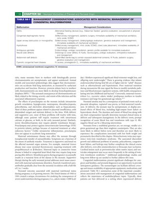
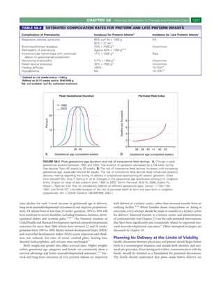
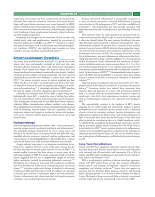
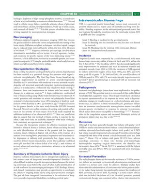
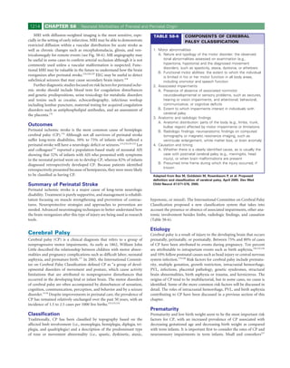
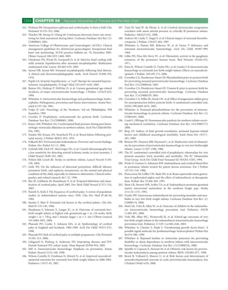
NEC may present slowly or as a sudden catastrophic event. Abdom- develop levels greater than 15 mg/dL.133 Risk factors for development
inal distention occurs early, with bloody stools present in 25% of of severe jaundice are outlined in Table 58-5.
cases.110 The radiographic hallmark is the presence of pneumatosis Several important risk factors have their origin in the prenatal and
intestinalis or portal venous gas (see Fig. 58-2). Progression may be perinatal environment. Hyperbilirubinemia is seen more frequently in
rapid, resulting in bowel perforation with evidence of free air on the infants of mothers who are diabetic (IDM). The pathogenesis of
radiograph. Early management consists of bowel decompression, increased bilirubin in IDM infants is uncertain but has been attributed
intravenous antibiotics, and respiratory and cardiovascular support as to polycythemia as well as increased red cell turnover.136,137 Prenatally,
indicated. The single absolute indication for surgical intervention is maternal blood group immunization may result from blood transfu-
pneumoperitoneum (Fig. 58-5). sion or fetal maternal hemorrhage. Although the prevalence of Rh(D)
For infants who survive NEC, morbidity is high, including high immunization has significantly decreased with the advent of preven-
rates of growth failure, chronic lung disease, and nosocomial infec- tion programs, including use of Rh immune globulin, antibodies to
tions.129-131 Lengths of stay and hospital costs are significantly length- other blood group antigens may still occur. ABO hemolytic disease, a
ened, particularly in surgical NEC.131 Long-term neurologic outcomes common cause of severe jaundice in the newborn, rarely causes hemo-
A B
FIGURE 58-5 Diagnosis and pathology of necrotizing enterocolitis. A, Typical radiographic appearance
of necrotizing enterocolitis, demonstrating pneumatosis and intramural gas. B, Intraoperative photograph of
the small bowel, which contains intramural gas.](https://image.slidesharecdn.com/4-u1-0-b978-1-4160-4224-2-50061-2-docpdf-120121090924-phpapp01/85/4-u1-0-b978-1-4160-4224-2-50061-2-docpdf-12-320.jpg)
![CHAPTER 58 Neonatal Morbidities of Prenatal and Perinatal Origin 1221
17. Smulian JC, Shen-Schwarz S, Vintzileos AM, et al: Clinical chorioamnio- 40. Haberland CA, Phibbs CS, Baker LC: Effect of opening midlevel
nitis and histologic placental inflammation. Obstet Gynecol 94:1000- neonatal intensive care units on the location of low birth weight births in
1005, 1999. California. Pediatrics 118:e1667-e1679, 2006.
18. Escobar GJ, Clark RH, Greene JD: Short-term outcomes of infants born 41. Bell EF: Noninitiation or withdrawal of intensive care for high-risk new-
at 35 and 36 weeks’ gestation: We need to ask more questions. Semin borns. Pediatrics 119:401-403, 2007.
Perinatol 30:28-33, 2006. 42. Committee on Bioethics: Ethics and care of critically ill infants and chil-
19. Stein RE, Siegel MJ, Bauman LJ: Are children of moderately low birth dren. Pediatrics 98:149-152, 2006.
weight at increased risk for poor health? A new look at an old question. 43. Dudell GG, Jain L: Hypoxic respiratory failure in the late preterm infant
Pediatrics 118:217-223, 2006. [abstract]. Clin Perinatol 33:803-830; viii-ix, 2006.
20. Kirkegaard I, Obel C, Hedegaard M, et al: Gestational age and birth weight 44. Jain L, Dudell GG: Respiratory transition in infants delivered by cesarean
in relation to school performance of 10-year-old children: A follow-up section. Semin Perinatol 30:296-304, 2006.
study of children born after 32 completed weeks. Pediatrics 118:1600- 45. Jain L, Eaton DC: Physiology of fetal lung fluid clearance and the effect
1606, 2006. of labor. Semin Perinatol 30:34-43, 2006.
21. Hulsey TC, Alexander GR, Robillard PY, et al: Hyaline membrane disease: 46. Riskin A, Abend-Weinger M, Riskin-Mashiah S, et al: Cesarean section,
The role of ethnicity and maternal risk characteristics. Am J Obstet gestational age, and transient tachypnea of the newborn: Timing is the
Gynecol 168:572-576, 1993. key. Am J Perinatol 22:377-382, 2005.
22. Mikkola K, Ritari N, Tommiska V, et al: Neurodevelopmental outcome 47. Kolas T, Saugstad OD, Daltveit AK, et al: Planned cesarean versus planned
at 5 years of age of a national cohort of extremely low birth weight infants vaginal delivery at term: Comparison of newborn infant outcomes. Am J
who were born in 1996-1997. Pediatrics 116:1391-1400, 2005. Obstet Gynecol 195:1538-1543, 2006.
23. Hintz SR, Kendrick DE, Vohr BR, et al: Changes in neurodevelopmental 48. Ronca AE, Abel RA, Ronan PJ, et al: Effects of labor contractions on cat-
outcomes at 18 to 22 months’ corrected age among infants of less than 25 echolamine release and breathing frequency in newborn rats. Behav Neu-
weeks’ gestational age born in 1993-1999. Pediatrics 115:1645-1651, rosci 120:1308-1314, 2006.
2005. 49. Lewis V, Whitelaw A: Furosemide for transient tachypnea of the newborn.
24. Ho S, Saigal S: Current survival and early outcomes of infants of border- Cochrane Database Syst Rev (1):CD003064, 2002.
line viability. Neoreviews 6:e123-e132, 2005. 50. ACOG Committee on Obstetric Practice and AAP Committee on Fetur
25. Wang ML, Dorer DJ, Fleming MP, et al: Clinical outcomes of near-term and Newborn: Intrapartum and postpartum care of the mother. In
infants. Pediatrics 114:372-376, 2004. Lockwood CJ, Lemmons JA (eds): Guidelines for Perinatal Care, 6th ed.
26. Davidoff MJ, Dias T, Damus K, et al: Changes in the gestational age dis- Elk Grove Village, IL, American Academy of Pediatrics and American
tribution among U.S. singleton births: Impact on rates of late preterm College of Obstetricians and Gynecologists, 2007, pp 139-174.
birth, 1992 to 2002. Semin Perinatol 30:8-15, 2006. 51. Lindner W, Pohlandt F, Grab D, et al: Acute respiratory failure and short-
27. Yudkin PL, Wood L, Redman CW: Risk of unexplained stillbirth at dif- term outcome after premature rupture of the membranes and oligohy-
ferent gestational ages. Lancet 1:1192-1194, 1987. dramnios before 20 weeks of gestation. J Pediatr 140:177-182, 2002.
28. Smith GC: Life-table analysis of the risk of perinatal death at term and post 52. Gerten KA, Coonrod DV, Bay RC, et al: Cesarean delivery and respiratory
term in singleton pregnancies. Am J Obstet Gynecol 184:489-496, 2001. distress syndrome: Does labor make a difference? Am J Obstet Gynecol
29. Nuffield Council on Bioethics: Critical Care Decisions in Fetal and Neo- 193(Pt 2):1061-1064, 2005.
natal Medicine: Ethical Issues. London, Nuffield Council on Bioethics, 53. Eckert Seitz E, Fiori HH, Luz JH, et al: Stable microbubble test on tracheal
2006. aspirate for the diagnosis of respiratory distress syndrome. Biol Neonate
30. MacDonald H: Perinatal care at the threshold of viability. Pediatrics 87:140-144, 2005.
110:1024-1027, 2002. 54. Kallapur S, Ikegami M: The surfactants. Am J Perinatol 17:335-343,
31. Committee on the Fetus and Newborn: Postnatal corticosteroids to treat 2000.
or prevent chronic lung disease in preterm infants. Pediatrics 109:330- 55. Halliday HL: Recent clinical trials of surfactant treatment for neonates.
338, 2002. Biol Neonate 89:323-329, 2006.
32. Wood NS, Marlow N, Costeloe K, et al: Neurologic and developmental 56. Ammari A, Suri M, Milisavljevic V, et al: Variables associated with the
disability after extremely preterm birth. EPICure Study Group. N Engl J early failure of nasal CPAP in very low birth weight infants. J Pediatr
Med 343:378-384, 2000. 147:341-347, 2005.
33. Costeloe K, Hennessy E, Gibson AT, et al: The EPICure study: Outcomes 57. Ho JJ, Henderson-Smart DJ, Davis PG: Early versus delayed initiation of
to discharge from hospital for infants born at the threshold of viability. continuous distending pressure for respiratory distress syndrome in
Pediatrics 106:659-671, 2000. preterm infants. Cochrane Database Syst Rev (2):CD002975, 2002.
34. Vanhaesebrouck P, Allegaert K, Bottu J, et al: The EPIBEL study: Out- 58. Stevens T, Harrington E, Blennow M, et al: Early surfactant administration
comes to discharge from hospital for extremely preterm infants in with brief ventilation vs. selective surfactant and continued mechanical
Belgium. Pediatrics 114:663-675, 2004. ventilation for preterm infants with or at risk for respiratory distress syn-
35. American Academy of Pediatrics, American Heart Association: Ethics and drome. Cochrane Database Syst Rev (4):CD003063, 2007.
care at the end of life. In Textbook of Neonatal Resuscitation Textbook, 59. Stevens TP, Blennow M, Soll RF: Early surfactant administration with brief
5th ed. Elk Grove Village, IL, American Academy of Pediatrics and Ameri- ventilation vs. selective surfactant and continued mechanical ventilation
can Heart Association, 2006, pp 9-5 to 9-6. for preterm infants with or at risk for respiratory distress syndrome.
36. Lucey JF, Rowan CA, Shiono P, et al: Fetal infants: The fate of 4172 infants Cochrane Database Syst Rev (3):CD003063, 2004.
with birth weights of 401 to 500 grams—the Vermont Oxford Network 60. Howlett A, Ohlsson A: Inositol for respiratory distress syndrome in
experience (1996-2000). Pediatrics 113:1559-1566, 2004. preterm infants. Cochrane Database Syst Rev (4):CD000366, 2003.
37. Vohr BR, Wright LL, Poole WK, et al: Neurodevelopmental outcomes of 61. Kinsella JP, Cutter GR, Walsh WF, et al: Early inhaled nitric oxide therapy
extremely low birth weight infants <32 weeks’ gestation between 1993 and in premature newborns with respiratory failure. N Engl J Med 355:354-
1998. Pediatrics 116:635-643, 2005. 364, 2006.
38. Cifuentes J, Bronstein J, Phibbs CS, et al: Mortality in low birth weight 62. Ballard RA, Truog WE, Cnaan A, et al: Inhaled nitric oxide in preterm
infants according to level of neonatal care at hospital of birth. Pediatrics infants undergoing mechanical ventilation. N Engl J Med 355:343-353,
109:745-751, 2002. 2006.
39. Warner B, Musial MJ, Chenier T, et al: The effect of birth hospital type on 63. Aghai ZH, Saslow JG, Nakhla T, et al: Synchronized nasal intermittent
the outcome of very low birth weight infants. Pediatrics 113(Pt 1):35-41, positive pressure ventilation (SNIPPV) decreases work of breathing
2004. (WOB) in premature infants with respiratory distress syndrome (RDS)](https://image.slidesharecdn.com/4-u1-0-b978-1-4160-4224-2-50061-2-docpdf-120121090924-phpapp01/85/4-u1-0-b978-1-4160-4224-2-50061-2-docpdf-25-320.jpg)
![1226 CHAPTER 58 Neonatal Morbidities of Prenatal and Perinatal Origin
258. Liu J, Li Z, Lin Q, et al: Cerebral palsy and multiple births in China. 285. Okusawa S, Gelfand JA, Ikejima T, et al: Interleukin 1 induces a shock-like
International journal of epidemiology 29:292-299, 2000. state in rabbits. Synergism with tumor necrosis factor and the effect of
259. Ellenberg JH, Nelson KB: Birth weight and gestational age in children with cyclooxygenase inhibition. J Clin Invest 81:1162-1172, 1988.
cerebral palsy or seizure disorders. Am J Dis Child 133:1044-1048, 1979. 286. Yoon BH, Romero R, Yang SH, et al: Interleukin-6 concentrations in
260. Blair E, Stanley F: Intrauterine growth and spastic cerebral palsy. I. Asso- umbilical cord plasma are elevated in neonates with white matter lesions
ciation with birth weight for gestational age. Am J Obstet Gynecol associated with periventricular leukomalacia. Am J Obstet Gynecol
162:229-237, 1990. 174:1433-1440, 1996.
261. Topp M, Langhoff-Roos J, Uldall P, et al: Intrauterine growth and gesta- 287. Nelson KB, Grether JK: Potentially asphyxiating conditions and spastic
tional age in preterm infants with cerebral palsy. Early Hum Dev 44:27-36, cerebral palsy in infants of normal birth weight. Am J Obstet Gynecol
1996. 179:507-513, 1998.
262. Uvebrant P, Hagberg G: Intrauterine growth in children with cerebral 288. Nelson KB, Dambrosia JM, Grether JK, Phillips TM: Neonatal cytokines
palsy. Acta Paediatr 81:407-412, 1992. and coagulation factors in children with cerebral palsy. Ann Neurol
263. Hadlock FP, Harrist RB, Martinez-Poyer J: In utero analysis of fetal 44:665-675, 1998.
growth: A sonographic weight standard. Radiology 181:129-133, 1991. 289. Wheater M, Rennie JM: Perinatal infection is an important risk factor for
264. Marsal K, Persson PH, Larsen T, et al: Intrauterine growth curves based cerebral palsy in very-low-birthweight infants. Dev Med Child Neurol
on ultrasonically estimated foetal weights. Acta Paediatr 85:843-848, 42:364-367, 2000.
1996. 290. Murphy DJ, Hope PL, Johnson A: Neonatal risk factors for cerebral palsy
265. Mongelli M, Gardosi J: Longitudinal study of fetal growth in subgroups in very preterm babies: Case-control study. BMJ 314:404-408, 1997.
of a low-risk population. Ultrasound Obstet Gynecol 6:340-344, 1995. 291. Redline RW: Placental pathology and cerebral palsy. Clin Perinatol
266. Jarvis S, Glinianaia SV, Blair E: Cerebral palsy and intrauterine growth. 33:503-516, 2006.
Clin Perinatol 33:285-300, 2006. 292. Redline RW: Severe fetal placental vascular lesions in term infants with
267. Yanney M, Marlow N: Paediatric consequences of fetal growth restriction. neurologic impairment. Am J Obstet Gynecol 192:452-457, 2005.
Semin Fetal Neonatal Med 9:411-418, 2004. 293. Redline RW, Patterson P: Patterns of placental injury. Correlations with
268. Surveillance of cerebral palsy in Europe: A collaboration of cerebral palsy gestational age, placental weight, and clinical diagnoses. Arch Pathol Lab
surveys and registers. Surveillance of Cerebral Palsy in Europe (SCPE). Med118:698-701, 1994.
Dev Med Child Neurol 42:816-824, 2000. 294. Wichers MJ, Odding E, Stam HJ, et al: Clinical presentation, associated
269. Jarvis S, Glinianaia SV, Arnaud C, et al: Case gender and severity in cere- disorders and aetiological moments in cerebral palsy: A Dutch popula-
bral palsy varies with intrauterine growth. Archives of disease in child- tion-based study. Disabil Rehabil 27:583-589, 2005.
hood 90:474-479, 2005. 295. Shy KK, Luthy DA, Bennett FC, et al: Effects of electronic fetal-heart-rate
270. Hermansen MC, Hermansen MG: Perinatal infections and cerebral palsy. monitoring, as compared with periodic auscultation, on the neurologic
Clin Perinatol 33:315-333, 2006. development of premature infants. N Engl J Med 322:588-593, 1990.
271. Goldenberg RL, Hauth JC, Andrews WW: Intrauterine infection and 296. Nelson KB, Dambrosia JM, Ting TY, Grether JK: Uncertain value of
preterm delivery. N Engl J Med 342:1500-1507, 2000. electronic fetal monitoring in predicting cerebral palsy. N Engl J Med
272. Goldenberg RL, Culhane JF, Johnson DC: Maternal infection and adverse 334:613-618, 1996.
fetal and neonatal outcomes. Clin Perinatol 32:523-559, 2005. 297. Martin JA, Hamilton BE, Sutton PD, et al: Births: Final data for 2003. Natl
273. Dammann O, Leviton A: Maternal intrauterine infection, cytokines, and Vital Stat Rep 54:1-116, 2005.
brain damage in the preterm newborn. Pediatr Res 42:1-8, 1997. 298. Thorngren-Jerneck K, Herbst A: Perinatal factors associated with cerebral
274. Dammann O, Leviton A: The role of perinatal brain damage in develop- palsy in children born in Sweden. Obstet Gynecol 108:1499-1505, 2006.
mental disabilities: An epidemiologic perspective. Ment Retard Dev 299. Crowley PA: Antenatal corticosteroid therapy: A meta-analysis of the ran-
Disabil Res Rev 3:13-21, 1997. domized trials, 1972 to 1994. Am J Obstet Gynecol 173:322-335, 1995.
275. Alexander JM, Gilstrap LC, Cox SM, et al: Clinical chorioamnionitis and 300. Yeh TF, Lin YJ, Huang CC, et al: Early dexamethasone therapy in preterm
the prognosis for very low birth weight infants. Obstet Gynecol 91(Pt infants: A follow-up study. Pediatrics 101:E7, 1998.
1):725-729, 1998. 301. O’Shea TM, Kothadia JM, Klinepeter KL, et al: Randomized placebo-con-
276. Wu YW, Colford JM Jr: Chorioamnionitis as a risk factor for cerebral trolled trial of a 42-day tapering course of dexamethasone to reduce the
palsy: A meta-analysis. JAMA 284:1417-1424, 2000. duration of ventilator dependency in very low birth weight infants:
277. Grafe MR: The correlation of prenatal brain damage with placental Outcome of study participants at 1-year adjusted age. Pediatrics 104(Pt
pathology. J Neuropathol Exp Neurol 53:407-415, 1994. 1):15-21, 1999.
278. Salafia CM, Minior VK, Rosenkrantz TS, et al: Maternal, placental, and 302. Shinwell ES, Karplus M, Reich D, et al: Early postnatal dexamethasone
neonatal associations with early germinal matrix/intraventricular hemor- treatment and increased incidence of cerebral palsy. Arch Dis Child Fetal
rhage in infants born before 32 weeks’ gestation. Am J Perinatol 12:429- Neonatal Ed 83:F177-F181, 2000.
436, 1995. 303. McDonald JW, Silverstein FS, Johnston MV: Magnesium reduces N-
279. De Felice C, Toti P, Parrini S, et al: Histologic chorioamnionitis and sever- methyl-D-aspartate (NMDA)–mediated brain injury in perinatal rats.
ity of illness in very low birth weight newborns. Pediatr Crit Care Med Neurosci Lett 109:234-238, 1990.
6:298-302, 2005. 304. Weglicki WB, Phillips TM, Freedman AM, et al: Magnesium-deficiency
280. Kraus FT: Cerebral palsy and thrombi in placental vessels of the fetus: elevates circulating levels of inflammatory cytokines and endothelin.
Insights from litigation. Hum Pathol 28:246-248, 1997. Molecular and cellular biochemistry 110:169-173, 1992.
281. Leviton A: Preterm birth and cerebral palsy: Is tumor necrosis factor the 305. Wiswell TE, Graziani LJ, Caddell JL, et al: Maternally administered mag-
missing link? Dev Med Child Neurol 35:553-558, 1993. nesium sulphate protects against early brain injury and long-term adverse
282. Adinolfi M: Infectious diseases in pregnancy, cytokines and neurological neurodevelopmental outcomes in preterm infants: A prospective study.
impairment: An hypothesis. Dev Med Child Neurol 35:549-558, 1993. Pediatr Res 39:253A, 1996.
283. Dammann O, Leviton A: Brain damage in preterm newborns: Might 306. Nelson KB, Grether JK: Can magnesium sulfate reduce the risk of cerebral
enhancement of developmentally regulated endogenous protection open palsy in very low birthweight infants? Pediatrics 95:263-269, 1995.
a door for prevention? Pediatrics 104(Pt 1):541-550, 1999. 307. Hauth JC, Goldenberg RL, Nelson KB, et al: Reduction of cerebral palsy
284. Chao CC, Hu S, Ehrlich L, et al: Interleukin-1 and tumor necrosis factor- with maternal MgSO4 treatment in newborns weighing 500-1000 g
alpha synergistically mediate neurotoxicity: Involvement of nitric oxide [abstract]. Am J Obstet Gynecol 172(Pt 2):419, 1995.
and of N-methyl-D-aspartate receptors. Brain Behav Immun 9:355-365, 308. Schendel DE, Berg CJ, Yeargin-Allsopp M, et al: Prenatal magnesium
1995. sulfate exposure and the risk for cerebral palsy or mental retardation](https://image.slidesharecdn.com/4-u1-0-b978-1-4160-4224-2-50061-2-docpdf-120121090924-phpapp01/85/4-u1-0-b978-1-4160-4224-2-50061-2-docpdf-30-320.jpg)

This document summarizes common neonatal morbidities that can result from complications during pregnancy and delivery. It discusses how conditions like diabetes, hypertension, infection, and nutritional imbalances in the mother can negatively impact the health of the newborn. The summary provides management considerations for treating infants born with various medical issues and outlines how close collaboration between obstetric and neonatal clinicians is important for counseling families and ensuring the best outcomes for both mother and baby.