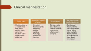
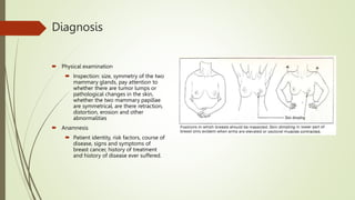
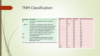
SlideShare a Scribd company logo
Breast cancer
By :
Saruveswaran R Rasasingam
Preceptopr :
dr. Adrioki Risa, Sp.B
Chapter 1:
Introduction
Introduction
Breast cancer
• Uncontrolled
growth of breast
tissue cells that can
invade and spread
to other parts of
the body
Breast cancer is the
most common cancer
in
developed/developin
g countries
• Fifth most
common cause of
death from cancer
The incidence rate in
developing countries
is 40/100,000
population
• Indonesia urutan
ke-8 di Asia
tenggara dan no-
23 di Asia
Introduction
 Research conducted at RSUP Dr. M. Jamil Padang on risk factors that can cause
breast cancer are non pregnant, very short duration of breastfeeding, menopause,
obesity, high fat intake, living in urban areas, family history of breast cancer and a
history of blunt breast trauma.
 For the prevention and control of cancer in Indonesia, the government has made
various efforts, including early detection of breast cancer in women aged 30-50
years using the Clinical Breast Examination (SADANIS) method.
Sindrome koroner Akut. Perki. 2018
Chapter 2:
Literature review
Anatomy of
breast
 Boundaries
 Superior : II-III ribs
 Inferior : VI-VII ribs
 Medial : linea
parasternalis
 Lateral : line anterior
axillae/ linea
mid axillae
Anatomy of
breast
 Each breast consists of 12-20
glandular lobules, each of which has
a duct called the lactiferous duct,
which opens into the mammary
papillae.
 Between the mammary glands and
the pectoral fascia, as well as
between the skin and the glands, is
adipose tissue.
 Between the lobules, there are
connective tissue called Cooper's
ligaments which form the framework
of the breast
Anatomy of breast
 Breast bleeding is mainly derived from branches of the anterior
perforating artery from the internal mammary artery, the lateral
thoracic artery which branches from the axillary artery, and several
intercostal arteries.
 About 75% of the lymphatic drainage of the breast drains into the
axillary lymphatic group
 Level I
 Located laterally / below the lower limit m. pectoralis minor
 Level II
 Located inside (deep) or behind m. pectoralis minor i.e. the
central group
 Level III
 Located medial to or above the upper limit of m. pectoralis
minor is the subclavicular group
Breast Cancer
• Breast cancer : a group of malignant tumors in breast tissue which can originate
from lactiferous duct epithelium (70%) or lobular epithelium (10%)
Definition
• Most common cancer in women
• The main cause of death from cancer
• The highest incidence is in developed countries, and there is an increase in
developing countries due to lifestyle changes
• Based on Riskesdas data, the prevalence of tumors/cancer in Indonesia shows an
increase from 1.4 per 1000 population in 2013 to 1.79 per 1000 population in
2018
Epidemiology
Etiology
 Gene mutation
1. BRCA-1 (locus 17q21)
2. Gen p53 (locus 17p13)
3. BRCA-2 at chromosome 13
 Mutasi tersebut diduga akibat paparan mutagen
 mutagen endogen : radikal bebas seperti lipid peroksidase
 mutagen eksogen : radiasi
 Virus (belum ada bukti pada manusia)
Risk factor
 Age
 It is rare under the age of 30 years, but the incidence
increases sharply until around the age of 50 years and
then it continues to increase but slowly --> ovarian
haemorrhagic effects
 Reproduction and hormones
 Age of menarche, age of menopause, 35th first birth,
hormonal contraception, postmenopausal hormone
replacement therapy
 Genetics and familial
 Approximately 5-10% of breast cancer occurs due to a
genetic predisposition. And the risk increases 2x if one of
the nuclear family members suffers from breast cancer
 Lifestyle
 Obesity, physical activity, smoking and alcohol
 Exogenous exposure
 Radiation, chemicals
Pathogenesis
• unevenly
distributed
proliferation of
polyclonal
epithelial cells with
overlapping nuclei
and disorganized
ductal lumens
Ductal hyperplasia
• the cytoplasm of the
cells becomes more
pronounced and
does not overlap the
regular duct lumen
hyperplasia
atypical
• cell proliferation
with cytological
features consistent
with malignancy.
Proliferation has not
invaded the stroma
or penetrated the
basement
membrane
Carcinoma in-situ
• Tumor cells have
penetrated the
basement
membrane and
invaded the stroma -
-> metastasized
Invasive
carcinoma
Clinical manifestation
Tumor Mass
• Most manifest as
a breast mass
that is not
painful. Most
common
location is the
upper lateral
quadrant
Mammary papilla
changes
• Retraction,
distortion of the
mammary
papillae,
papillary
discharge,
eczematoid
changes
Skin changes
• Dimple marks,
peau d'orange,
satellite nodules,
ulcerations,
inflammatory
changes
Regional lymph
node enlargement
• Pembesaran
kelenjar limfe
aksilar ipsilateral
dapat soliter
atau multiple.
kelenjar limfe
supraklavikular
juga dapat
membesar
Diagnosis
 Physical examination
 Inspection: size, symmetry of the two
mammary glands, pay attention to
whether there are tumor lumps or
pathological changes in the skin,
whether the two mammary papillae
are symmetrical, are there retraction,
distortion, erosion and other
abnormalities
 Anamnesis
 Patient identity, risk factors, course of
disease, signs and symptoms of
breast cancer, history of treatment
and history of disease ever suffered.
Palpation
 Breast
 Lymp nodes
Supporting investigation
 Mammography
 Breast ultrasound
 MRIs
 Immunohistochemistry
 FNAB
 Core biopsy
 Open biopsy
 Sentinel node biopsy
 Bone Scan, Chest Photo and Ultrasound Abdomen
 Laboratory Examination: tumor markers such as CA-15-3 and CEA
TNM Classification
TNM Classification
Classification
Non invasive carcinoma
• Ductal carcinoma in situ (DCIS)
• Lobular carcinoma in situ (LCIS)
Invasive carcinoma
• Invasive ductal carcinoma
• Adenocarcinoma with productive fibrosis (scirrhous, simplex, NST)
• Medullary carcinoma (4%)
• Mucinous (colloid) carcinoma (2%)
• Papillary carcinoma (2%)
• Tubular carcinoma (2%)
• Invasive lobular carcinoma (10%)
Paget’s disease
Procedures
• Classic Radical Mastectomy
• Modified Radical Mastectomy
• Simple Mastectomy
• BCS (Breast Conserving Surgery)
Operation
• adjuvant therapy
• Neoadjuvant therapy
• Primary therapy
Chemotherapy(systemic)
• DNA damage by interrupting the replication process and reducing the risk of local recurrence
Radiotherapy
• in the breast that shows positive expression of estrogen receptor (ER) or progesterone receptor (PR)
Hormanal therapy
• menghambat proses yang berperan dalam pertumbuhan sel-sel kanker
Target therapy
Prognosis
 The prognosis of breast cancer
is indicated by the life
expectancy or disease-free
interval
Chapter 3: Case
illustration
Patient identity
 Name : Miss Feb.Oz
 Gender : Female
 Age : 20 years old
 Address : Ombilin
 Occupation : Student
 Marital status : Single
Anamnesis
 A 20-year-old female patient came to the Policlinic of General Hospital
(RSU) Prof. Dr. M Ali Hanafiah with:
Chief Complaint
 Pain and Lump on the superior lateral of the right breast
History of Present Disease
- The lump in the right breast was initially felt since 1 years ago, the lump was felt as large as a marble and was
not painful. Then the lump felt increasingly enlarged to the size of a ping pong ball and with increasing pain
since 1 month . The patient did not complain of pus-like discharge and blood from the nipple, the nipple
wasn’t pulled inward and the skin around the breast had no changes in the breast skin resembling orange peel.
- Patient complaints that the pain gets worse while she’s resting.,
- Slight fever 2 days prior to getting admitted.
- There is no bony pain.
- There is no headache.
- Shortness of breath not present
- Decrease in appetite not present.
- No history of serious weight loss in this 1 year.
- Nausea and vomiting are absent.
- There is no complaint about bowel movements.
 There is no complaint about urination
Past medical history
- History of tumor on right breast inferior subareolar region.
- History of lumpectomy in the year 2019
- Diabetes Mellitus (-)
- Hypertension (-)
- Heart Disease (-)
- Kidney Disease (-)
- History of asthma (-)
- History of drug, food, and other substance allergies (-)
Family history of disease
 Patient’s aunty (mom’s sister) had similar issues before marriage and has gone through
lumpectomy thrice throughout till today
Social history, psychology, habits
- The patient is now a student.
- The patient had her menarche at the age of 15.
- Menstrual cycle 28-30 days, regular, 2-5 days in length, 3 sanitary pads usually used,
no serious pain.
- The patient has no children.
- No history of hormonal contraception
- There is no history of radiation to the chest wall.
Physical examination
 General condition: Appears moderately ill
Consciousness: Composmentis cooperative
 Vital Sign :
 -Blood Pressure: 110/70 mmHg
 -Pulse : 82 beats/minute
 -Breathing : 20 beats/minute
 -Temperature : 36,5°C
General Status
 -Head : normocephalic, no abnormalities
 -Eyes : non anemic conjunctiva, non-icteric sclera
 -Skin : good skin turgor
 -Nose : no abnormalities
 -Ears : no abnormalities
 -Mouth : no abnormalities
 -Neck : no abnormalities
 -KGB : no abnormalities
Cont.
Lungs
• Inspection:
normochest,
symmetrical chest
wall movement
• Palpation: left
fremitus equal to
right
• Percussion: sonor
• Auscultation:
vesicular breath
sounds, rhonki -/-,
wheezing -/-
Heart
• Inspection: ictus
cordis not visible
• Palpation: palpable
ictus cordis 1 finger
medial LMCS RIC V
• Percussion: heart
limits within normal
limits
• Auscultation:
regular heart sound,
normal S1 S2,
murmur (-), gallop
(-)
Abdomen
• Inspection:
distention (-)
• Auscultation: bowel
sounds (+) normal
• Palpation: sociable,
NT (-), NL (-), liver
and spleen not
palpable
• Percussion: timpani
Ekstremitas
• Udem -/-
• Warm acral
• CRT < 2 seconds
Local status
 Right mammary region
 Inspection: A mass in the upper outer quadrant, the mammary gland the size of a
chicken egg, peau d'orange (-), satellite nodules (-), nipple retraction (-), nipple
discharge (-), abscess (-), blood (-),
 Palpation: palpable solitary mass with a size of 4 x 4 cm in the region of upper outer
quadrant of the right breast, hard consistency, flat surface, firm boundaries, free to
move, and sensitive to touch
 Left Mammary Region
 Inspection: No visible lump
 Palpation: 1 corn seed sized palpable mass on the inferior subareolar region. Size of 0.5
x 0.5cm, hardconsistency, smooth surface, firm borders, free to move and sensitive to
touch
Cont.
 Right Axillary Region
 Inspection: no visible mass
 Palpation: palpable mass with a size of 1 x 1 cm, hard consistency, smooth surface, firm boundaries, fixed, and no pain.
 Left Axillary Region
 Inspection: no visible mass
 Palpation: no palpable mass
 Dextra supraclavicular region
 Inspection: no visible mass
 Palpation: 1 corn seed sized palpable mass on the inferior subareolar region. Size of 0.5 x 0.5cm, hard consistency,
smooth surface, firm borders, free to move and sensitive to touch.
 Left supraclavicular region
 Inspection: no visible mass
 Palpation: no palpable mass
Cont.
 Left Infraclavicular Region
 Inspection: no visible mass
 Palpation: no palpable mass
 Dextra infraclavicular region
 Inspection: no visible mass
 Palpation: no palpable mass
Diagnosis
 Right Mammary tumor Susp mammary fibroadenoma.
Supporting examination
 Laboratory examination
 Chest x-ray
 Anatomical pathology
 Abdominal ultrasound
 Immunohistochemistry
Procedures
 Lumpectomy
 Post lumpectomy lab test
Prognosis
 Quo ad vitam : Dubia ad malam
 Quo ad functionam : Dubia ad malam
 Quo ad sanationam : Dubia ad malam
Discussion
Discussion
 A patient Miss Feboz, a 20-year-old woman came to the Outpatient of General Hospital Prof.
Dr.M Ali Hanafiah with complaints of a lump the size of a table tenis ball in the right breast.
The patient felt a lump in the right breast initially felt in the last 1 year, the lump was felt as big
as a pea and painless. Then thelump is got bigger and denser in the past month. The patient does
not have previous breast discharge such as pus or blood from the nipple, and the the nipple
wasn’t pulled inward. If there was a retraction of the breast,it indicates tumor invasion into the
subpapillary tissue. There is no change in the breast skin that resembles orange peel. This is
because the tumor in this patient did not cause any drainage obstruction to the subcuticular
lymphatic vasa, causing this to occur.10
Discussion
 The patient has not complained of symptoms such as decreased appetite and drastic weight loss,
in this past year. The patient is 20 years old, where the age factor plays an important role in
causing breast cancer. With increasing age, the incidence of breast cancer will increase. After the
age of 50 years, the frequency continues to increase but slowly. This malignancy is very rare in
women under the age of 20. A patient's risk factors for mammary malignancy are sought by
asking about age at menarche, menstrual history, pregnancy, breastfeeding, hormonal
contraceptive use, and radiation exposure. Among these risk factors, the patient had never been
pregnant (reproductive) and never breastfed. According to research, breastfeeding reduces the
risk of developing breast cancer especially if the breastfeeding period is carried out for 27-52
weeks. This reduction in risk is thoughtto be because breastfeeding reduces a person's
menstrual period.3
Discussion
 On physical examination, a solitary mass with a size of 4 cm x 4 cm in the upper outer
quadrant, region of the mammary gland was palpated, hard consistency, flat surface,
well-defined, freely moving , painless, nipple discharge (-) peau consistency, flat surface,
well-defined, fixed, painless, nipple discharge (-) peau d'orange (-), nipple retraction (-).
 The presence of a mass with a hard consistency, fixed and painless is a description of a
malignancy. The presence of a mass with the diameter of 4cm x 4cm is classified to be
T2. There was no regional metastasis on the particular lump and it was mobile so it was
classified to be N0. There was no metastases to distant organs (M0). Therefore the lump
on the right breast of this patient was classified to be Stage 2A.
Discussion
 On examination of the left breast, a mass with a size of 0.5 x 0.5 cm, hard consistency,
smooth surface, firm boundaries, non-fixed and painless was palpated. This is the basis
for upholding the N0 classification in the diagnosis. The patient did not have any signs
of distant metastases, so M0 was established.
 Based on history, physical examination and support, this patient was diagnosed with
mammary tumor right suspected mammary fibroadenoma. In this patient, the
lumpectomy was performed and the lump the was removed was sent for lab
examinations to identify the type of tissues involved.
Discussion
 If the patient was histopathologically proven as mammary carcinoma, the therapy plan
will be neoadjuvant chemotherapy and MRM because the above 3 has to be
accompanied with tumor stage chemotherapy treatment. Chemotherapy is systemic,
serves to destroy cancer cells, works by inhibiting or disrupting DNA synthesis in the
cell cycle.3 However on this particular patient the lump wasn’t easily bleeding, was
freely moving and not fixated to any tissues. Therefore, its suspected to be a
Fibroadenoma and being removed early prevent complivations and preserves life
quality.
Discussion
 The prognosis of this patient's 5-year survival rate is high (Stage 2A, 92%). Education
that can be given to the family is education about the patient's disease and
management aimed at improving quality of life, and education to family members
about risk factors for breast cancer and breast cancer screening in family members at
risk. 3
Thank you

More Related Content

Similar to 3.pptx

Ovarian cancer
Ovarian cancerOvarian cancer
Ovarian cancer
Uma Chidiebere
 
cancer.pdf
cancer.pdfcancer.pdf
cancer.pdf
AbiVill
 
Care of patient with cancer thyroid and parathyroid
Care of patient with cancer thyroid and parathyroidCare of patient with cancer thyroid and parathyroid
Care of patient with cancer thyroid and parathyroid
Saumya Srivastava
 
Breast cancer epidemiology
Breast cancer epidemiology Breast cancer epidemiology
Breast cancer epidemiology
D.A.B.M
 
Breast disease
Breast diseaseBreast disease
Breast disease
Izza Abid
 
Breast Cancer.pptx
Breast Cancer.pptxBreast Cancer.pptx
Breast Cancer.pptx
AdrielCabo
 
Ovarian & endometrial cancer
Ovarian & endometrial cancerOvarian & endometrial cancer
Ovarian & endometrial cancer
Isha Jaiswal
 
Cancer 2018
Cancer 2018Cancer 2018
Cancer 2018
EsserHealth
 
Breast Cancer for public awareness by Dr Rubz
Breast Cancer for public awareness by Dr  RubzBreast Cancer for public awareness by Dr  Rubz
Breast Cancer for public awareness by Dr Rubz
Dr. Rubz
 
EMPOWER-pp (1).pptx
EMPOWER-pp (1).pptxEMPOWER-pp (1).pptx
EMPOWER-pp (1).pptx
ssuser1de5b01
 
Ovary Anatomy and Malignancy: A Guide to surgical Management
Ovary Anatomy and Malignancy: A Guide to surgical ManagementOvary Anatomy and Malignancy: A Guide to surgical Management
Ovary Anatomy and Malignancy: A Guide to surgical Management
AmiyArnav1
 
Breast cancer by Waweru and Kavuka.pptptx
Breast cancer by Waweru and Kavuka.pptptxBreast cancer by Waweru and Kavuka.pptptx
Breast cancer by Waweru and Kavuka.pptptx
venusodero
 
breast cancer case study
breast cancer case studybreast cancer case study
breast cancer case study
ahmedalgzali1
 
Fgt uterus and cervix
Fgt   uterus and cervixFgt   uterus and cervix
Fgt uterus and cervix
Janani Mathialagan
 
Ovarian Malignancy ppt
Ovarian Malignancy  pptOvarian Malignancy  ppt
Ovarian Malignancy ppt
Abhishek Kothule
 
Wilm's tumor
Wilm's tumorWilm's tumor
Wilm's tumor
Ganesh naik
 
Pelvic inflammatory disease ppt
Pelvic inflammatory disease pptPelvic inflammatory disease ppt
Pelvic inflammatory disease ppt
Meenakshi Kaushik
 
Carcinoma of uterine &amp; cervix
Carcinoma of uterine &amp; cervixCarcinoma of uterine &amp; cervix
Carcinoma of uterine &amp; cervix
اثير عبد الحسن
 
Oncology (Uterine Cancer)
Oncology (Uterine Cancer) Oncology (Uterine Cancer)
Oncology (Uterine Cancer)
dralialhayali
 
Breast cancer presentation
Breast cancer presentationBreast cancer presentation
Breast cancer presentation
Sajeer Muliyathil
 

Similar to 3.pptx (20)

Ovarian cancer
Ovarian cancerOvarian cancer
Ovarian cancer
 
cancer.pdf
cancer.pdfcancer.pdf
cancer.pdf
 
Care of patient with cancer thyroid and parathyroid
Care of patient with cancer thyroid and parathyroidCare of patient with cancer thyroid and parathyroid
Care of patient with cancer thyroid and parathyroid
 
Breast cancer epidemiology
Breast cancer epidemiology Breast cancer epidemiology
Breast cancer epidemiology
 
Breast disease
Breast diseaseBreast disease
Breast disease
 
Breast Cancer.pptx
Breast Cancer.pptxBreast Cancer.pptx
Breast Cancer.pptx
 
Ovarian & endometrial cancer
Ovarian & endometrial cancerOvarian & endometrial cancer
Ovarian & endometrial cancer
 
Cancer 2018
Cancer 2018Cancer 2018
Cancer 2018
 
Breast Cancer for public awareness by Dr Rubz
Breast Cancer for public awareness by Dr  RubzBreast Cancer for public awareness by Dr  Rubz
Breast Cancer for public awareness by Dr Rubz
 
EMPOWER-pp (1).pptx
EMPOWER-pp (1).pptxEMPOWER-pp (1).pptx
EMPOWER-pp (1).pptx
 
Ovary Anatomy and Malignancy: A Guide to surgical Management
Ovary Anatomy and Malignancy: A Guide to surgical ManagementOvary Anatomy and Malignancy: A Guide to surgical Management
Ovary Anatomy and Malignancy: A Guide to surgical Management
 
Breast cancer by Waweru and Kavuka.pptptx
Breast cancer by Waweru and Kavuka.pptptxBreast cancer by Waweru and Kavuka.pptptx
Breast cancer by Waweru and Kavuka.pptptx
 
breast cancer case study
breast cancer case studybreast cancer case study
breast cancer case study
 
Fgt uterus and cervix
Fgt   uterus and cervixFgt   uterus and cervix
Fgt uterus and cervix
 
Ovarian Malignancy ppt
Ovarian Malignancy  pptOvarian Malignancy  ppt
Ovarian Malignancy ppt
 
Wilm's tumor
Wilm's tumorWilm's tumor
Wilm's tumor
 
Pelvic inflammatory disease ppt
Pelvic inflammatory disease pptPelvic inflammatory disease ppt
Pelvic inflammatory disease ppt
 
Carcinoma of uterine &amp; cervix
Carcinoma of uterine &amp; cervixCarcinoma of uterine &amp; cervix
Carcinoma of uterine &amp; cervix
 
Oncology (Uterine Cancer)
Oncology (Uterine Cancer) Oncology (Uterine Cancer)
Oncology (Uterine Cancer)
 
Breast cancer presentation
Breast cancer presentationBreast cancer presentation
Breast cancer presentation
 

Recently uploaded

BRACHYTHERAPY OVERVIEW AND APPLICATORS
BRACHYTHERAPY OVERVIEW  AND  APPLICATORSBRACHYTHERAPY OVERVIEW  AND  APPLICATORS
BRACHYTHERAPY OVERVIEW AND APPLICATORS
Krishan Murari
 
Lung Cancer: Artificial Intelligence, Synergetics, Complex System Analysis, S...
Lung Cancer: Artificial Intelligence, Synergetics, Complex System Analysis, S...Lung Cancer: Artificial Intelligence, Synergetics, Complex System Analysis, S...
Lung Cancer: Artificial Intelligence, Synergetics, Complex System Analysis, S...
Oleg Kshivets
 
Ozempic: Preoperative Management of Patients on GLP-1 Receptor Agonists
Ozempic: Preoperative Management of Patients on GLP-1 Receptor Agonists  Ozempic: Preoperative Management of Patients on GLP-1 Receptor Agonists
Ozempic: Preoperative Management of Patients on GLP-1 Receptor Agonists
Saeid Safari
 
Physiology of Special Chemical Sensation of Taste
Physiology of Special Chemical Sensation of TastePhysiology of Special Chemical Sensation of Taste
Physiology of Special Chemical Sensation of Taste
MedicoseAcademics
 
ANATOMY AND PHYSIOLOGY OF URINARY SYSTEM.pptx
ANATOMY AND PHYSIOLOGY OF URINARY SYSTEM.pptxANATOMY AND PHYSIOLOGY OF URINARY SYSTEM.pptx
ANATOMY AND PHYSIOLOGY OF URINARY SYSTEM.pptx
Swetaba Besh
 
basicmodesofventilation2022-220313203758.pdf
basicmodesofventilation2022-220313203758.pdfbasicmodesofventilation2022-220313203758.pdf
basicmodesofventilation2022-220313203758.pdf
aljamhori teaching hospital
 
How STIs Influence the Development of Pelvic Inflammatory Disease.pptx
How STIs Influence the Development of Pelvic Inflammatory Disease.pptxHow STIs Influence the Development of Pelvic Inflammatory Disease.pptx
How STIs Influence the Development of Pelvic Inflammatory Disease.pptx
FFragrant
 
heat stroke and heat exhaustion in children
heat stroke and heat exhaustion in childrenheat stroke and heat exhaustion in children
heat stroke and heat exhaustion in children
SumeraAhmad5
 
Hemodialysis: Chapter 3, Dialysis Water Unit - Dr.Gawad
Hemodialysis: Chapter 3, Dialysis Water Unit - Dr.GawadHemodialysis: Chapter 3, Dialysis Water Unit - Dr.Gawad
Hemodialysis: Chapter 3, Dialysis Water Unit - Dr.Gawad
NephroTube - Dr.Gawad
 
Couples presenting to the infertility clinic- Do they really have infertility...
Couples presenting to the infertility clinic- Do they really have infertility...Couples presenting to the infertility clinic- Do they really have infertility...
Couples presenting to the infertility clinic- Do they really have infertility...
Sujoy Dasgupta
 
Phone Us ❤85270-49040❤ #ℂall #gIRLS In Surat By Surat @ℂall @Girls Hotel With...
Phone Us ❤85270-49040❤ #ℂall #gIRLS In Surat By Surat @ℂall @Girls Hotel With...Phone Us ❤85270-49040❤ #ℂall #gIRLS In Surat By Surat @ℂall @Girls Hotel With...
Phone Us ❤85270-49040❤ #ℂall #gIRLS In Surat By Surat @ℂall @Girls Hotel With...
Savita Shen $i11
 
Cervical & Brachial Plexus By Dr. RIG.pptx
Cervical & Brachial Plexus By Dr. RIG.pptxCervical & Brachial Plexus By Dr. RIG.pptx
Cervical & Brachial Plexus By Dr. RIG.pptx
Dr. Rabia Inam Gandapore
 
BENIGN PROSTATIC HYPERPLASIA.BPH. BPHpdf
BENIGN PROSTATIC HYPERPLASIA.BPH. BPHpdfBENIGN PROSTATIC HYPERPLASIA.BPH. BPHpdf
BENIGN PROSTATIC HYPERPLASIA.BPH. BPHpdf
DR SETH JOTHAM
 
Non-respiratory Functions of the Lungs.pdf
Non-respiratory Functions of the Lungs.pdfNon-respiratory Functions of the Lungs.pdf
Non-respiratory Functions of the Lungs.pdf
MedicoseAcademics
 
Knee anatomy and clinical tests 2024.pdf
Knee anatomy and clinical tests 2024.pdfKnee anatomy and clinical tests 2024.pdf
Knee anatomy and clinical tests 2024.pdf
vimalpl1234
 
Charaka Samhita Sutra sthana Chapter 15 Upakalpaniyaadhyaya
Charaka Samhita Sutra sthana Chapter 15 UpakalpaniyaadhyayaCharaka Samhita Sutra sthana Chapter 15 Upakalpaniyaadhyaya
Charaka Samhita Sutra sthana Chapter 15 Upakalpaniyaadhyaya
Dr KHALID B.M
 
New Drug Discovery and Development .....
New Drug Discovery and Development .....New Drug Discovery and Development .....
New Drug Discovery and Development .....
NEHA GUPTA
 
Prix Galien International 2024 Forum Program
Prix Galien International 2024 Forum ProgramPrix Galien International 2024 Forum Program
Prix Galien International 2024 Forum Program
Levi Shapiro
 
The Normal Electrocardiogram - Part I of II
The Normal Electrocardiogram - Part I of IIThe Normal Electrocardiogram - Part I of II
The Normal Electrocardiogram - Part I of II
MedicoseAcademics
 
Maxilla, Mandible & Hyoid Bone & Clinical Correlations by Dr. RIG.pptx
Maxilla, Mandible & Hyoid Bone & Clinical Correlations by Dr. RIG.pptxMaxilla, Mandible & Hyoid Bone & Clinical Correlations by Dr. RIG.pptx
Maxilla, Mandible & Hyoid Bone & Clinical Correlations by Dr. RIG.pptx
Dr. Rabia Inam Gandapore
 

Recently uploaded (20)

BRACHYTHERAPY OVERVIEW AND APPLICATORS
BRACHYTHERAPY OVERVIEW  AND  APPLICATORSBRACHYTHERAPY OVERVIEW  AND  APPLICATORS
BRACHYTHERAPY OVERVIEW AND APPLICATORS
 
Lung Cancer: Artificial Intelligence, Synergetics, Complex System Analysis, S...
Lung Cancer: Artificial Intelligence, Synergetics, Complex System Analysis, S...Lung Cancer: Artificial Intelligence, Synergetics, Complex System Analysis, S...
Lung Cancer: Artificial Intelligence, Synergetics, Complex System Analysis, S...
 
Ozempic: Preoperative Management of Patients on GLP-1 Receptor Agonists
Ozempic: Preoperative Management of Patients on GLP-1 Receptor Agonists  Ozempic: Preoperative Management of Patients on GLP-1 Receptor Agonists
Ozempic: Preoperative Management of Patients on GLP-1 Receptor Agonists
 
Physiology of Special Chemical Sensation of Taste
Physiology of Special Chemical Sensation of TastePhysiology of Special Chemical Sensation of Taste
Physiology of Special Chemical Sensation of Taste
 
ANATOMY AND PHYSIOLOGY OF URINARY SYSTEM.pptx
ANATOMY AND PHYSIOLOGY OF URINARY SYSTEM.pptxANATOMY AND PHYSIOLOGY OF URINARY SYSTEM.pptx
ANATOMY AND PHYSIOLOGY OF URINARY SYSTEM.pptx
 
basicmodesofventilation2022-220313203758.pdf
basicmodesofventilation2022-220313203758.pdfbasicmodesofventilation2022-220313203758.pdf
basicmodesofventilation2022-220313203758.pdf
 
How STIs Influence the Development of Pelvic Inflammatory Disease.pptx
How STIs Influence the Development of Pelvic Inflammatory Disease.pptxHow STIs Influence the Development of Pelvic Inflammatory Disease.pptx
How STIs Influence the Development of Pelvic Inflammatory Disease.pptx
 
heat stroke and heat exhaustion in children
heat stroke and heat exhaustion in childrenheat stroke and heat exhaustion in children
heat stroke and heat exhaustion in children
 
Hemodialysis: Chapter 3, Dialysis Water Unit - Dr.Gawad
Hemodialysis: Chapter 3, Dialysis Water Unit - Dr.GawadHemodialysis: Chapter 3, Dialysis Water Unit - Dr.Gawad
Hemodialysis: Chapter 3, Dialysis Water Unit - Dr.Gawad
 
Couples presenting to the infertility clinic- Do they really have infertility...
Couples presenting to the infertility clinic- Do they really have infertility...Couples presenting to the infertility clinic- Do they really have infertility...
Couples presenting to the infertility clinic- Do they really have infertility...
 
Phone Us ❤85270-49040❤ #ℂall #gIRLS In Surat By Surat @ℂall @Girls Hotel With...
Phone Us ❤85270-49040❤ #ℂall #gIRLS In Surat By Surat @ℂall @Girls Hotel With...Phone Us ❤85270-49040❤ #ℂall #gIRLS In Surat By Surat @ℂall @Girls Hotel With...
Phone Us ❤85270-49040❤ #ℂall #gIRLS In Surat By Surat @ℂall @Girls Hotel With...
 
Cervical & Brachial Plexus By Dr. RIG.pptx
Cervical & Brachial Plexus By Dr. RIG.pptxCervical & Brachial Plexus By Dr. RIG.pptx
Cervical & Brachial Plexus By Dr. RIG.pptx
 
BENIGN PROSTATIC HYPERPLASIA.BPH. BPHpdf
BENIGN PROSTATIC HYPERPLASIA.BPH. BPHpdfBENIGN PROSTATIC HYPERPLASIA.BPH. BPHpdf
BENIGN PROSTATIC HYPERPLASIA.BPH. BPHpdf
 
Non-respiratory Functions of the Lungs.pdf
Non-respiratory Functions of the Lungs.pdfNon-respiratory Functions of the Lungs.pdf
Non-respiratory Functions of the Lungs.pdf
 
Knee anatomy and clinical tests 2024.pdf
Knee anatomy and clinical tests 2024.pdfKnee anatomy and clinical tests 2024.pdf
Knee anatomy and clinical tests 2024.pdf
 
Charaka Samhita Sutra sthana Chapter 15 Upakalpaniyaadhyaya
Charaka Samhita Sutra sthana Chapter 15 UpakalpaniyaadhyayaCharaka Samhita Sutra sthana Chapter 15 Upakalpaniyaadhyaya
Charaka Samhita Sutra sthana Chapter 15 Upakalpaniyaadhyaya
 
New Drug Discovery and Development .....
New Drug Discovery and Development .....New Drug Discovery and Development .....
New Drug Discovery and Development .....
 
Prix Galien International 2024 Forum Program
Prix Galien International 2024 Forum ProgramPrix Galien International 2024 Forum Program
Prix Galien International 2024 Forum Program
 
The Normal Electrocardiogram - Part I of II
The Normal Electrocardiogram - Part I of IIThe Normal Electrocardiogram - Part I of II
The Normal Electrocardiogram - Part I of II
 
Maxilla, Mandible & Hyoid Bone & Clinical Correlations by Dr. RIG.pptx
Maxilla, Mandible & Hyoid Bone & Clinical Correlations by Dr. RIG.pptxMaxilla, Mandible & Hyoid Bone & Clinical Correlations by Dr. RIG.pptx
Maxilla, Mandible & Hyoid Bone & Clinical Correlations by Dr. RIG.pptx
 

3.pptx

  • 1. Breast cancer By : Saruveswaran R Rasasingam Preceptopr : dr. Adrioki Risa, Sp.B
  • 3. Introduction Breast cancer • Uncontrolled growth of breast tissue cells that can invade and spread to other parts of the body Breast cancer is the most common cancer in developed/developin g countries • Fifth most common cause of death from cancer The incidence rate in developing countries is 40/100,000 population • Indonesia urutan ke-8 di Asia tenggara dan no- 23 di Asia
  • 4. Introduction  Research conducted at RSUP Dr. M. Jamil Padang on risk factors that can cause breast cancer are non pregnant, very short duration of breastfeeding, menopause, obesity, high fat intake, living in urban areas, family history of breast cancer and a history of blunt breast trauma.  For the prevention and control of cancer in Indonesia, the government has made various efforts, including early detection of breast cancer in women aged 30-50 years using the Clinical Breast Examination (SADANIS) method. Sindrome koroner Akut. Perki. 2018
  • 6. Anatomy of breast  Boundaries  Superior : II-III ribs  Inferior : VI-VII ribs  Medial : linea parasternalis  Lateral : line anterior axillae/ linea mid axillae
  • 7. Anatomy of breast  Each breast consists of 12-20 glandular lobules, each of which has a duct called the lactiferous duct, which opens into the mammary papillae.  Between the mammary glands and the pectoral fascia, as well as between the skin and the glands, is adipose tissue.  Between the lobules, there are connective tissue called Cooper's ligaments which form the framework of the breast
  • 8. Anatomy of breast  Breast bleeding is mainly derived from branches of the anterior perforating artery from the internal mammary artery, the lateral thoracic artery which branches from the axillary artery, and several intercostal arteries.  About 75% of the lymphatic drainage of the breast drains into the axillary lymphatic group  Level I  Located laterally / below the lower limit m. pectoralis minor  Level II  Located inside (deep) or behind m. pectoralis minor i.e. the central group  Level III  Located medial to or above the upper limit of m. pectoralis minor is the subclavicular group
  • 9. Breast Cancer • Breast cancer : a group of malignant tumors in breast tissue which can originate from lactiferous duct epithelium (70%) or lobular epithelium (10%) Definition • Most common cancer in women • The main cause of death from cancer • The highest incidence is in developed countries, and there is an increase in developing countries due to lifestyle changes • Based on Riskesdas data, the prevalence of tumors/cancer in Indonesia shows an increase from 1.4 per 1000 population in 2013 to 1.79 per 1000 population in 2018 Epidemiology
  • 10. Etiology  Gene mutation 1. BRCA-1 (locus 17q21) 2. Gen p53 (locus 17p13) 3. BRCA-2 at chromosome 13  Mutasi tersebut diduga akibat paparan mutagen  mutagen endogen : radikal bebas seperti lipid peroksidase  mutagen eksogen : radiasi  Virus (belum ada bukti pada manusia)
  • 11. Risk factor  Age  It is rare under the age of 30 years, but the incidence increases sharply until around the age of 50 years and then it continues to increase but slowly --> ovarian haemorrhagic effects  Reproduction and hormones  Age of menarche, age of menopause, 35th first birth, hormonal contraception, postmenopausal hormone replacement therapy  Genetics and familial  Approximately 5-10% of breast cancer occurs due to a genetic predisposition. And the risk increases 2x if one of the nuclear family members suffers from breast cancer  Lifestyle  Obesity, physical activity, smoking and alcohol  Exogenous exposure  Radiation, chemicals
  • 12. Pathogenesis • unevenly distributed proliferation of polyclonal epithelial cells with overlapping nuclei and disorganized ductal lumens Ductal hyperplasia • the cytoplasm of the cells becomes more pronounced and does not overlap the regular duct lumen hyperplasia atypical • cell proliferation with cytological features consistent with malignancy. Proliferation has not invaded the stroma or penetrated the basement membrane Carcinoma in-situ • Tumor cells have penetrated the basement membrane and invaded the stroma - -> metastasized Invasive carcinoma
  • 13. Clinical manifestation Tumor Mass • Most manifest as a breast mass that is not painful. Most common location is the upper lateral quadrant Mammary papilla changes • Retraction, distortion of the mammary papillae, papillary discharge, eczematoid changes Skin changes • Dimple marks, peau d'orange, satellite nodules, ulcerations, inflammatory changes Regional lymph node enlargement • Pembesaran kelenjar limfe aksilar ipsilateral dapat soliter atau multiple. kelenjar limfe supraklavikular juga dapat membesar
  • 14. Diagnosis  Physical examination  Inspection: size, symmetry of the two mammary glands, pay attention to whether there are tumor lumps or pathological changes in the skin, whether the two mammary papillae are symmetrical, are there retraction, distortion, erosion and other abnormalities  Anamnesis  Patient identity, risk factors, course of disease, signs and symptoms of breast cancer, history of treatment and history of disease ever suffered.
  • 16. Supporting investigation  Mammography  Breast ultrasound  MRIs  Immunohistochemistry  FNAB  Core biopsy  Open biopsy  Sentinel node biopsy  Bone Scan, Chest Photo and Ultrasound Abdomen  Laboratory Examination: tumor markers such as CA-15-3 and CEA
  • 19. Classification Non invasive carcinoma • Ductal carcinoma in situ (DCIS) • Lobular carcinoma in situ (LCIS) Invasive carcinoma • Invasive ductal carcinoma • Adenocarcinoma with productive fibrosis (scirrhous, simplex, NST) • Medullary carcinoma (4%) • Mucinous (colloid) carcinoma (2%) • Papillary carcinoma (2%) • Tubular carcinoma (2%) • Invasive lobular carcinoma (10%) Paget’s disease
  • 20. Procedures • Classic Radical Mastectomy • Modified Radical Mastectomy • Simple Mastectomy • BCS (Breast Conserving Surgery) Operation • adjuvant therapy • Neoadjuvant therapy • Primary therapy Chemotherapy(systemic) • DNA damage by interrupting the replication process and reducing the risk of local recurrence Radiotherapy • in the breast that shows positive expression of estrogen receptor (ER) or progesterone receptor (PR) Hormanal therapy • menghambat proses yang berperan dalam pertumbuhan sel-sel kanker Target therapy
  • 21. Prognosis  The prognosis of breast cancer is indicated by the life expectancy or disease-free interval
  • 23. Patient identity  Name : Miss Feb.Oz  Gender : Female  Age : 20 years old  Address : Ombilin  Occupation : Student  Marital status : Single
  • 24. Anamnesis  A 20-year-old female patient came to the Policlinic of General Hospital (RSU) Prof. Dr. M Ali Hanafiah with: Chief Complaint  Pain and Lump on the superior lateral of the right breast
  • 25. History of Present Disease - The lump in the right breast was initially felt since 1 years ago, the lump was felt as large as a marble and was not painful. Then the lump felt increasingly enlarged to the size of a ping pong ball and with increasing pain since 1 month . The patient did not complain of pus-like discharge and blood from the nipple, the nipple wasn’t pulled inward and the skin around the breast had no changes in the breast skin resembling orange peel. - Patient complaints that the pain gets worse while she’s resting., - Slight fever 2 days prior to getting admitted. - There is no bony pain. - There is no headache. - Shortness of breath not present - Decrease in appetite not present. - No history of serious weight loss in this 1 year. - Nausea and vomiting are absent. - There is no complaint about bowel movements.  There is no complaint about urination
  • 26. Past medical history - History of tumor on right breast inferior subareolar region. - History of lumpectomy in the year 2019 - Diabetes Mellitus (-) - Hypertension (-) - Heart Disease (-) - Kidney Disease (-) - History of asthma (-) - History of drug, food, and other substance allergies (-)
  • 27. Family history of disease  Patient’s aunty (mom’s sister) had similar issues before marriage and has gone through lumpectomy thrice throughout till today
  • 28. Social history, psychology, habits - The patient is now a student. - The patient had her menarche at the age of 15. - Menstrual cycle 28-30 days, regular, 2-5 days in length, 3 sanitary pads usually used, no serious pain. - The patient has no children. - No history of hormonal contraception - There is no history of radiation to the chest wall.
  • 29. Physical examination  General condition: Appears moderately ill Consciousness: Composmentis cooperative  Vital Sign :  -Blood Pressure: 110/70 mmHg  -Pulse : 82 beats/minute  -Breathing : 20 beats/minute  -Temperature : 36,5°C
  • 30. General Status  -Head : normocephalic, no abnormalities  -Eyes : non anemic conjunctiva, non-icteric sclera  -Skin : good skin turgor  -Nose : no abnormalities  -Ears : no abnormalities  -Mouth : no abnormalities  -Neck : no abnormalities  -KGB : no abnormalities
  • 31. Cont. Lungs • Inspection: normochest, symmetrical chest wall movement • Palpation: left fremitus equal to right • Percussion: sonor • Auscultation: vesicular breath sounds, rhonki -/-, wheezing -/- Heart • Inspection: ictus cordis not visible • Palpation: palpable ictus cordis 1 finger medial LMCS RIC V • Percussion: heart limits within normal limits • Auscultation: regular heart sound, normal S1 S2, murmur (-), gallop (-) Abdomen • Inspection: distention (-) • Auscultation: bowel sounds (+) normal • Palpation: sociable, NT (-), NL (-), liver and spleen not palpable • Percussion: timpani Ekstremitas • Udem -/- • Warm acral • CRT < 2 seconds
  • 32. Local status  Right mammary region  Inspection: A mass in the upper outer quadrant, the mammary gland the size of a chicken egg, peau d'orange (-), satellite nodules (-), nipple retraction (-), nipple discharge (-), abscess (-), blood (-),  Palpation: palpable solitary mass with a size of 4 x 4 cm in the region of upper outer quadrant of the right breast, hard consistency, flat surface, firm boundaries, free to move, and sensitive to touch  Left Mammary Region  Inspection: No visible lump  Palpation: 1 corn seed sized palpable mass on the inferior subareolar region. Size of 0.5 x 0.5cm, hardconsistency, smooth surface, firm borders, free to move and sensitive to touch
  • 33. Cont.  Right Axillary Region  Inspection: no visible mass  Palpation: palpable mass with a size of 1 x 1 cm, hard consistency, smooth surface, firm boundaries, fixed, and no pain.  Left Axillary Region  Inspection: no visible mass  Palpation: no palpable mass  Dextra supraclavicular region  Inspection: no visible mass  Palpation: 1 corn seed sized palpable mass on the inferior subareolar region. Size of 0.5 x 0.5cm, hard consistency, smooth surface, firm borders, free to move and sensitive to touch.  Left supraclavicular region  Inspection: no visible mass  Palpation: no palpable mass
  • 34. Cont.  Left Infraclavicular Region  Inspection: no visible mass  Palpation: no palpable mass  Dextra infraclavicular region  Inspection: no visible mass  Palpation: no palpable mass
  • 35. Diagnosis  Right Mammary tumor Susp mammary fibroadenoma.
  • 36. Supporting examination  Laboratory examination  Chest x-ray  Anatomical pathology  Abdominal ultrasound  Immunohistochemistry
  • 37. Procedures  Lumpectomy  Post lumpectomy lab test
  • 38. Prognosis  Quo ad vitam : Dubia ad malam  Quo ad functionam : Dubia ad malam  Quo ad sanationam : Dubia ad malam
  • 40. Discussion  A patient Miss Feboz, a 20-year-old woman came to the Outpatient of General Hospital Prof. Dr.M Ali Hanafiah with complaints of a lump the size of a table tenis ball in the right breast. The patient felt a lump in the right breast initially felt in the last 1 year, the lump was felt as big as a pea and painless. Then thelump is got bigger and denser in the past month. The patient does not have previous breast discharge such as pus or blood from the nipple, and the the nipple wasn’t pulled inward. If there was a retraction of the breast,it indicates tumor invasion into the subpapillary tissue. There is no change in the breast skin that resembles orange peel. This is because the tumor in this patient did not cause any drainage obstruction to the subcuticular lymphatic vasa, causing this to occur.10
  • 41. Discussion  The patient has not complained of symptoms such as decreased appetite and drastic weight loss, in this past year. The patient is 20 years old, where the age factor plays an important role in causing breast cancer. With increasing age, the incidence of breast cancer will increase. After the age of 50 years, the frequency continues to increase but slowly. This malignancy is very rare in women under the age of 20. A patient's risk factors for mammary malignancy are sought by asking about age at menarche, menstrual history, pregnancy, breastfeeding, hormonal contraceptive use, and radiation exposure. Among these risk factors, the patient had never been pregnant (reproductive) and never breastfed. According to research, breastfeeding reduces the risk of developing breast cancer especially if the breastfeeding period is carried out for 27-52 weeks. This reduction in risk is thoughtto be because breastfeeding reduces a person's menstrual period.3
  • 42. Discussion  On physical examination, a solitary mass with a size of 4 cm x 4 cm in the upper outer quadrant, region of the mammary gland was palpated, hard consistency, flat surface, well-defined, freely moving , painless, nipple discharge (-) peau consistency, flat surface, well-defined, fixed, painless, nipple discharge (-) peau d'orange (-), nipple retraction (-).  The presence of a mass with a hard consistency, fixed and painless is a description of a malignancy. The presence of a mass with the diameter of 4cm x 4cm is classified to be T2. There was no regional metastasis on the particular lump and it was mobile so it was classified to be N0. There was no metastases to distant organs (M0). Therefore the lump on the right breast of this patient was classified to be Stage 2A.
  • 43. Discussion  On examination of the left breast, a mass with a size of 0.5 x 0.5 cm, hard consistency, smooth surface, firm boundaries, non-fixed and painless was palpated. This is the basis for upholding the N0 classification in the diagnosis. The patient did not have any signs of distant metastases, so M0 was established.  Based on history, physical examination and support, this patient was diagnosed with mammary tumor right suspected mammary fibroadenoma. In this patient, the lumpectomy was performed and the lump the was removed was sent for lab examinations to identify the type of tissues involved.
  • 44. Discussion  If the patient was histopathologically proven as mammary carcinoma, the therapy plan will be neoadjuvant chemotherapy and MRM because the above 3 has to be accompanied with tumor stage chemotherapy treatment. Chemotherapy is systemic, serves to destroy cancer cells, works by inhibiting or disrupting DNA synthesis in the cell cycle.3 However on this particular patient the lump wasn’t easily bleeding, was freely moving and not fixated to any tissues. Therefore, its suspected to be a Fibroadenoma and being removed early prevent complivations and preserves life quality.
  • 45. Discussion  The prognosis of this patient's 5-year survival rate is high (Stage 2A, 92%). Education that can be given to the family is education about the patient's disease and management aimed at improving quality of life, and education to family members about risk factors for breast cancer and breast cancer screening in family members at risk. 3