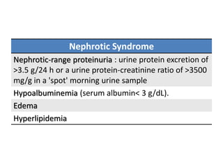
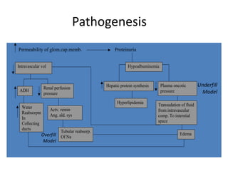
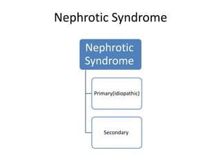
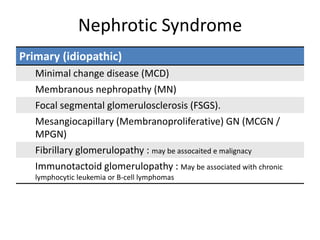
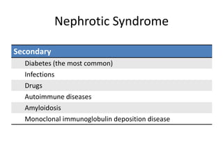
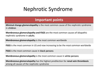
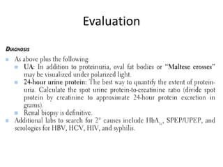
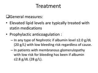
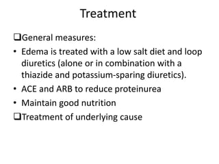
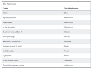
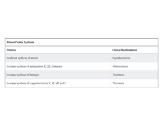
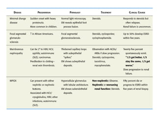
Nephrotic Syndrome
Abdulrahman Mashi
Nephrotic Syndrome
Nephrotic-range proteinuria : urine protein excretion of
>3.5 g/24 h or a urine protein-creatinine ratio of >3500
mg/g in a 'spot' morning urine sample
Hypoalbuminemia (serum albumin< 3 g/dL).
Edema
Hyperlipidemia
Pathogenesis
Permeability of glom.cap.memb. Proteinuria
Intravascular vol
ADH
Renal perfusion
pressure
Water
Reabsorptn
In
Collecting
ducts
Actv. reinin
Ang. ald. sys
Tubular reabsorp.
Of Na
Hypoalbuminemia
Hepatic protein synthesis Plasma oncotic
pressure
Hyperlipidemia Transudation of fluid
from intravascular
comp. To interstial
space
Edema
Underfill
Model
Overfill
Model
• Elevated lipids occur from a combination of
– increased hepatic apolipoprotein synthesis in
response to a low plasma oncotic pressure
– decreased activity of key enzymes such as
lipoprotein lipase and lecithin-cholesterol
acyltransferase.
Nephrotic Syndrome
Nephrotic
Syndrome
Primary(Idiopathic)
Secondary
Nephrotic Syndrome
Primary (idiopathic)
Minimal change disease (MCD)
Membranous nephropathy (MN)
Focal segmental glomerulosclerosis (FSGS).
Mesangiocapillary (Membranoproliferative) GN (MCGN /
MPGN)
Fibrillary glomerulopathy : may be assocaited e malignacy
Immunotactoid glomerulopathy : May be associated with chronic
lymphocytic leukemia or B-cell lymphomas
Nephrotic Syndrome
Secondary
Diabetes (the most common)
Infections
Drugs
Autoimmune diseases
Amyloidosis
Monoclonal immunoglobulin deposition disease
Nephrotic Syndrome
Important points
Minimal change glomerulopathy is the most common cause of the nephrotic syndrome
in children
Membranous glomerulopathy and FSGS are the most common causes of idiopathic
nephrotic syndrome in adults.
Membranous glomerulopathy is the most common worldwide
FSGS is the most common in US and now increasing to be the most common worldwide
FSGS is the most common cause in black persons
Membranous glomerulopathy is the most common cause in white persons
Membranous glomerulopathy has the highest predilection for renal vein thrombosis
among all causes of the nephrotic syndrome
Evaluation
Treatment
General measures:
• Elevated lipid levels are typically treated with
statin medications
• Prophylactic anticoagulation :
– In any type of Nephrotic if albumin level ≤2.0 g/dL
(20 g/L) with low bleeding risk regardless of cause.
– In patients with membranous glomerulopathy
with low risk for bleeding has been if albumin
≤2.8 g/dL (28 g/L).
Treatment
General measures:
• Edema is treated with a low salt diet and loop
diuretics (alone or in combination with a
thiazide and potassium-sparing diuretics).
• ACE and ARB to reduce proteinurea
• Maintain good nutrition
Treatment of underlying cause
Complications
FSGS
• Causes:
– Idiopathic : Some cases of idiopathic FSGS are thought to be related to a
circulating plasma factor because a significant number of patients show
recurrence after kidney transplantation.
– Genetic mutations to podocyte proteins
– Secondary :
• Hyperfiltration injury to the glomerulus (DM, HTN
&Morbid obesity )
• Reduced kidney mass: progressive kidney disease, obesity,
sickle cell disease, reflux nephropathy, congenital small kidneys
• Direct podocyte injury: infections (HIV) and drugs
(pamidronate, interferon and NSAID). Lymphoma
FSGS
Five subtypes
not otherwise specified
perihilar variant
tip variant
cellular variant
collapsing variant (may be associated with HIV infection,
heroin use, parvovirus infection, or pamidronate exposure)
FSGS
Clinical Manifestations :
• Either asymptomatic proteinuria or edema.
• More than two thirds of patients are fully nephrotic
at presentation
• Subnephrotic proteinuria may occur, especially with
secondary FSGS from hyperfiltration injury.
• Hypertension, microscopic hematuria, and varying
degrees of kidney failure are common.
FSGS
Diagnosis
The hallmark of FSGS is the presence of segmental
scars in some glomeruli.
Electron microscopy shows visceral epithelial cell foot
process effacement
no immune deposits.
Treatment and Prognosis
• In idiopathic FSGS, only a minority of patients
experience a spontaneous remission.
• Therefore, treatment is indicated in most patients.
• Therapy is usually with glucocorticoids or calcineurin
inhibitors, both at the time of initial presentation and
for relapsing disease.
• A complete or partial remission may be seen in up to
40% to 60% of patients using these treatments.
• FSGS recurs in the transplanted kidney in 30% or more
of cases.
• In secondary FSGS caused by infection or drugs,
treatment of the infection or removal of the
offending agent may halt progression of the
disease and improve symptoms.
• In obese patients with likely secondary FSGS,
weight loss is sometimes associated with a drop
in proteinuria, as is the use of ACE inhibitors or
angiotensin receptor blockers (ARBs), and is the
preferred initial therapy.
Membranous Glomerulopathy
• Primary or secondary
• Primary form (most common worldwide):
antiphospholipase A2 receptor autoantibodies
can be found in 75% of cases lead to activation
of complement and damage the GBM.
Membranous Glomerulopathy
• May be associated with or secondary to:
– SLE
– Infections: hepatitis B and C virus infections; syphilis;
malaria
– Medication exposure: penicillamine; NSAIDs; TNF-α
inhibitors; tiopronin
– Mercury or gold exposure
– Malignancies: Solid organ Malignancy (bladder, breast,
colon, lung, pancreas, prostate, stomach carcinoma);
carcinoid; sarcomas; lymphomas; leukemias
– Thyroid disease
Clinical Manifestations
• Like other causes of the nephrotic syndrome
(edema, hypertension, microhematuria), but
the propensity to thromboembolic events
(particularly renal vein thrombosis) is much
higher.
• Secondary causes should be sought,
particularly occult malignancy in older
patients.
Membranous Glomerulopathy
Diagnosis
Light microscopy shows glomerular capillary loops that
often appear thickened without any proliferative
lesions.
Immunofluorescence and electron microscopy show
subepithelial immune dense deposits.
Where available, PLA2R antibodies should be measured.
Treatment
• Up to one third of patients with idiopathic MG remit
spontaneously in 6 to 12 months.
• Conservative management is appropriate during this
period.
• In patients with idiopathic MG who have persistent
disease after 6 to 12 months or who have worsening
kidney function or a thromboembolic event, regimens
containing alternating glucocorticoids with
cyclophosphamide or calcineurin inhibitors
(cyclosporine or tacrolimus) may be used.
Treatment
• Other options for relapsing or refractory
disease include mycophenolate mofetil,
adrenocorticotropic hormone, and the anti–B-
cell antibody rituximab.
• For secondary causes of MG treat the
underlying cause like treatment of hepatitis B
virus infection with antiviral agents
Minimal Change Glomerulopathy
• Minimal change disease is the most common cause of
idiopathic nephrotic syndrome in children and accounts for
approximately 10% of cases in adults.
• The pathogenesis of MCG is not fully understood but is
thought to be related to production of cytokines by immune
cells that lead to podocyte dysfunction.
• A history of viral respiratory infection, atopy, or
immunization preceding the onset of edema may be present.
Minimal Change Glomerulopathy
• Primary form (Idiopathic)
• secondary :
– Medications (such as NSAIDs, lithium,
pamidronate, rifampin and the interferons)
– Malignancies (such as Hodgkin lymphoma , other
lymphoproliferative disorders and thymoma)
– Infections: Infectious Mononucleosis and TB
Clinical Manifestations
• Patients with MCG typically present with
acute onset of edema and weight gain due to
fluid retention.
• Urine protein levels tend to be significantly
elevated (urine protein-creatinine ratio
typically 5000-10,000 mg/g).
MCD
Diagnosis
Normal glomeruli on both light and immunofluorescence
microscopy.
The tubules may show lipid accumulation.
On electron microscopy, the GBM is normal with
extensive effacement of visceral epithelial foot processes.
Treatment
• Patients typically respond to glucocorticoids (1mg/kg/d or
2mg/kg every other day )within 8 to 16 weeks.
• Complete remission achieved in around 70 %
• Relapse is common, and in up to 40% of patients, the
course of MCG is one of remission followed by relapse.
• For frequently relapsing or glucocorticoid-dependent
disease, treatment options include cyclophosphamide,
calcineurin inhibitors (tacrolimus or cyclosporine),
mycophenolate mofetil, and rituximab.
Diabetic Nephropathy
• Diabetic nephropathy (DN) is the leading
cause of ESKD in the United States
• The strongest clinical indicator for progressive
kidney disease is the level of urine albumin.
Risk factors for developing DN
older age
race (American Indian, Mexican American, and black)
poor glycemic control
hypertension
cigarette smoking
family history of kidney disease
Clinical Manifestations
• Moderately increased albuminuria (formerly known as
microalbuminuria), defined as a urine albumin-creatinine
ratio of 30 to 300 mg/g, is typically the first abnormality
seen in patients with type 1 and type 2 diabetes.
• Overt nephropathy (formerly known as overt proteinurea)
(urine protein-creatinine ratio >300 mg/g) occurs around 10
to 15 years from disease onset in approximately 50% of
patients with moderately increased albuminuria and
progresses to ESKD in most patients.
Diagnosis
Annual testing for albuminuria should begin at the time
of diagnosis in type 2 diabetes and 5 years after diagnosis
in type 1 diabetes.
Kidney biopsy is not indicated unless there is a suspicion
of another glomerular disease.
On electron microscopy, the GBM is normal with
extensive effacement of visceral epithelial foot processes.
Moderately increased albuminuria defined as a urine
albumin-creatinine ratio of 30 to 300 mg/g
Overt nephropathy (urine protein-creatinine ratio >300
mg/g)
Indications for kidney biopsy
acute onset of the nephrotic syndrome
findings of another systemic disease
short duration from onset of diabetes to onset of
proteinuria
abnormal serologies
rapid rate of progression of kidney dysfunction
Pathology
• DN affects every compartment of the kidney.
• In the glomerulus, there is expansion of the
mesangium and thickening of the basement
membrane, followed by focal (nodular) sclerosis
(the Kimmelstiel-Wilson lesion) then global
sclerosis of the glomerulus.
• Interstitial fibrosis, tubular atrophy with
thickened tubular basement membranes, and
arteriolosclerosis are also seen.
Treatment
• Achieving targets of glycemic control
(hemoglobin A1c <7%) and blood pressure
(<140/90 mm Hg.
• In patients who have diabetes with
moderately increased albuminuria or severely
increased albuminuria , ACE inhibitors or ARBs
have been shown to slow progression.
Read about
• MPGN
• Amylodosis
• Multiple Myloma
• HIV associated nephropathy
• Hepatitis B associated kidney disease
References
317081254-Nephrotic-Syndrome-2016.pptx

317081254-Nephrotic-Syndrome-2016.pptx

  • 1.
  • 2.
    Nephrotic Syndrome Nephrotic-range proteinuria: urine protein excretion of >3.5 g/24 h or a urine protein-creatinine ratio of >3500 mg/g in a 'spot' morning urine sample Hypoalbuminemia (serum albumin< 3 g/dL). Edema Hyperlipidemia
  • 3.
    Pathogenesis Permeability of glom.cap.memb.Proteinuria Intravascular vol ADH Renal perfusion pressure Water Reabsorptn In Collecting ducts Actv. reinin Ang. ald. sys Tubular reabsorp. Of Na Hypoalbuminemia Hepatic protein synthesis Plasma oncotic pressure Hyperlipidemia Transudation of fluid from intravascular comp. To interstial space Edema Underfill Model Overfill Model
  • 4.
    • Elevated lipidsoccur from a combination of – increased hepatic apolipoprotein synthesis in response to a low plasma oncotic pressure – decreased activity of key enzymes such as lipoprotein lipase and lecithin-cholesterol acyltransferase.
  • 5.
  • 6.
    Nephrotic Syndrome Primary (idiopathic) Minimalchange disease (MCD) Membranous nephropathy (MN) Focal segmental glomerulosclerosis (FSGS). Mesangiocapillary (Membranoproliferative) GN (MCGN / MPGN) Fibrillary glomerulopathy : may be assocaited e malignacy Immunotactoid glomerulopathy : May be associated with chronic lymphocytic leukemia or B-cell lymphomas
  • 7.
    Nephrotic Syndrome Secondary Diabetes (themost common) Infections Drugs Autoimmune diseases Amyloidosis Monoclonal immunoglobulin deposition disease
  • 8.
    Nephrotic Syndrome Important points Minimalchange glomerulopathy is the most common cause of the nephrotic syndrome in children Membranous glomerulopathy and FSGS are the most common causes of idiopathic nephrotic syndrome in adults. Membranous glomerulopathy is the most common worldwide FSGS is the most common in US and now increasing to be the most common worldwide FSGS is the most common cause in black persons Membranous glomerulopathy is the most common cause in white persons Membranous glomerulopathy has the highest predilection for renal vein thrombosis among all causes of the nephrotic syndrome
  • 9.
  • 10.
    Treatment General measures: • Elevatedlipid levels are typically treated with statin medications • Prophylactic anticoagulation : – In any type of Nephrotic if albumin level ≤2.0 g/dL (20 g/L) with low bleeding risk regardless of cause. – In patients with membranous glomerulopathy with low risk for bleeding has been if albumin ≤2.8 g/dL (28 g/L).
  • 11.
    Treatment General measures: • Edemais treated with a low salt diet and loop diuretics (alone or in combination with a thiazide and potassium-sparing diuretics). • ACE and ARB to reduce proteinurea • Maintain good nutrition Treatment of underlying cause
  • 12.
  • 15.
    FSGS • Causes: – Idiopathic: Some cases of idiopathic FSGS are thought to be related to a circulating plasma factor because a significant number of patients show recurrence after kidney transplantation. – Genetic mutations to podocyte proteins – Secondary : • Hyperfiltration injury to the glomerulus (DM, HTN &Morbid obesity ) • Reduced kidney mass: progressive kidney disease, obesity, sickle cell disease, reflux nephropathy, congenital small kidneys • Direct podocyte injury: infections (HIV) and drugs (pamidronate, interferon and NSAID). Lymphoma
  • 16.
    FSGS Five subtypes not otherwisespecified perihilar variant tip variant cellular variant collapsing variant (may be associated with HIV infection, heroin use, parvovirus infection, or pamidronate exposure)
  • 17.
    FSGS Clinical Manifestations : •Either asymptomatic proteinuria or edema. • More than two thirds of patients are fully nephrotic at presentation • Subnephrotic proteinuria may occur, especially with secondary FSGS from hyperfiltration injury. • Hypertension, microscopic hematuria, and varying degrees of kidney failure are common.
  • 18.
    FSGS Diagnosis The hallmark ofFSGS is the presence of segmental scars in some glomeruli. Electron microscopy shows visceral epithelial cell foot process effacement no immune deposits.
  • 19.
    Treatment and Prognosis •In idiopathic FSGS, only a minority of patients experience a spontaneous remission. • Therefore, treatment is indicated in most patients. • Therapy is usually with glucocorticoids or calcineurin inhibitors, both at the time of initial presentation and for relapsing disease. • A complete or partial remission may be seen in up to 40% to 60% of patients using these treatments. • FSGS recurs in the transplanted kidney in 30% or more of cases.
  • 20.
    • In secondaryFSGS caused by infection or drugs, treatment of the infection or removal of the offending agent may halt progression of the disease and improve symptoms. • In obese patients with likely secondary FSGS, weight loss is sometimes associated with a drop in proteinuria, as is the use of ACE inhibitors or angiotensin receptor blockers (ARBs), and is the preferred initial therapy.
  • 21.
    Membranous Glomerulopathy • Primaryor secondary • Primary form (most common worldwide): antiphospholipase A2 receptor autoantibodies can be found in 75% of cases lead to activation of complement and damage the GBM.
  • 22.
    Membranous Glomerulopathy • Maybe associated with or secondary to: – SLE – Infections: hepatitis B and C virus infections; syphilis; malaria – Medication exposure: penicillamine; NSAIDs; TNF-α inhibitors; tiopronin – Mercury or gold exposure – Malignancies: Solid organ Malignancy (bladder, breast, colon, lung, pancreas, prostate, stomach carcinoma); carcinoid; sarcomas; lymphomas; leukemias – Thyroid disease
  • 23.
    Clinical Manifestations • Likeother causes of the nephrotic syndrome (edema, hypertension, microhematuria), but the propensity to thromboembolic events (particularly renal vein thrombosis) is much higher. • Secondary causes should be sought, particularly occult malignancy in older patients.
  • 24.
    Membranous Glomerulopathy Diagnosis Light microscopyshows glomerular capillary loops that often appear thickened without any proliferative lesions. Immunofluorescence and electron microscopy show subepithelial immune dense deposits. Where available, PLA2R antibodies should be measured.
  • 25.
    Treatment • Up toone third of patients with idiopathic MG remit spontaneously in 6 to 12 months. • Conservative management is appropriate during this period. • In patients with idiopathic MG who have persistent disease after 6 to 12 months or who have worsening kidney function or a thromboembolic event, regimens containing alternating glucocorticoids with cyclophosphamide or calcineurin inhibitors (cyclosporine or tacrolimus) may be used.
  • 26.
    Treatment • Other optionsfor relapsing or refractory disease include mycophenolate mofetil, adrenocorticotropic hormone, and the anti–B- cell antibody rituximab. • For secondary causes of MG treat the underlying cause like treatment of hepatitis B virus infection with antiviral agents
  • 28.
    Minimal Change Glomerulopathy •Minimal change disease is the most common cause of idiopathic nephrotic syndrome in children and accounts for approximately 10% of cases in adults. • The pathogenesis of MCG is not fully understood but is thought to be related to production of cytokines by immune cells that lead to podocyte dysfunction. • A history of viral respiratory infection, atopy, or immunization preceding the onset of edema may be present.
  • 29.
    Minimal Change Glomerulopathy •Primary form (Idiopathic) • secondary : – Medications (such as NSAIDs, lithium, pamidronate, rifampin and the interferons) – Malignancies (such as Hodgkin lymphoma , other lymphoproliferative disorders and thymoma) – Infections: Infectious Mononucleosis and TB
  • 30.
    Clinical Manifestations • Patientswith MCG typically present with acute onset of edema and weight gain due to fluid retention. • Urine protein levels tend to be significantly elevated (urine protein-creatinine ratio typically 5000-10,000 mg/g).
  • 31.
    MCD Diagnosis Normal glomeruli onboth light and immunofluorescence microscopy. The tubules may show lipid accumulation. On electron microscopy, the GBM is normal with extensive effacement of visceral epithelial foot processes.
  • 32.
    Treatment • Patients typicallyrespond to glucocorticoids (1mg/kg/d or 2mg/kg every other day )within 8 to 16 weeks. • Complete remission achieved in around 70 % • Relapse is common, and in up to 40% of patients, the course of MCG is one of remission followed by relapse. • For frequently relapsing or glucocorticoid-dependent disease, treatment options include cyclophosphamide, calcineurin inhibitors (tacrolimus or cyclosporine), mycophenolate mofetil, and rituximab.
  • 34.
    Diabetic Nephropathy • Diabeticnephropathy (DN) is the leading cause of ESKD in the United States • The strongest clinical indicator for progressive kidney disease is the level of urine albumin.
  • 35.
    Risk factors fordeveloping DN older age race (American Indian, Mexican American, and black) poor glycemic control hypertension cigarette smoking family history of kidney disease
  • 37.
    Clinical Manifestations • Moderatelyincreased albuminuria (formerly known as microalbuminuria), defined as a urine albumin-creatinine ratio of 30 to 300 mg/g, is typically the first abnormality seen in patients with type 1 and type 2 diabetes. • Overt nephropathy (formerly known as overt proteinurea) (urine protein-creatinine ratio >300 mg/g) occurs around 10 to 15 years from disease onset in approximately 50% of patients with moderately increased albuminuria and progresses to ESKD in most patients.
  • 38.
    Diagnosis Annual testing foralbuminuria should begin at the time of diagnosis in type 2 diabetes and 5 years after diagnosis in type 1 diabetes. Kidney biopsy is not indicated unless there is a suspicion of another glomerular disease. On electron microscopy, the GBM is normal with extensive effacement of visceral epithelial foot processes. Moderately increased albuminuria defined as a urine albumin-creatinine ratio of 30 to 300 mg/g Overt nephropathy (urine protein-creatinine ratio >300 mg/g)
  • 39.
    Indications for kidneybiopsy acute onset of the nephrotic syndrome findings of another systemic disease short duration from onset of diabetes to onset of proteinuria abnormal serologies rapid rate of progression of kidney dysfunction
  • 40.
    Pathology • DN affectsevery compartment of the kidney. • In the glomerulus, there is expansion of the mesangium and thickening of the basement membrane, followed by focal (nodular) sclerosis (the Kimmelstiel-Wilson lesion) then global sclerosis of the glomerulus. • Interstitial fibrosis, tubular atrophy with thickened tubular basement membranes, and arteriolosclerosis are also seen.
  • 41.
    Treatment • Achieving targetsof glycemic control (hemoglobin A1c <7%) and blood pressure (<140/90 mm Hg. • In patients who have diabetes with moderately increased albuminuria or severely increased albuminuria , ACE inhibitors or ARBs have been shown to slow progression.
  • 42.
    Read about • MPGN •Amylodosis • Multiple Myloma • HIV associated nephropathy • Hepatitis B associated kidney disease
  • 44.