Download to read offline
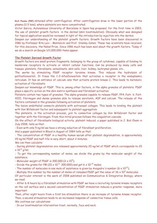
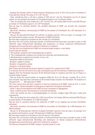


This document discusses regenerating facial tissues through platelet-derived growth factor (PDGF) injections rather than surgical facelifts. It provides calculations showing that the number of PDGF molecules released during injections is sufficient to stimulate the fibroblasts in the treated facial areas. Research studies cited found that PDGF injections led to angiogenesis within a week, maximum fibroblast activation within a month, and new collagen formation within two months, regenerating skin tissues. The technique aims to biologically rejuvenate facial tissues rather than just achieving aesthetic youth through damaging surgical procedures.


