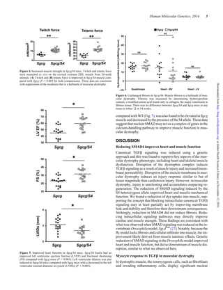
1) Muscle injury in mice lacking the protein γ-sarcoglycan causes an increase in SMAD signaling, which contributes to transforming growth factor β (TGFβ) signaling.
2) Reducing SMAD signaling through introducing a heterozygous mutation of the gene SMAD4 in γ-sarcoglycan mice led to improved body mass, cardiac function, and muscle force compared to γ-sarcoglycan mice.
3) While fibrosis was not reduced, membrane damage was decreased, suggesting intracellular SMAD4 targets are important for mediating muscle and heart dysfunction in muscular dystrophy.
![background were used (20). Mice were selected to be homozy-
gous for the Ltbp4 deletion (15) to avoid the modifier effect
of the insertion/deletion in the Ltbp4. SBE-luc mice, formally
B6.Cg-Tg(SBE/TK-luc)7Twc/J (17) (Jackson Laboratory strain
005999), were used to detect SMAD activity. The SMAD4f
allele used in these studies flanked exon 2 of SMAD4 with loxP
sites (18). These mice were bred with Mox2-cre mice, which
express Cre recombinase in the germline (32) (Jackson Labora-
tory strain 003755). The resulting SMADN/+
mice were out-
crossed to remove the Mox2-cre transgene, bred with Sgcg
mice, and then bred to additional Sgcg null mice to generate
appropriate genotypes (Sgcg and Sgcg/S4 indicating SMAD4
heterozygous mice).
Mice were analyzed at 8,12or24weeksasindicatedand com-
pared with littermate controls. Mice were housed under the aus-
pices of the Animal Care and Use Committee and Animal
Resources Center at the University of Chicago. The order of
harvest was chosen to minimize interference of one assay with
the next. Two days prior to harvest, mice were weighed and
injected Evan’s blue. On the subsequent day, grip strength meas-
urement and echocardiography were performed. On the day of
harvest, serum was collected immediately prior to sacrifice,
and collection of the muscles for ex vivo measurement occurred
immediately after. Tissues were weighed at the time of collec-
tion and the tibia length was measured using a millimeter ruler
after being stripped of muscle.
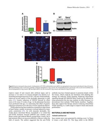
Antibodies
Goat anti-phosphorylated SMAD2/3 (pSMAD2/3), # sc11769
was from Santa Cruz Biotechnology or from Abcam to
pSMAD3 (ab51451). The sorcin antibody was a generous gift
from Hector Valdivia (33). Rabbit anti-laminin # L9393 was
from Sigma. Goat anti-rabbit IgG # 111-035-003 was from
Jackson Immunoresearch. Donkey anti-rabbit IgG Alexa 488
conjugate (DaR-488), # A-21208 and Donkey anti-goat IgG
Alexa 594 conjugate (DaG-594), # A-11058 were from Invitro-
gen. Anti-TGFb antibody (1D11) (12) and control 13C4 were
obtained from Genzyme Corporation (Framingham, MA, USA).
Ex vivo muscle contraction
Intact extensor EDL muscles were excised in regular rodent
Ringer’s solution (in mM: 146 NaCl, 5 KCl, 2 CaCl2, 1 MgCl2
and 10 HEPES, pH 7.4) essentially according to the
TREAT-NMD standard operating procedure (34). Muscles
were suspended in a vertical tissue bath containing oxygenated
Kreb’s solution (in mM: 121 NaCl, 5 KCl, 1.8 CaCl2, 0.5
MgCl2, 0.1 EDTA, 0.4 NaH2PO4, 24 NaHCO3 and 5 glucose,
pH 7.4, with continuous bubbling 95% O2/5% CO2). The distal
tendon was attached to a fixed support with 6-0 silk suture,
while the proximal tendon was tied to the lever arm of a force
transducer (Aurora Scientific, 300C-LR). The isolated muscles
were stimulated with 0.5 ms pulses at 1 A via two parallel plat-
inum electrodes flaking the muscle. The length of each EDL
was adjusted to that which produced the maximal twitch force
(L0), which was measured using fine calipers. Maximal twitch
and tetanic force were elicited with a series of 3 trains, 3 min
apart. The data were combined from three separate animals of
each genotype. Males were used for these studies since there
were baseline differences between sexes in Sgcg mice. Subse-
quently, each EDL was subjected to an eccentric contraction
protocol, which consisted of five 700 ms, 80 Hz tetani with a
lengthening by 10% of L0 during the last 200 ms of each
tetanus. At the conclusion of each experiment, each muscle
was blotted and immediately weighed. All data were analyzed
offline following data import into Clampfit software (Molecular
Devices, Clampfit v10.2.0.14). All force measurements (P0)
were normalized to cross-sectional area and expressed as specif-
ic force (sP0; mN/mm2
), which was calculated according to the
following equation:
sP0 =
P0
{muscle mass/[(L0 × 0.44) × 1.06 g/m2]}
,
where 0.44 is the muscle length to fiber length ratio for EDL, and
1.06 is the density of muscle (34).
Immunofluorescence microscopy
After eccentric contraction, EDL muscles were maintained in
oxygenated nutrient back for 1 h to allow for signaling, then
snap frozen in liquid nitrogen-chilled isopentane. Samples
were processed similarly to a previously published method
(35). Ten micrometers sections were cut and sections were
fixed for 10 min in ethanol on ice. Sections were blocked for
1 h at 48C in 10% fetal bovine serum (FBS), 0.03% Triton X-
100 in phosphate-buffered saline (PBS). Samples were incu-
bated overnight at 48C in 1% FBS, 0.03% Triton X-100, PBS
with 1 : 100 dilution of anti-pSMAD2/3 and 1:200 anti-laminin.
Sampleswerewashedthreetimeswith0.03%TritonX-100,PBS
on ice for 15 min. DaG-594 and DaR-488 were applied for 3 h at
48C in 1% FBS 0.03% Triton X-100, PBS. Slides were again
washed, and mounted using Vectashield with DAPI (Vector
Laboratories). Images werecollected at ×40 originalmagnifica-
tion using an Axiophot microscope with iVision software and
manipulated within NIH guidelines using Adobe Photoshop
CS4 and ImageJ.
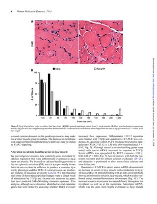
Histology
A cross-sectional disc of muscle was harvested from the
mid-belly of the quadriceps muscle. The disc was fixed in forma-
lin, dehydrated, paraffin embedded, sectioned at 6 mm thickness
and stained using hematoxylin and eosin (H&E). For analysis of
central nuclei, five random fields each were collected from five
mice of each genotype (total 25 sections). The number of
fibers and number of fibers with at least one non-peripheral
nucleus were counted and the ratio calculated. For fiber size,
fibers were outlined using Adobe Photoshop CS4 or the GNU
Image Manipulation Program 2.0.2. Fiber size was determined
using ImageJ. Two hundred fibers were counted for each
genotype.
Echocardiography
Mice were depilated, anesthetized with isoflurane and placed
on a 378C warming pad. Anesthesia was titrated to produce a
heart rate of 350–450 beats/min. Left ventricular dimensions
were quantified by transthoracic echocardiography using a
8 Human Molecular Genetics, 2014
atGalterHealthSciencesLibraryonNovember12,2014http://hmg.oxfordjournals.org/Downloadedfrom](https://image.slidesharecdn.com/f9c8a3ad-aa4c-4f8e-8b13-58253d6f430d-150413155938-conversion-gate01/85/Hum-Mol-Genet-2014-Goldstein-hmg-ddu390-8-320.jpg)


![Pells et al [2015] PLoS ONE 10[7] e0131102](https://cdn.slidesharecdn.com/ss_thumbnails/f79bb09e-8eb1-41e7-8042-28c0aa4a48c6-150720142907-lva1-app6891-thumbnail.jpg?width=640&height=640&fit=bounds)


