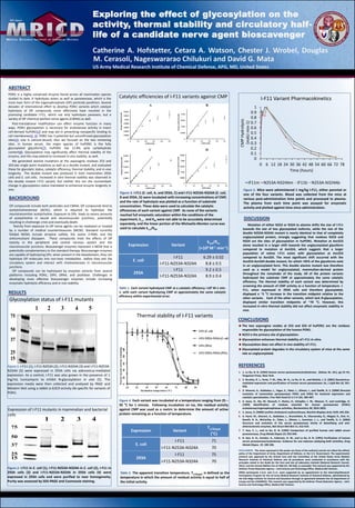
Exploring the effect of glycosylation on the
activity, thermal stability and circulatory half-
life of a candidate nerve agent bioscavenger
Catherine A. Hofstetter, Cetara A. Watson, Chester J. Wrobel, Douglas
M. Cerasoli, Nageswararao Chilukuri and David G. Mata
US Army Medical Research Institute of Chemical Defense, APG, MD, United States
BACKGROUND
ABSTRACT
OP compounds include both pesticides and CWNA. OP compounds bind to
acetylcholinesterase (AChE), which is required to hydrolyze the
neurotransmitter acetylcholine. Exposure to OPs leads to excess amounts
of acetylcholine in neural and neuromuscular junctions, potentially
resulting in cholinergic crisis and eventually death.
Toxicity from exposure to OP nerve agents can be mediated or treated
by a number of medical countermeasures (MCM). Standard currently
fielded MCMs include atropine sulfate, the oxime 2-PAM, and the
anticonvulsant diazapam. These compounds treat the effects of OP
toxicity in the peripheral and central nervous system and the
neuromuscular junctions. Bioscavenger enzymes represent a MCM that is
potentially complementary to the standard therapy. Bioscavenger enzymes
are capable of hydrolyzing OPs; when present in the bloodstream, they can
hydrolyze OP molecules into non-toxic metabolites before they exit the
circulatory system and interact with cholinesterases in neuromuscular
junctions.
OP compounds can be hydrolyzed by enzymes variants from several
platforms including PON1, OPH, OPAA, and prolidase. Challenges in
developing more effective bioscavenger enzymes include increasing
enzymatic hydrolytic efficiency and in vivo stability.
Figure 3. text
PON1 is a highly conserved enzyme found across all mammalian species
studied to date. It hydrolyzes esters as well as paraoxonase, which is the
more toxic form of the organophosphate (OP) pesticide parathion. Several
decades of international effort to develop PON1 variants which catalyze
hydrolysis of OP compounds more effectively have resulted in the
promising candidate I-F11, which not only hydrolyzes paraoxon, but a
variety of OP chemical warfare nerve agents (CWNA) as well.
Post-translational modification can affect enzyme function in many
ways. PON1 glycosylation is necessary for arylesterase activity in insect
cell-derived huPON1(2) and may aid in preventing nonspecific binding to
cell membranes(5, 6). PON1 has 3 potential but unconfirmed glycosylation
sites(1); one is calcium-bound, thus we focused on the two remaining
sites. In human serum, the major species of huPON1 is the fully
glycosylated glycoform(7); huPON1 has 15.8% w/w carbohydrate
content(8). Glycosylation may significantly affect thermal stability of the
enzyme, and this may extend to increased in vivo stability as well.
We generated alanine mutations at the asparagine residues 253 and
324,two single point mutations as well as a double mutant, and evaluated
these for glycolytic status, catalytic efficiency, thermal stability, and in vivo
longevity. The double mutant was produced in both mammalian 293A
cells and E. coli cells. Increased in vitro thermal stability was observed in
the double mutant I-F11 variant, but neither this nor the concomitant
change in glycosylation status translated to enhanced enzyme longevity in
vivo.
RESULTS
1: I-F11
2: I-F11-N253A
3: I-F11-N324A
4: I-F11-N253A-N324A
5: I-F11 + 1 mg/mL tunicamycin
1 2 3 4 5
75
50
37
25
15
Glycosylation status of I-F11 mutants
Figure 1. I-F11 (1), I-F11-N253A (2), I-F11-N324A (3) and I-F11-N253A-
N324A (5) were expressed in 293A cells via adenovirus-mediated
expression. As a control, I-F11 was also grown in the presence of 1
mg/mL tunicamycin to inhibit N-glycosylation in vivo (5). The
expression media were then collected and analyzed by PAGE and
Western blot using a rabbit α-G3C9 activity Ab specific for variants of
PON1.
Table 1. Each variant hydrolyzed CMP at a catalytic efficiency >106 M-1 min-
1, with each variant hydrolyzing CMP at approximately the same catalytic
efficiency within experimental error.
Expression Variant
kcat/Km
(×106 M-1 min-1)
E. coli
I-F11 6.29 ± 0.02
I-F11-N253A-N324A 8.8 ± 0.5
293A
I-F11 9.2 ± 0.5
I-F11-N253A-N324A 8.9 ± 0.4
1 2 3 4
75
50
37
25
15
1: I-F11 (E. coli)
2: I-F11-N253A-N324A (E. coli)
3: I-F11 (293A)
4: I-F11-N253A-N324A (293A)
Expression of I-F11 mutants in mammalian and bacterial
cells
Figure 2. I-F11 in E. coli (1), I-F11-N253A-N324A in E. coli (2), I-F11 in
293A cells (3) and I-F11-N253A-N324A in 293A cells (4) were
expressed in 293A cells and were purified to near homogeneity.
Purity was assessed by SDS-PAGE and Coomassie staining.
Catalytic efficiencies of I-F11 variants against CMP
A B
C D
Figure 3. I-F11 (E. coli, A, and 293A, C) and I-F11-N253A-N324A (E. coli,
B and 293A, D) were incubated with increasing concentrations of CMP,
and the rate of hydrolysis was plotted as a function of substrate
concentration. These data were used to calculate the catalytic
efficiencies of each variant against CMP. As none of the variants
reached full enzymatic saturation within the conditions of the
experiment, kcat and KM were not able to be accurately determined
separately, and the linear portion of the Michaelis-Menten curve was
used to calculate kcat/KM.
CONCLUSIONS
The two asparagine resides at 253 and 324 of huPON1 are the residues
responsible for glycosylation of the human PON1.
N253 is the primary site of glycosylation.
Glycosylation enhances thermal stability of I-F11 in vitro.
Glycosylation does not affect in vivo stability of I-F11.
Glycosylated protein degrades in the circulatory system of mice at the same
rate as unglycosylated.
DISCLAIMERS. The views expressed in this poster are those of the author(s) and do not reflect the official
policy of the Department of Army, Department of Defense, or the U.S. Government. The experimental
protocol was approved by the Animal Care and Use Committee at the United States Army Medical
Research Institute of Chemical Defense and all procedures were conducted in accordance with the
principles stated in the Guide for the Care and Use of Laboratory Animals (National Research Council,
2011), and the Animal Welfare Act of 1966 (P.L. 89-544), as amended. This research was supported by the
Defense Threat Reduction Agency – Joint Science and Technology Office, Medical S&T Division.
ORISE participants C.A.H. and C.L.C. were supported by an appointment to the Internship/Research
Participation Program for the US Army Medical Research Institute of Chemical Defense, administered by
the Oak Ridge Institute for Science and Education through an agreement between the US Department of
Energy and the USAMRICD. This research was supported by the Defense Threat Reduction Agency – Joint
Science and Technology Office, Medical S&T Division.
Mutation of either N253 or N324 to alanine shifts the size of I-F11
towards the size of less glycosylated isoforms, while the size of the
double N253A-N324A mutant is nearly identical to that of completely
unglycosylated protein, strongly suggesting that residues N253 and
N324 are the sites of glycosylation in huPON1. Mutation at Asn253
alone resulted in a larger shift towards the unglycosylated glycoform
compared to mutation at Asn324, which suggests that a larger
population of native I-F11 exists with glycosylation at Asn253
compared to Asn324. The most significant shift occurred with the
Asn253-Asn324 double mutant, for which >95% of the glycoforms exist
in an unglycosylated form. This double alanine mutant was therefore
used as a model for unglycosylated, mammalian-derived protein
throughout the remainder of the study. All of the protein variants
hydrolyzed the substrate CMP at approximately the same catalytic
efficiency. The thermal stability of each variant was determined by
screening the amount of CMP activity as a function of temperature. I-
F11, when expressed in 293A cells and therefore glycosylated,
displayed a ~5 °C increase in the transition midpoint relative to the
other variants. Each of the other variants, which lack N-glycosylation,
displayed similar transition midpoints of ~70 °C. However, this
increased in vitro thermal stability did not affect enzymatic stability in
vivo.
DISCUSSION
REFERENCES
1. 1. La Du, B. N. (1992) Human serum paraoxonase/arylesterase, (Kalow, W., Ed.), pp 51–91,
Pergamon Press, New York.
2. 2. Brushia, R. J., Forte, T. M., Oda, M. N., La Du, B. N., and Bielicki, J. K. (2001) Baculovirus-
mediated expression and purification of human serum paraoxonase 1A, J Lipid Res 42, 951-
958.
3. 3. Aharoni, A., Gaidukov, L., Yagur, S., Toker, L., Silman, I., and Tawfik, D. S. (2004) Directed
evolution of mammalian paraoxonases PON1 and PON3 for bacterial expression and
catalytic specialization, Proc Natl Acad Sci U S A 101, 482-487.
4. 4. Josse, D., Xie, W., Renault, F., Rochu, D., Schopfer, L. M., Masson, P., and Lockridge, O.
(1999) Identification of residues essential for human paraoxonase (PON1)
arylesterase/organophosphatase activities, Biochemistry 38, 2816-2825.
5. 5. Jonas, A. (2000) Lecithin cholesterol acyltransferase, Biochim Biophys Acta 1529, 245-256.
6. 6. Harel, M., Aharoni, A., Gaidukov, L., Brumshtein, B., Khersonsky, O., Meged, R., Dvir, H.,
Ravelli, R. B., McCarthy, A., Toker, L., Silman, I., Sussman, J. L., and Tawfik, D. S. (2004)
Structure and evolution of the serum paraoxonase family of detoxifying and anti-
atherosclerotic enzymes, Nat Struct Mol Biol 11, 412-419.
7. 7. Kuo, C. L., and La Du, B. N. (1995) Comparison of purified human and rabbit serum
paraoxonases, Drug Metab Dispos 23, 935-944.
8. 8. Gan, K. N., Smolen, A., Eckerson, H. W., and La Du, B. N. (1991) Purification of human
serum paraoxonase/arylesterase. Evidence for one esterase catalyzing both activities, Drug
Metab Dispos. 19, 100-106.
0
0.1
0.2
0.3
0.4
0.5
0.6
0.7
0.8
0.9
1
0 6 12 18 24 30 36 42 48 54 60 66 72 78
CMPHydrolysis
(mAUmin-1)
Time (hours)
I-F11 Variant Pharmacokinetics
IF11m N253A-N324Am IF11b N253A-N324Ab
Figure 5. Mice were administered 1 mg/kg I-F11, either parental or
one of the four variants. Blood was collected from the mice at
various post-administration time points and processed to plasma.
The plasma from each time point was assayed for enzymatic
activity and plotted against time since administration.
25
!"
!#$"
!#%"
!#&"
!#' "
( "
( #$"
$)" *) " %) " ) )" &) " +) " ' )" , ) "
-./0123"24"5361/7"80196:;"
<=> ?=./:@.="
5-( (A" 5-( (BC$) *8BC*$%8A" 5-( (> " 5-(( BC$) *8BC*$%8> "
I-F11 (E. coli)
I-F11 (293A)
I-F11-N253A-N324A (293A)
I-F11-N253A-N324A (E. coli)
25 35 45 55 65 75 85 95
Incubation temperature (°C)
Percentenzymeactivityremaining
020406080100
Figure 4. Each variant was incubated at a temperature ranging from 25 –
95 °C for 1 minute. Following incubation on ice, the residual activity
against CMP was used as a metric to determine the amount of active
protein remaining as a function of temperature.
Thermal stability of I-F11 variants
Expression Variant
T1/2(app)
(°C)
E. coli
I-F11 71
I-F11-N253A-N324A 70
293A
I-F11 75
I-F11-N253A-N324A 70
Table 2. The apparent transition temperature, T1/2(app), is defined as the
temperature in which the amount of residual activity is equal to half of
the initial activity.

Spain PON1 IF11 poster

  • 1.
    Exploring the effectof glycosylation on the activity, thermal stability and circulatory half- life of a candidate nerve agent bioscavenger Catherine A. Hofstetter, Cetara A. Watson, Chester J. Wrobel, Douglas M. Cerasoli, Nageswararao Chilukuri and David G. Mata US Army Medical Research Institute of Chemical Defense, APG, MD, United States BACKGROUND ABSTRACT OP compounds include both pesticides and CWNA. OP compounds bind to acetylcholinesterase (AChE), which is required to hydrolyze the neurotransmitter acetylcholine. Exposure to OPs leads to excess amounts of acetylcholine in neural and neuromuscular junctions, potentially resulting in cholinergic crisis and eventually death. Toxicity from exposure to OP nerve agents can be mediated or treated by a number of medical countermeasures (MCM). Standard currently fielded MCMs include atropine sulfate, the oxime 2-PAM, and the anticonvulsant diazapam. These compounds treat the effects of OP toxicity in the peripheral and central nervous system and the neuromuscular junctions. Bioscavenger enzymes represent a MCM that is potentially complementary to the standard therapy. Bioscavenger enzymes are capable of hydrolyzing OPs; when present in the bloodstream, they can hydrolyze OP molecules into non-toxic metabolites before they exit the circulatory system and interact with cholinesterases in neuromuscular junctions. OP compounds can be hydrolyzed by enzymes variants from several platforms including PON1, OPH, OPAA, and prolidase. Challenges in developing more effective bioscavenger enzymes include increasing enzymatic hydrolytic efficiency and in vivo stability. Figure 3. text PON1 is a highly conserved enzyme found across all mammalian species studied to date. It hydrolyzes esters as well as paraoxonase, which is the more toxic form of the organophosphate (OP) pesticide parathion. Several decades of international effort to develop PON1 variants which catalyze hydrolysis of OP compounds more effectively have resulted in the promising candidate I-F11, which not only hydrolyzes paraoxon, but a variety of OP chemical warfare nerve agents (CWNA) as well. Post-translational modification can affect enzyme function in many ways. PON1 glycosylation is necessary for arylesterase activity in insect cell-derived huPON1(2) and may aid in preventing nonspecific binding to cell membranes(5, 6). PON1 has 3 potential but unconfirmed glycosylation sites(1); one is calcium-bound, thus we focused on the two remaining sites. In human serum, the major species of huPON1 is the fully glycosylated glycoform(7); huPON1 has 15.8% w/w carbohydrate content(8). Glycosylation may significantly affect thermal stability of the enzyme, and this may extend to increased in vivo stability as well. We generated alanine mutations at the asparagine residues 253 and 324,two single point mutations as well as a double mutant, and evaluated these for glycolytic status, catalytic efficiency, thermal stability, and in vivo longevity. The double mutant was produced in both mammalian 293A cells and E. coli cells. Increased in vitro thermal stability was observed in the double mutant I-F11 variant, but neither this nor the concomitant change in glycosylation status translated to enhanced enzyme longevity in vivo. RESULTS 1: I-F11 2: I-F11-N253A 3: I-F11-N324A 4: I-F11-N253A-N324A 5: I-F11 + 1 mg/mL tunicamycin 1 2 3 4 5 75 50 37 25 15 Glycosylation status of I-F11 mutants Figure 1. I-F11 (1), I-F11-N253A (2), I-F11-N324A (3) and I-F11-N253A- N324A (5) were expressed in 293A cells via adenovirus-mediated expression. As a control, I-F11 was also grown in the presence of 1 mg/mL tunicamycin to inhibit N-glycosylation in vivo (5). The expression media were then collected and analyzed by PAGE and Western blot using a rabbit α-G3C9 activity Ab specific for variants of PON1. Table 1. Each variant hydrolyzed CMP at a catalytic efficiency >106 M-1 min- 1, with each variant hydrolyzing CMP at approximately the same catalytic efficiency within experimental error. Expression Variant kcat/Km (×106 M-1 min-1) E. coli I-F11 6.29 ± 0.02 I-F11-N253A-N324A 8.8 ± 0.5 293A I-F11 9.2 ± 0.5 I-F11-N253A-N324A 8.9 ± 0.4 1 2 3 4 75 50 37 25 15 1: I-F11 (E. coli) 2: I-F11-N253A-N324A (E. coli) 3: I-F11 (293A) 4: I-F11-N253A-N324A (293A) Expression of I-F11 mutants in mammalian and bacterial cells Figure 2. I-F11 in E. coli (1), I-F11-N253A-N324A in E. coli (2), I-F11 in 293A cells (3) and I-F11-N253A-N324A in 293A cells (4) were expressed in 293A cells and were purified to near homogeneity. Purity was assessed by SDS-PAGE and Coomassie staining. Catalytic efficiencies of I-F11 variants against CMP A B C D Figure 3. I-F11 (E. coli, A, and 293A, C) and I-F11-N253A-N324A (E. coli, B and 293A, D) were incubated with increasing concentrations of CMP, and the rate of hydrolysis was plotted as a function of substrate concentration. These data were used to calculate the catalytic efficiencies of each variant against CMP. As none of the variants reached full enzymatic saturation within the conditions of the experiment, kcat and KM were not able to be accurately determined separately, and the linear portion of the Michaelis-Menten curve was used to calculate kcat/KM. CONCLUSIONS The two asparagine resides at 253 and 324 of huPON1 are the residues responsible for glycosylation of the human PON1. N253 is the primary site of glycosylation. Glycosylation enhances thermal stability of I-F11 in vitro. Glycosylation does not affect in vivo stability of I-F11. Glycosylated protein degrades in the circulatory system of mice at the same rate as unglycosylated. DISCLAIMERS. The views expressed in this poster are those of the author(s) and do not reflect the official policy of the Department of Army, Department of Defense, or the U.S. Government. The experimental protocol was approved by the Animal Care and Use Committee at the United States Army Medical Research Institute of Chemical Defense and all procedures were conducted in accordance with the principles stated in the Guide for the Care and Use of Laboratory Animals (National Research Council, 2011), and the Animal Welfare Act of 1966 (P.L. 89-544), as amended. This research was supported by the Defense Threat Reduction Agency – Joint Science and Technology Office, Medical S&T Division. ORISE participants C.A.H. and C.L.C. were supported by an appointment to the Internship/Research Participation Program for the US Army Medical Research Institute of Chemical Defense, administered by the Oak Ridge Institute for Science and Education through an agreement between the US Department of Energy and the USAMRICD. This research was supported by the Defense Threat Reduction Agency – Joint Science and Technology Office, Medical S&T Division. Mutation of either N253 or N324 to alanine shifts the size of I-F11 towards the size of less glycosylated isoforms, while the size of the double N253A-N324A mutant is nearly identical to that of completely unglycosylated protein, strongly suggesting that residues N253 and N324 are the sites of glycosylation in huPON1. Mutation at Asn253 alone resulted in a larger shift towards the unglycosylated glycoform compared to mutation at Asn324, which suggests that a larger population of native I-F11 exists with glycosylation at Asn253 compared to Asn324. The most significant shift occurred with the Asn253-Asn324 double mutant, for which >95% of the glycoforms exist in an unglycosylated form. This double alanine mutant was therefore used as a model for unglycosylated, mammalian-derived protein throughout the remainder of the study. All of the protein variants hydrolyzed the substrate CMP at approximately the same catalytic efficiency. The thermal stability of each variant was determined by screening the amount of CMP activity as a function of temperature. I- F11, when expressed in 293A cells and therefore glycosylated, displayed a ~5 °C increase in the transition midpoint relative to the other variants. Each of the other variants, which lack N-glycosylation, displayed similar transition midpoints of ~70 °C. However, this increased in vitro thermal stability did not affect enzymatic stability in vivo. DISCUSSION REFERENCES 1. 1. La Du, B. N. (1992) Human serum paraoxonase/arylesterase, (Kalow, W., Ed.), pp 51–91, Pergamon Press, New York. 2. 2. Brushia, R. J., Forte, T. M., Oda, M. N., La Du, B. N., and Bielicki, J. K. (2001) Baculovirus- mediated expression and purification of human serum paraoxonase 1A, J Lipid Res 42, 951- 958. 3. 3. Aharoni, A., Gaidukov, L., Yagur, S., Toker, L., Silman, I., and Tawfik, D. S. (2004) Directed evolution of mammalian paraoxonases PON1 and PON3 for bacterial expression and catalytic specialization, Proc Natl Acad Sci U S A 101, 482-487. 4. 4. Josse, D., Xie, W., Renault, F., Rochu, D., Schopfer, L. M., Masson, P., and Lockridge, O. (1999) Identification of residues essential for human paraoxonase (PON1) arylesterase/organophosphatase activities, Biochemistry 38, 2816-2825. 5. 5. Jonas, A. (2000) Lecithin cholesterol acyltransferase, Biochim Biophys Acta 1529, 245-256. 6. 6. Harel, M., Aharoni, A., Gaidukov, L., Brumshtein, B., Khersonsky, O., Meged, R., Dvir, H., Ravelli, R. B., McCarthy, A., Toker, L., Silman, I., Sussman, J. L., and Tawfik, D. S. (2004) Structure and evolution of the serum paraoxonase family of detoxifying and anti- atherosclerotic enzymes, Nat Struct Mol Biol 11, 412-419. 7. 7. Kuo, C. L., and La Du, B. N. (1995) Comparison of purified human and rabbit serum paraoxonases, Drug Metab Dispos 23, 935-944. 8. 8. Gan, K. N., Smolen, A., Eckerson, H. W., and La Du, B. N. (1991) Purification of human serum paraoxonase/arylesterase. Evidence for one esterase catalyzing both activities, Drug Metab Dispos. 19, 100-106. 0 0.1 0.2 0.3 0.4 0.5 0.6 0.7 0.8 0.9 1 0 6 12 18 24 30 36 42 48 54 60 66 72 78 CMPHydrolysis (mAUmin-1) Time (hours) I-F11 Variant Pharmacokinetics IF11m N253A-N324Am IF11b N253A-N324Ab Figure 5. Mice were administered 1 mg/kg I-F11, either parental or one of the four variants. Blood was collected from the mice at various post-administration time points and processed to plasma. The plasma from each time point was assayed for enzymatic activity and plotted against time since administration. 25 !" !#$" !#%" !#&" !#' " ( " ( #$" $)" *) " %) " ) )" &) " +) " ' )" , ) " -./0123"24"5361/7"80196:;" <=> ?=./:@.=" 5-( (A" 5-( (BC$) *8BC*$%8A" 5-( (> " 5-(( BC$) *8BC*$%8> " I-F11 (E. coli) I-F11 (293A) I-F11-N253A-N324A (293A) I-F11-N253A-N324A (E. coli) 25 35 45 55 65 75 85 95 Incubation temperature (°C) Percentenzymeactivityremaining 020406080100 Figure 4. Each variant was incubated at a temperature ranging from 25 – 95 °C for 1 minute. Following incubation on ice, the residual activity against CMP was used as a metric to determine the amount of active protein remaining as a function of temperature. Thermal stability of I-F11 variants Expression Variant T1/2(app) (°C) E. coli I-F11 71 I-F11-N253A-N324A 70 293A I-F11 75 I-F11-N253A-N324A 70 Table 2. The apparent transition temperature, T1/2(app), is defined as the temperature in which the amount of residual activity is equal to half of the initial activity.