This document presents an improved method for watershed and MRI image segmentation. The watershed transformation is a powerful tool for image segmentation but often results in oversegmentation. The author proposes a marker-controlled watershed segmentation approach to address this issue. The method involves computing internal and external markers to distinguish foreground objects from background. This approach is tested on MRI brain images and results in clear segmentation of regions of interest while reducing user interaction and speeding up the segmentation process overall. The method provides a robust tool for medical image segmentation that could benefit applications like diagnosis, teaching, and research.


![International Journal of Advance Research In Science And Engineering http://www.ijarse.com
IJARSE, Vol. No.3, Special Issue (01), September 2014 ISSN-2319-8354(E)
285 | P a g e
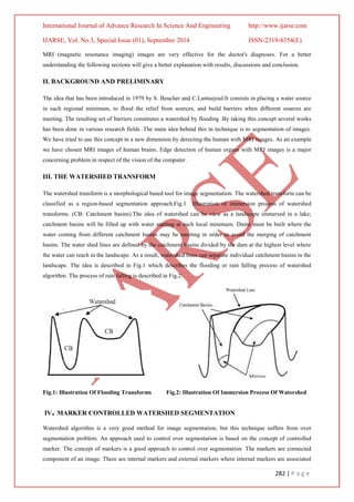
Fig. Illustrations Using Marker Foreground and Background Methods
REFERENCES
[1] Serra, J., Image analysis and mathematical morphology, Academic Press, New York, 1982.
[2] S.Beucher, F.Meyer,"The Morphological Approach to segmentation: The Watershed transform", in
Mathematical Morphology Image Processing, E.R. Dougherty, Ed. New York Marcel Dekker, vol.12,
pp.433-481, 1993.
[3] Beucher, S. "The Watershed Transform Applied to Image Segmentation", Proceedings of the pfefferkorn
Conference on Signal and Image Processing in Microscopy and Microanalysis, pp. 299-314, September
1991.
[4] Rafael C. Gonzalez, Richard E. Woods, "Digital Image processing:, 2nd edition, Pearson Education, pp.
589-656, 2007.
[5] Malik Khan, "Modified Watershed Algorithm for Segmentation of 2D Images", Journal of Information
Science & Information Technology, 6, No.3, pp.546-552,2009.
[6] S. Beucher,Watershed, hierarchial segmentation and water fall algorithm"' in Mathematical Morphology
and its Application to Image Processing, Dordecht, The Netherlands: Kluwer, pp.69-76,1994.
[7] R.M. Haralick and L.G. Shapiro,"Survey: Image Segmentation Technique", Comput. vis. Graph. Im.
Proc,29 (1985) 100-132.
[8] Rivest, J., Beucher, S., delhomme. J, "Marker-controlled segmentation: an application to electrical borehole
imaging", Journal of Electronic Imaging, April pp.136-142,1992.
[9] MRI image segmentation by Dibyendu Goshal and Pinaki Pratim Acharjya, journal of Emerging
Technology and advanced engineering.
[10] An improved watershed image segmentation by Anju Bala , Journal of Scientific and Engineering Research](https://image.slidesharecdn.com/f2482d6c-7785-4704-9430-c1f46a626f4c-170106042032/85/1412194747_414ijarse-5-320.jpg)