SlideShare a Scribd company logo
1 of 13
Biological Molecules Carbohydrates Carbohydrates include sugars, starch, glycogen and cellulose. There are Monosaccharides disaccharides and polysaccharides. Monosaccharides consist of 3 separate groups trioses pentoses and hexose's. These are all types of soluble reducing sugars with the exception of sucrose. Disaccharides are carbohydrates whose molecules are made of two monosaccharides linked together. E.g., glucose + glucose = maltose. An example of a hexose (6carbons) molecule, (Glucose) in alpha (right) and beta (left) form. These ring forms are produced when a bond forms between the oxygen attached to carbon 5 and carbon 1. An example of a disaccharide below in a condensation reaction Aldehydes and ketones Aldehydes are reducing agents. This means that aldose sugars are reducing agents, and are called reducing sugars. When a reducing agent reduces anotehr compound, the reducing agent is oxidised. Ketose sugars are not reducing agents but they react to form reducing agents in alkaline conditions used to test for reducing sugars. This means that Ketoses such as fructose are not as strong reducing agents as aldoses such as glucose. All monosaccharides as shown above are either aldoses or ketoses, this also applies to most disaccharides. Sucrose is the only exception. Proteins Proteins are polymers who's molecules are made from many amino acid molecules linked together. Examples of proteins can be seen in the form of enzymes, antibodies and haemoglobin. 3 groups Amino group – NH2 Carboxyl group – COOH R group – always different determining the amino acid. Protein Structure Primary Structure -  Sequence of amino acids in a polypeptide chain. Secondary Structure -  Repeating three dimensional structure of the polypeptide backbone. The two common structures are alpha helix and beta strand. Tertiary structure  -   the chain. The three dimensional shape of the polypeptide chain. Quaternary structure -  Many proteins or more polypeptide chains are bonded to make a complete molecules. Disulphide links and hydrogen bonds hold the chains together. Phospholipids have a similar structure to triglycerides, but with a phosphate group in place of one fatty acid chain. There may also be other groups attached to the phosphate. Phospholipids have a polar hydrophilic "head" (the negatively-charged phosphate group) and two non-polar hydrophobic "tails" (the fatty acid chains). As shown below Triglycerides Triglyceride molecules are made from one molecule of glycerol linked to three fatty acids, glycerol is an alcohol. Fatty acids are organic acids also called carboxylic acids and always have a COOH group. By condensation this group combines with the - OH group of a glycerol molecule forming an Ester bond. Triglycerides functions are storage molecules, long chains of fat can be broken down for energy. Bonds Proteins –  Hydrogen form between groups with dipoles (unevenly charged molecules). Ionic bonds forms between groups with opposite charges, disulphide links form between cysteine side chains. Peptide bonds join 2 peptides to make a dipeptide or more to make a poly peptide Carbohydrates –  Glycosidic bonds covalent and formed between c1 and c4, (1 – 4) bond. Phospholipids and triglycerides are joined by ester bonds. A saturated fatty acid contains only single bonds between carbons. Unsaturated contain one or more double bonds between carbon atoms.
Introduction – What is a cell? A cell is a basic unit from which living organisms are made. All living organisms are made up of one or more cells. A cell is surrounded by a cell surface membrane, and contains a solution of proteins and other substances in water. This solution is called cytoplasm. Within this, there are many structures called organelles which make up the cell. Most cell are very small, which can only be seen by using a microscope. However, some are big enough to be seen by the naked eye. Bacteria cells and many protoctists are made up of just one cell, and are known as unicellular organisms. Others, including all plants and animals are made up of many cells, and these are called multicellular organisms.  There are two main cells: Eukaryotic Prokaryotic References: AQA AS Biology. Module 1: Molecule, Cells and Systems www.biologymad.com www.swarthmore/edu/NatSci/cpurrin1/posteradvice.htm ‘ Tool Box’ AS Biology CD Organelles Cell wall –  layer of tough cellulose fibrils which surround the plant cells. It allows cells to become turgid without bursting, maintaining the shape of a cell. Cell membrane  – Thin boundary between cell and environment, controlling the passage of materials in and out of the cell. Proteins are also embedded within the membrane for cell recognition in the immune system. Nucleus  – one of the main organelles found in all eukaryotic cells. Usually large,  about 10 цm in diameter, bounded by a large double membrane with many pores. It is the site of DNA, arranged in chromosomes which are responsible for the cell division and protein synthesis. Nucleolus  – Darkly staining region found in the nucleus, which synthesizes ribosomes. Mitochondrion  – Elongated with a smooth outer membrane, and a folded inner membrane, ranging from 1цm to 10цm in size, in the cytoplasm of the cell. Their function is the site of aerobic respiration, and most of the cell’s ATP is made here. The main use of ATP is for the process of photosynthesis. Chloroplast  – Biconcave disc ‘lens shaped’ with many internal membranes. There are numerous in the cytoplasm, and use the ATP made for photosynthesis. Containing large amounts of chlorophyll, this is the pigment which colours the leaves and is the site of photosynthesis. Vacuole  – Large central organelle in most plant cells, which is surrounded by a membrane called the tonoplast, full of fluid. Contains cell sap, and stores compounds like sucrose and sugar beet.  Rough Endoplasmic Reticulum  – This is an extensive membrane system with many cavities and tubes, with ribosomes attached. It is found throughout the cytoplasm, connected to the nuclear membrane. It transports system, collecting, storing, packaging and transporting proteins made on the ribosomes. Smooth Endoplasmic Reticulum  – Membrane system with small cavities, and no ribosomes attached. Unlike the RER, it is only found in small patches in the cytoplasm, and synthesizes lipids and some steroids. Golgi body  – Stack of flattened membranes, resembling stack of pancakes. The size varies, and is close to the RER. The function of this organelle is to receive vesicles from the RER and appears to modify chemicals before their secretion from the cell. Ribosome  – Small and dense structure results in them being last out of the main organelles to be the last sediment, acting like a giant enzyme. In eukaryotic cells, they are large, 80s compared to 70s in a prokaryotic cell. Ribosomes are where the genetic code is used to build proteins, process called translation Microscopy The two main aspects of microscopy are magnification, and resolution Magnification is a measure of enlargement, calculated by the eyepiece lens x the lens Resolution refers to the detail seen There are three main types of microscopes used to look at cells and their organelles. Each show a different level of resolution. The light microscope uses lenses to focus a beam of light coming from the object, and the magnification and resolution are limited by the wavelength of light. This causes the magnification to be lower, only maximising x 1500, compared to x 500,000 for an electron microscope. Also the limited wavelength results in a lower resolving power, of 200 nm. This means that light microscopes cannot resolve objects closer than 200nm into two separate images. Transmitting Electron microscopes use magnets to focus a beam of electrons onto the object, transmitting them trough. This means that sections have to be thin, and preparation is complex. The preparation process could result in artefacts, which appear on the specimen, that would not be there naturally in a living organism. However, the magnification level is much higher of x 500,000 with a resolution power of 1nm, making the observation very detailed. In Scanning Electron microscopes the electron beam bounces off the surface of the object, allowing 3D objects to be seen in detail. Here, there is no need for a thin section of the specimen, making the preparation easier. Like the TEM, these microscopes have a high magnification and resolution, enabling better detailed objects to be seen. Size and Scaling 1mm = 1000 цm 1цm = 1000nm Cells, Organelles and Microscopes Carys Norman Fig. 3.  Photographs of mitochondria (top right), RER (bottom right) and nucleus and nucleolus (left) ,[object Object],[object Object],[object Object],[object Object],[object Object],[object Object],[object Object],[object Object],[object Object],[object Object],[object Object],[object Object],[object Object],Fig. 4. Light microscope Prokaryotic and Eukaryotic Cells Eukaryotic cells have a ‘true nucleus’ referring to a large nucleus which contains linear DNA inside a membrane. Plant, animal and fungi cells are all types of eukaryotic cells.  Under an optical or light microscope organelles within an animal cell which can be seen include;  Nucleus – a large circular organelle that contains DNA Nucleolus – a dark feature inside the nucleus Plasma membrane – very thin membrane surrounding the cell Cytoplasm – ‘cell fluid’, containing different organelles which cannot be distinguished at this level of magnification. Figure 1.  Photograph from an electron micrograph of an eukaryotic cell, showing the different organelles. Features of a prokaryotic cell: Complex cell wall which is coarse, tough and made out of murein (protein) Plasma membrane – main boundary between cell and the environment Mesosomes – inner folding of the cell membrane, giving the cell a large surface area for the attachment of enzymes used in metabolic processes Genetic material – found in the nucleoid, but there is no nuclear membrane. Have a single circular piece of DNA Ribosomes – protein synthesis  Figure 2 . Photograph of a prokaryotic cell taken from an electron micrograph, showing the different organelles.
Introduction Life on Earth evolved in the water, and all life still depends on water. At least 80% of the mass of living organisms is water, and almost all the chemical reactions of life take place in aqueous solution. The other chemicals that make up living things are mostly organic macromolecules belonging to the four groups proteins, nucleic acids, carbohydrates or lipids. These macromolecules are made up from specific monomers as shown in the table below. Between them these four groups make up 93% of the dry mass of living organisms, the remaining 7% comprising small organic molecules (like vitamins) and inorganic ions. Biochemical Tests  These five tests identify the main biologically important chemical compounds: Starch  (iodine test). To approximately 2 cm³ of test solution add two drops of iodine/potassium iodide solution. A blue-black colour indicates the presence of starch as a starch-polyiodide complex is formed. Starch is only slightly soluble in water, but the test works well in a suspension or as a solid.  Reducing Sugars  (Benedict's test). All monosaccharides and most disaccharides (except sucrose) will reduce copper (II) sulphate, producing a precipitate of copper (I) oxide on heating, so they are called reducing sugars. Benedict’s reagent is an aqueous solution of copper (II) sulphate, sodium carbonate and sodium citrate. To approximately 2 cm³ of test solution add an equal quantity of Benedict’s reagent. Shake, and heat for a few minutes at 95°C in a water bath. A precipitate indicates reducing sugar. The colour and density of the precipitate gives an indication of the amount of reducing sugar present, so this test is semi-quantitative. The original pale blue colour means no reducing sugar, a green precipitate means relatively little sugar; a brown or red precipitate means progressively more sugar is present.  Non-reducing Sugars  (Benedict's test). Sucrose is called a non-reducing sugar because it does not reduce copper sulphate, so there is no direct test for sucrose. However, if it is first hydrolysed (broken down) to its constituent monosaccharides (glucose and fructose), it will then give a positive Benedict's test. So sucrose is the only sugar that will give a negative Benedict's test before hydrolysis and a positive test afterwards. First test a sample for reducing sugars, to see if there are any present before hydrolysis. Then, using a separate sample, boil the test solution with dilute hydrochloric acid for a few minutes to hydrolyse the glycosidic bond. Neutralise the solution by gently adding small amounts of solid sodium hydrogen carbonate until it stops fizzing, then test as before for reducing sugars. Lipids  (emulsion test). Lipids do not dissolve in water, but do dissolve in ethanol. This characteristic is used in the emulsion test. Do not start by dissolving the sample in water, but instead shake some of the test sample with about 4 cm³ of ethanol. Decant the liquid into a test tube of water, leaving any undissolved substances behind. If there are lipids dissolved in the ethanol, they will precipitate in the water, forming a cloudy white emulsion. The test can be improved by adding the dye Sudan III, which stains lipids red.  Protein  (biuret test). To about 2 cm³ of test solution add an equal volume of biuret solution, down the side of the test tube. A blue ring forms at the surface of the solution, which disappears on shaking, and the solution turns lilac-purple, indicating protein. The colour is due to a complex between nitrogen atoms in the peptide chain and Cu2+ ions, so this is really a test for peptide bonds.  Carbohydrates    Carbohydrates contain only the elements carbon, hydrogen and oxygen. The group includes monomers, dimers and polymers, as shown in this diagram:  Monosaccharides (simple sugars)    These all have the formula (CH2O)n, where n can be 3-7. The most common and important monosaccharide is glucose, which is a six-carbon or hexose sugar, so has the formula C6H12O6. Its structure is: Glucose forms a six-sided ring, although in three-dimensions it forms a structure that looks a bit like a chair. The six carbon atoms are numbered as shown, so we can refer to individual carbon atoms in the structure. In animals glucose is the main transport sugar in the blood, and its concentration in the blood is carefully controlled. There are many isomers of glucose, with the same chemical formula (C6H12O6), but different structural formulae. These isomers include fructose and galactose.  Disaccharides (double sugars)   Disaccharides are formed when two monosaccharides are joined together by a glycosidic bond. This reaction involves the formation of a molecule of water (H2O) when two glucose molecules join together to form the disaccharide maltose. This bond is between carbon 1 of one molecule and carbon 4 of the other molecule it is called a 1-4 glycosidic bond. A reaction, where H2O is formed, is called a condensation reaction. The reverse process, when bonds are broken by the addition of water is called a hydrolysis reaction. There are three common disaccharides: Maltose  (or malt sugar) is glucose 1-4 glucose. It is formed on digestion of starch by amylase, because this enzyme breaks starch down into two-glucose units. Brewing beer starts with malt, which is a maltose solution made from germinated barley. Maltose is the structure shown above.  Sucrose  (or cane sugar) is glucose 1-2 fructose. It is common in plants because it is less reactive than glucose, and it is their main transport sugar. It is the common table sugar.  Lactose  (or milk sugar) is galactose 1-4 glucose. It is found only in mammalian milk, and is the main source of energy for infant mammals.  Polysaccharides   Polysaccharides are long chains of many monosaccharides joined together by glycosidic bonds. There are three important polysaccharides:  Starch  is the plant storage polysaccharide. It is insoluble and forms starch granules inside many plant cells. Being insoluble means starch does not change the water potential of cells, so does not cause the cells to take up water by osmosis (more on osmosis later). It is not a pure substance, but is a mixture of amylose and amylopectin.  Amylose  is simply poly-(1-4) glucose, so is a straight chain. In fact the chain is floppy, and it tends to coil up into a helix. Amylopectin  is poly(1-4) glucose with about 4% (1-6) branches. This gives it a more open molecular structure than amylose. Because it has more ends, it can be broken more quickly than amylose by amylase enzymes. Glycogen  is similar in structure to amylopectin. It is poly (1-4) glucose with 9% (1-6) branches. It is made by animals as their storage polysaccharide, and is found mainly in muscle and liver. Because it is so highly branched, it can be mobilised (broken down to glucose for energy) very quickly. Cellulose  is only found in plants, where it is the main component of cell walls. Starch and glycogen contain a-glucose, in which the hydroxyl group on carbon 1 sticks down from the ring, while cellulose contains b-glucose, in which the hydroxyl group on carbon 1 sticks up. The b-glycosidic bond cannot be broken by amylase, but requires a specific cellulase enzyme. The only organisms that possess a cellulase enzyme are bacteria, so herbivorous animals, like cows and termites whose diet is mainly cellulose, have mutualistic bacteria in their guts so that they can digest cellulose. Humans cannot digest cellulose, and it is referred to as fibre. Tertiary Structure   This is the compact globular structure formed by the folding up of a whole polypeptide chain. Every protein has a unique tertiary structure, which is responsible for its properties and function. For example the shape of the active site in an enzyme is due to its tertiary structure. The tertiary structure is held together by bonds between the R groups of the amino acids in the protein, and so depends on what the sequence of amino acids is. There are three kinds of bonds involved: hydrogen bonds, which are weak.  ionic bonds between R-groups with positive or negative charges, which are quite strong.  sulphur bridges - covalent S-S bonds between two cysteine amino acids, which are strong.  So the secondary structure is due to backbone interactions and is thus largely independent of primary sequence, while tertiary structure is due to side chain interactions and thus depends on the amino acid sequence.  Quaternary Structure  This structure is found in proteins containing more than one polypeptide chain, and simply means how the different polypeptide chains are arranged together. The individual polypeptide chains are usually globular, but can arrange themselves into a variety of quaternary shapes. This final three-dimensional shape of a protein can be classified as globular or fibrous. The vast majority of proteins are globular, including enzymes, membrane proteins, receptors, storage proteins, etc. Fibrous proteins look like ropes and tend to have structural roles such as collagen (bone), keratin (hair), tubulin (cytoskeleton) and actin (muscle). They are usually composed of many polypeptide chains. A few proteins have both structures: the muscle protein myosin has a long fibrous tail and a globular head, which acts as an enzyme.  Biochemistry  –  Biological molecules, their uses and their chemical tests Chris Blackhall 12PK Lipids Lipids are a mixed group of hydrophobic compounds composed of the elements carbon, hydrogen and oxygen. Triglycerides   Triglycerides are commonly called fats or oils. They are made of glycerol and fatty acids. Glycerol  is a small, 3-carbon molecule with three alcohol groups. Fatty acids  are long molecules with a polar, hydrophilic end and a non-polar, hydrophobic "tail". The hydrocarbon chain can be from 14 to 22 CH2 units long, but it is always an even number because of the way fatty acids are made. The hydrocarbon chain is sometimes called an R group, so the formula of a fatty acid can be written as R-COO-. If there are no C=C double bonds in the hydrocarbon chain, then it is a saturated fatty acid (i.e. saturated with hydrogen). These fatty acids form straight chains, and have a high melting point.  If there are C=C double bonds in the hydrocarbon chain, then it is an unsaturated fatty acid (i.e. unsaturated with hydrogen). These fatty acids form bent chains, and have a low melting point. Fatty acids with more than one double bond are called poly-unsaturated fatty acids (PUFAs).  One molecule of glycerol joins togther with three fatty acid molecules to form a triglyceride molecule:  Phospholipids    Phospholipids have a similar structure to triglycerides, but with a phosphate group in place of one fatty acid chain. There may also be other groups attached to the phosphate. Phospholipids have a polar hydrophilic "head" (the negatively-charged phosphate group) and two non-polar hydrophobic "tails" (the fatty acid chains). This mixture of properties is fundamental to biology, for phospholipids are the main components of cell membranes. When mixed with water, phospholipids form droplet spheres with the hydrophilic heads facting the water and the hydrophobic tails facing each other. Alternatively, they may form a double-layered phospholipid bilayer. This traps a compartment of water in the middle separated from the external water by the hydrophobic sphere. This naturally-occurring structure is called a liposome, and is similar to a membrane surrounding a cell.  ,[object Object],[object Object],[object Object],[object Object],[object Object],[object Object],[object Object],[object Object],[object Object],[object Object],[object Object],[object Object],[object Object],[object Object],[object Object],[object Object],[object Object],[object Object],[object Object],[object Object],[object Object],[object Object],[object Object],[object Object],[object Object],[object Object],[object Object],[object Object],[object Object],[object Object],[object Object],Group name  monomers  polymers   % dry mass   Proteins amino acids polypeptides 50 nucleic acids nucleotides polynucleotides 18 carbohydrates monosaccharides polysaccharides 15         Group name  components  largest unit  % dry mass  lipids   fatty acids + glycerol Triglycerides 10
Introduction Organisms are mad from a huge variety of organic and inorganic compounds which can be classified into a few major types. The main organic molecules are carbohydrates, proteins, and lipids.  Carbohydrate, protein and lipid molecules are all based on the elements carbon, hydrogen and oxygen. Proteins also contain nitrogen and sometimes sulphur.  There is a huge variety of carbohydrates, proteins and lipids, but these are made from a small number of similar molecules. These building blocks are joined together by condensation reactions which link substances together and also produce water. Large organic molecules are broken down by the process of hydrolysis, which literally means splitting using water. ,[object Object],[object Object],[object Object],[object Object],[object Object],[object Object],[object Object],[object Object],[object Object],[object Object],[object Object],[object Object],[object Object],[object Object],[object Object],[object Object],[object Object],[object Object],[object Object],[object Object],[object Object],[object Object],[object Object],[object Object],[object Object],[object Object],[object Object],[object Object],[object Object],[object Object],[object Object],[object Object],[object Object],[object Object],[object Object],[object Object],[object Object],[object Object],Biological molecules Chris Kelly 12WH South Bromsgrove High School, A Specialist Technology College, Bromsgrove, Worcestershire, B60 3NL .  When these chains lie parallel to each other, many hydrogen bonds  form along the length so that the individual cellulose chains are bound  together into strong fibrils. These fibrils are glued together to form cell  walls. Cellulose is the most abundant polysaccharide on earth, but most  animals cannot digest it because they do not possess the enzyme  cellulase. Proteins  Are very important in living organism. Amino acids are the monomers  of a protein. Amino acids differ on the basis of their functional group.  e.g. Amine, Carboxyl, functional. Amino acids are joined together by peptide bonds. The reaction  involves the formation of a molecule of water in another condensation  polymerisation reaction. (Peptide bonds form between adjacent amino  acids. This is a condensation reaction). The structure of proteins is complex and can be described at four level: Primary Secondary Tertiary Quaternary  ,[object Object],[object Object],[object Object],[object Object],[object Object],[object Object],[object Object],[object Object],[object Object],[object Object],[object Object],[object Object],[object Object],[object Object],[object Object],[object Object],[object Object],[object Object],[object Object],[object Object],[object Object],[object Object],[object Object],[object Object],[object Object],[object Object],[object Object],[object Object],[object Object],[object Object],[object Object],[object Object],[object Object],[object Object]
Introduction Organisms are made of a huge variety of compounds. The main organic compounds are carbohydrates, proteins, lipids, and nucleic acids. I will write about these first three groups. The molecules of these groups are mainly made of  Carbon, Oxygen and Hydrogen. There are many different lipids, proteins and carbohydrates, they are all made of a small number of simpler molecules joined together. The joining of these smaller molecules happens in a condensation reaction. In which H 2 O is produced. And the opposite can happen by adding water, this is called hydrolysis and is used to break large molecules into smaller ones. E.g. a disaccharide into two monosaccharides.  But the first subject topic is lipids. Lipids Lipids are fats, oils and waxes. They are insoluble in water, this is because they have no dipoles and so no charges to attract water molecules. Lipids are made of Carbon oxygen and hydrogen.  Most lipids are composed of compounds called  triglycerides .  Triglycerides are a glycerol bonded to three fatty acid chains.  (see fig. 1) They are made from a special type of condensation reaction, called esterfication. Which forms ester bonds between the glycerol and fatty acid.  (see fig. 2) Fig. 1 Carbohydrates Carbohydrates are separated into three categories. Monosaccharides - one, single sugar Disaccharides - two, double sugar Polysaccharides - many, multiple sugars. And sugars are separated into reducing sugars, and non-reducing sugars.  Reducing sugars can donate electrons to other molecules, all monosaccharides, and some disaccharides are reducing sugars. The Most common monosaccharide is glucose. There are two types of glucose, alpha (   and beta (    .  Glucose is a hexose sugar, because it has a structure like a hexagon  (see fig.3).  The difference between Alpha and Beta glucose is the 1 st  and 4 th  carbons.   Fig. 3 Chromatography Chromatography is used to separate, then identify the individual chemicals in a mixture. It works by drawing a line on a piece of chromatography paper (in pencil, because ink would run) called the origin. Then dropping a very small amount of liquid onto the origin. You then allow this spot to dry, then repeat the process about 4times, until you have a concentrated spot on the origin line.  (see fig. 7)  You then put the piece of paper into a solvent, but ensure the line of origin is above the solvent line.  Fig. 7 Biological Molecules Holly Stockham Biochemical tests There are tests that you can do to find out what a substance contains. These tests are below: Starch.   Adding Iodine to the solution, if it turns blue/black it contains starch.  Protein.   By adding biurets solution and heating up the solution gently you can see if it contains protein. A positive result would turn the solution lilac. Lipids.  You do the emulsion test, which is adding ethanol, shaking, then adding the solution to water. It is called the emulsion test because if the solution contains lipids a white emulsion will be formed. Reducing sugars.  Add Benedicts solution, then heat up to near boiling pt. the colour will go from  Blue  to  green  to  yellow  to   orange  to  red .  Depending how much reducing sugar it contains. (red containing the most reducing sugar.) Non-reducing sugars.  First you must add it to dilute HCL, which hydrolyses the non-reducing sugars to reducing sugars. Then it must be neutralised with NaHCO 3 , then there should be a positive result from the benedicts test (as above) References Advanced Biology by Jones and Jones. CGP AS-level Biology Collins AQA AS Biology Biology toolbox CD Proteins  Proteins are made from long chains of amino acids. (see fig. 5) And like lipids and carbohydrates, they are formed by condensation reactions, and broken down by hydrolysis. But the bonds between the amino acids, making the proteins are called peptide bonds  (see Fig. 6). Fig.5 An amino acid Fig. 6 Solvent front origin Spot of pigment The chemicals can then be identified comparing Rf values to a table of already known Rf values. The Rf value is calculated by this formula:  Rf value =  Distance from origin to top of spot Distance from origin to solvent front (Note: The Rf Value will be between 0 and 1.) But sometime the solvent doesn’t completely separate out all the chemicals properly, so you need to do  two way chromatography . Where you turn the paper 90 o  and place it into another solvent. Which should completely separate out the chemicals. glycerol Fatty acid chain Fig. 2 Ester bonds triglyceride Fatty acids can be saturated or unsaturated. The difference is that saturated fatty acids are completely “covered” in hydrogen, and they have no double bonds. Whereas unsaturated fatty acids have double bonds, and the capability to bond with more hydrogen. If an unsaturated lipid has more than one double bond it is called polyunsaturated. Saturated lipids are normally found in animals, and unsaturated, in plants. There is another type of lipid, a  phospholipid .  Phospholipids are used in cell membranes because they have a hydrophobic tail and hydrophilic head. So they control what can come in and out. The structure of a phospholipid varies from that of a triglyceride, but not by much. A phospholipid still has the same glycerol backbone as a triglyceride, only instead of having three fatty acid chains, it has two, and a phosphate group, which is ionised to attract water (that’s why the head is hydrophilic).  To break up a lipid you need to add water, hydrolysis. Which is normally done by adding a dilute acid, and heating it up. Then you get glycerol and chains of fatty acids again. The main use of triglycerides in the body is as an energy source. They contain much more energy per gram than either carbohydrates or proteins. Triglycerides can be broken down and oxidised in respiration and the energy from them is used to make ATP.  Alpha glucose  Beta Glucose To form a  Disaccharide  you must join two monosaccharides together in a condensation reaction  (see fig.4) . The monosaccharides bond at carbon 1 and 4, creating a 1-4 glycosidic bond. And produce water. That creates a disaccharide, some key examples of disaccharides are maltose, sucrose, and lactose. Maltose is formed from two alpha glucose molecules, and is the sugar produced when starch is broken down. To break a disaccharide into a monosaccharide/ break down a polysaccharide to a disaccharide you use hydrolysis, it basically means adding water. It reverses the condensation reaction, and is usually done by adding a warm dilute acid. Polysaccharides  are formed when 2 or more disaccharides join together.  The three main polysaccharides are: Starch  (amylose and amylopectin)  Amylose is a long unbranched chain, it’s compact, and is therefore good for storage.  Amylopectin is branched, which means it is easy to break down the side branches to get at the glucose.  Glycogen It’s similar to amylopectin, only with a lot more side branches, so it looks a bit like a brush. This is good for quick energy release, as glycogen is stored in animals. Cellulose Is a straight chain, (a bit like a brick wall) with hydrogen bonds linking the chains together. This is good because it makes the structure strong, as it is used for cell wall. Also enzymes can’t break it, because they can’t reach the glycosidic bonds. Fig. 4 Amine group Functional group Carboxyl group The formation of a protein. Showing the peptide bond The functional group is shown as “R” because it changes in different amino acids. For instance: The  Primary  structure of a protein is the order of the amino acids. The  Secondary  structure is the shape e.g. beta pleated sheet or a helix. The  Tertiary  structure is a globular protein. It’s made up of lots of helixes and beta pleated sheets. It's 3D, enzymes are globular proteins. Globular proteins are therefore affected by all the things you associate with enzymes, like temperature and pH. It is held together by many different bonds, like disulphide, hydrogen bonds, van der waals forces, and ionic bonds. The  Quaternary  structure is 2 or more polypeptides, so a big group of globular proteins. Beta pleated sheet helix C 4 H 9 leucine C 4 H 9 isoleucine C 3 H 7 valine CH 3 alanine H glycine Functional Group Amino Acid
Introduction-   A cell is the basic unit from which all living organisms are made. Some cells are big enough to be seen with the naked eye, however most cells are microscopic and can only be seen with microscopes. Bacteria tend to have the smallest cells, around 0.5um across, whereas human cells are between 10um and 30um in diameter. Despite the variations in size, cells have certain features in common.  For further information References – www.biologymad.com http://library.thinkquest.org Collins AQA AS Biology Module 1: Molecules, cells and systems Mary Jones and Geoff Jones Advanced Biology  Letts Biology Revision guide  Organelles Organelles are small structures within cells that perform dedicated functions. As the name implies, you can think of organelles as small organs. Organelles were historically identified through the use of microscopy, and were also identified through the use of cell fractionation.   There are many different types of organelles: Eukaryotic cells contain: Cell wall  – A layer of tough cellulose fibrils (made of a variety of polysaccharides) that surrounds plant cells to give support and protection. The cell wall allows the cell to become turgid without bursting and maintains the shape of the cell.  Cell membrane  – A thin boundary around all cells and most organelles that controls what passes in and out of the cell. Proteins are embedded in the cell membrane and are also involved in cell recognition in the immune system.  Nucleus  – Large, usually round, bounded by double membrane with many pores. The most important part of the cell, controls growth, contains DNA, responsible for cell division, regulates metabolism and protein synthesis. Quite simply, the total control of the cell. Nucleolus  – The darkly stained region in the nucleus that is involved with the synthesis of ribosomes.  Mitochondrion  – Usually round and elongated with a smooth outer membrane and a folded inner membrane. The mitochondria are contained in the cytoplasm of cells and are the site of aerobic respiration and where most of the cells ATP is produced. Chloroplast  – A biconcave disc (the perfect compromise between a sphere and a disc) with many internal membranes. Chloroplasts are the site of photosynthesis as they contain the green pigment chlorophyll.  Vacuole  – A large central organelle in most plant cells, surrounded by a membrane called the tonoplast, full of fluid. Vacuoles store compounds such as sucrose and   the difference in water potential compared to the surrounding fluids enables the cell to produce turgor pressure.  Rough endoplasmic reticulum (rough ER)  – The extensive membrane system that has many cavities and tubes with ribosomes attached. Rough ER is found throughout the cytoplasm connected to nuclear membrane and acts as the transport system in the cytoplasm, collecting, storing, packaging and transporting the proteins made in the ribosomes.  Smooth endoplasmic reticulum (smooth ER)  – The membrane system with small cavities and no ribosomes, found in small patches in the cytoplasm. The smooth ER is involved in the synthesis of lipids and some steroids.  Golgi body  – A stack of flattened membrane discs found free in the cytoplasm near to the rough endoplasmic reticulum. The golgi body receives packages (vesicules) of protein from the rough endoplasmic reticulum and is involved in the synthesis and modifying of chemicals before their secretion from the cell.  Ribosome  – A small and dense structure that acts as an enzyme. The ribosome is attached to the rough endoplasmic reticulum or free in the cytoplasm and is where the genetic code is used to build protein (translation).  Prokaryotic cells only contain a cell membrane, cell wall, ribosomes, DNA and a cilia or a flagella (as shown previously).  Cell fractionation – Cell fractionation is a process which allows scientists to extract pure samples of a particular organelle. Cell fractionation is a combination of various methods used to separate a cell’s organelles and components. There are two phases of cell fractionation: homogenization and centrifugation.  Homogenization is the process of breaking open the cells. Cells are broken apart by chemicals, enzymes, or sound waves. Some scientists even force the cells through small spaces at high pressure to break them apart. Centrifugation is the isolation of the cell organelles.  The tissue is chopped up and when the cells are broken apart substances that would normally be kept separate by membranes mix together and can begin to react, so a fluid is added to prevent this which is ice cold, contains a buffer and isotonic. The mixture is then put in a homogeniser (blender) to break up the cells. The resulting mixture is then centrifuged at high speed. The spinning greatly increases the gravitational field, and the organelles separate out according to density and shape. The first most heaviest organelle to sediment out is the nuclei. The remaining fluid, the supernatant, is poured off the centrifuged again to collect other organelles. The order that organelles sediment is: nuclei, mitochondria, chloroplasts (if using plant preparations), rough endoplasmic reticulum, plasma membranes, smooth endoplasmic reticulum and finally ribosomes.  Prokaryotic and eukaryotic cells, organelles and microscopy. Sarah Townsend South Bromsgrove High School Microscopy There are two main concepts in microscopy and these are resolution and magnification. Resolution is the ability to distinguish between two clear dots. Magnification is how much bigger a sample appears to be than it is in real life.  Light microscope -   This microscope uses light to see and magnify objects. The light rays are focused on to a transparent specimen by condenser rays. The rays pass through the specimen and are focused  Prokaryotic Cell –  (From the Greek meaning ‘before nuclei’ – the DNA is not enclosed by a membrane). All bacteria and cyanobacteria (blue –green algae) are prokaryotes as they lack a membrane – bound nucleus. They are usually smaller than eukaryotic cells and are similar in size to a mitochondrion or a chloroplast. In fact, they probably were prokaryotic cells which came to live inside the larger eukaryotic cells millions of years ago. They have few internal organs that are distinguishable. All prokaryotic cells are surrounded by a cell wall. However this is different from Figure 1 . A prokaryotic cell taken from an electron micrograph a plant cell wall because it contains different polysaccharides. Instead they contain peptidoglycans which are made up of molecules in which peptides and sugars are combined. The cell walls are very strong and important because they prevent the cell from bursting when the cell absorbs water and help to protect against invasion by viruses. Many prokaryotic cells have a thick layer of jelly surrounding them called a capsule which is made of polysaccharides and protects from viruses and antibodies. Beneath the cell wall is a cell surface membrane that is made up of a phospholipid bilayer and the cytoplasm that contains ribosomes that are approximately 20 nm in diameter. The DNA is not chromosomal but in a circular loop called a plasmid. Some contain a flagellum which is used for movement. They divide by binary fission and have three major shapes: rod, spherical and spiral.  Figure 2.  A eukaryotic cell taken from an electron micrograph. Eukaryotic cell –  (From the Greek meaning ‘truly nuclear’ – the larger nucleus contains the DNA inside a membrane). Organisms with complex cells are eukaryotes and are found in plants, animals and fungi. They can be easily distinguished through a membrane bound nucleus (nuclear envelope) and contain membrane bound structures (organelles). The organelles such as the mitrochondrion or chloroplast are there to perform metabollic functions and energy conversion. Other organelles provide structural support and cellular motility. They are generally much larger than prokaryotic cells. Not all eukaryotic cells contain cell walls, only plant cells, not animals cells. The plant cell wall contains pectin, hemicellulose and cellulose (different from a prokaryotic cell). The DNA is linear (chromosomal), complex, organised into chromosomes and combined with histone proteins. They divide by replication (making copies of themselves). The eukaryotic cell is clearly developed from a prokaryotic cell. This is because  eukaryotes formed through the merger of prokaryotes, which predate them in the fossil record by some 2 billion years.  again by two more lenses – the objective lens and the eyepiece lens. These two lenses produce a magnified image. As many specimens are colourless and nearly transparent, stains are used to make different parts of the specimen show up clearly. Some stains can be added to living cells, but others must be ‘fixed’ by adding a chemical known as a fixative. These chemicals react with substances such as proteins in the cell, making them insoluble and so anchoring them in position. The cells are killed when the fixative is added. Stains may be added either before or after the fixing process. So long as the specimen is thin enough to allow light to pass through,  Figure 3. A light microscope they can be used to see major organelles such as the nucleus or mitochondrion. The fluorescent light microscope is the most common and widely used light microscope. This microscope is used to visualise stained cells with fluorescent dyes. However, limitations occur as the microscope can not see beyond 200 nm resolution and x1500 magnification due to the long wavelength of light dissipating if the light is refracted too far. They are relatively cheap.    Electron microscope -  This microscope uses electrons to see instead of light. They are focused using electromagnets rather than glass lenses. As electrons are easily stopped by air molecules, the space inside must be a vacuum. As our eyes do not respond to electrons, the electrons are allowed to hit a fluorescent screen which emits visible light where the electrons hit. Electrons cannot penetrate materials as well as light, so specimens must be much thinner. This places great limitations on what can be viewed, in particular it is not possible to look at living material, all specimens musty be dead. Specimens are stained using heavy metal ions such as lead or osmium and are taken up by particular parts of the cells. Atoms of these ions have large, positively charged nuclei which scatter electrons rather than letting them pass straight through. These  Figure 4. An electron microscope  electrons therefore do not arrive on the screen, so leaving a dark area in the image. The structures in a cell which have taken up these heavy metal stains therefore appear dark. Scanning electron microscopes work in a very different way. The electrons do not pass through the specimen but are reflected off its surface. Scanning electron microscopes are used to provide three dimensional images of surfaces. Because the wavelength of electrons are shorter than light, they have a resolution of 1nm and a magnification of x500000. However, they are very expensive.  Figure 5.  cell fractionation
Introduction There are two main types of cells, Eukaryotic and Prokaryotic. They are very different and contain different organelles which help them to do their specific jobs.  An Organelle is a discrete structure of a cell with specialized functions. They are similar to the organs in a body because they are vital in the survival of the cell.  There are many ways to identify organelles such as microscopy which can be done using different microscopes to give different resolutions and better images. Cell fractionation can also be used as it is a method of splitting up the cell so that all the different organelles are separate.  There are two different types of microscopes, light and electron with there being two more different types of electron microscopes. Transition and Scanning which both do different jobs and see different things along with the light microscope. Microscopy There are two different types of Microscopes, Light and Electron microscopes they or both very different with there also being two different types of electron microscopes. Light Microscopes are is a very simple microscope with not a very high resolution or magnification. Resolution being the ability to distinguish between two points on an image and Magnification being how much bigger a sample appears to under the microscope. The resolving power of a microscope is limited by the wavelength of light which is 400 to 600 mm for visible light. To improve the resolving power of a light microscope a shorter wavelength of light is need that is why some microscopes have blue filters as blue has the shortest wavelength of visible light. Light microscopes cost between £100 and £500 compared to the cost of an electron microscope which is over £1,000,000. They are cheep to operate as well as being small and portable. The samples are simple and easy to prepare with little damage done to the samples during this process. A vacuum is not required unlike in the electron microscopes meaning that the specimen has to be dead. The natural colour is maintained in a light microscope even though stains are often needed to make the cells visible but the maximum magnification is only up to 2000 times.  Electron microscopes are much more advanced as they have a magnification of over 500,000 times. They use an electron beam which is very expensive and can damage the specimens which also must be stained with an electron dense chemical.  Electron microscopes can also be split into Scanning electron microscopes and Transmission electron microscopes.  Scanning electron microscopes pas the scanning beam over the surface of the specimen and electrons reflect of it as it has been coated in heavy metals. The reflected electron beams are focused on a florescent screen to make up the image which is linked to a computer to make the image clearer. It is similar to echo location. Transmission electro microscopes pas a beam of electrons through the thin specimen and focusing the image on a screen or film. The TEM is very big, expensive and not very clear.  Cells and Microscopy By Abby Pask Organelles There are many different Organelles with many different functions Eukaryotic cells Nucleus- Where DNA is kept and where RNA is transcribed Ribosome's- The site of protein synthesis, where RNA is translated into proteins Mitochondria- The site or aerobic respiration Chloroplasts- the site of photosynthesis Endoplasmic reticulum- There are two types the rough ER and the smooth ER.  The ER is the transport network for molecules targeted for certain modifications and specific final destinations. Golgi Apparatus - The ‘post box’ of the cell as it modifies molecules and packages them into small membrane bound sacs called vesicles. Prokaryotic Cells  Vacuole-  transport and storing nutrients, metabolites, and waste products. Cytoplasm- contains all the enzymes needed for metabolic reactions Nucleoid- The region of the cytoplasm that contains DNA   Meosome- A tightly folded region of the cell membrane containing proteins required for respiration and photosynthesis. Capsule- A thick polysaccharide layer outside of the cell wall used for protection and a food reserve. Flagellum- A rigid rotating helical shaped tail used for propulsion. Both types of Cells Cell Membrane- Made of phospholipids and proteins which is used for protection and transport. Cell Wall- Keeps a cell rigid and gives protection. Eukaryotic Cells Eukaryotic cells are the larger of the two types of cell and can be more than 10µm in diameter. They have a true nucleus and contain many organelles that can either be double or single. This makes them very complex cells which have a number or different jobs. The genetics of a Eukaryotic cell is made up of linear DNA and is associated with proteins to form chromatin. It has a true nucleus which means it has a nuclear envelope and is one of the main difference between a eukaryotic cell and a Prokaryotic cell. Another main difference is the number of ribosome's in the cell with the eukaryotic cell having a large number (80S) The cells walls are made up of either chitin in fungi or cellulose in plants but animal cells do not have a cell wall Figure 1.  This is a basic diagram of a eukaryotic cell with all the main organelles that it contains including the nucleus and the endoplasmic reticulum Figure 2 . These two photos are different eukaryotic cells that have been magnified using a microscope. The main recognisable organelles have been labels  Prokaryotic Cells Prokaryotic cells are much smaller than Eukaryotic cells being less than 5 µm in diameter. They have smaller ribosome's (70s) and less organelles which are all not membrane bound.  The genetics of a prokaryotic are also very different as there is circular DNA and no proteins. There is no true nucleus which means that the DNA is free to move within the cytoplasm. The cell walls are made from Glycoprotein's or other polysaccharides, but not cellulose or chitin as these are found in Eukaryotic cells. Prokaryotic cells include bacteria and blue-green algae  as they are very small and not very complex Figure 3.  This is a basic diagram of a Prokaryotic cell with all the main organelles that it contains including the ribosome's and the Nucleoid Figure 4.  This is a prokaryotic cell seen through a microscope it shows the cell wall and cell membrane which outline the cell.
Phospholipids have a similar structure to triglycerides, but with a  phosphate group in place of one fatty acid chain. There may also be  other groups attached to the phosphate. Phospholipids have a polar  hydrophilic "head" (the negatively-charged phosphate group) and two  non-polar hydrophobic "tails" (the fatty acid chains). This mixture of  properties is fundamental to biology, for phospholipids are the main  components of cell membranes. RECAP OF LIPIDS What's the difference between a phospholipids and triglyceride  Molecule?  Triglycerides are the familiar fats and oils. The molecules are made  from one molecule of glycerol linked to three fatty acids.  Glycerol is  an alcohol. Fatty acids are organic acids (also called carboxylic acids)  and also have a -COOH group. BY condensation, this group combines  with the -OH group of a glycerol molecule, forming an ester bond. Because triglycerides are fat, they acts as a storage molecule.   Phospholipids have two fatty acids and a phosphate molecule attaches  where as a triglyceride molecule only has three fatty acid chains. There  function is to form membranes. Biochemical tests These five tests identify the main biologically important chemical  compounds. For each test take a small amount of the substance to test,  and shake it in water in a test tube. If the sample is a piece of food,  then grind it with some water in a pestle and mortar to break up the  cells and release the cell contents. Many of these compounds are  insoluble, but the tests work just as well on a fine suspension. Starch  (iodine test). To approximately 2 cm³ of test solution add two  drops of iodine/potassium iodide solution. A blue-black colour  indicates the presence of starch as a starch-polyiodide complex is  formed.  Reducing Sugars  (Benedict's test). To approximately 2 cm³ of test  solution add an equal quantity of Benedict’s reagent. Shake, and heat  for a few minutes at 95°C in a water bath. A precipitate indicates  reducing sugar. The colour and density of the precipitate gives an  indication of the amount of reducing sugar present, so this test is semi- quantitative. A blue colour means no reducing sugar, a green  precipitate means relatively little sugar; a brown or red precipitate  means progressively more sugar is present.    Non-reducing Sugars  (Benedict's test). Using a separate sample, boil  the test solution with dilute hydrochloric acid for a few minutes to  hydrolyse the glycosidic bond. Neutralise the solution by gently  adding small amounts of solid sodium hydrogen carbonate until it  stops fizzing, then test as before for reducing sugars.  Biological molecules Chris Kelly 12WH South Bromsgrove High School, A Specialist Technology College, Bromsgrove, Worcestershire, B60 3NL .  Lipids  (emulsion test). Lipids do not dissolve in water, but do dissolve  in ethanol. This characteristic is used in the emulsion test. Do not start  by dissolving the sample in water, but instead shake some of the test  sample with about 4 cm³ of ethanol. Decant the liquid into a test tube  of water, leaving any undissolved substances behind. If there are lipids  dissolved in the ethanol, they will precipitate in the water, forming a  cloudy white emulsion. The test can be improved by adding the dye  Sudan III, which stains lipids red.  Protein  (biuret test). To about 2 cm³ of test solution add an equal  volume of biuret solution, down the side of the test tube. A blue  ring forms at the surface of the solution, which disappears on shaking,  and the solution turns lilac-purple, indicating protein.  Conclusion Biochemistry is a particularly difficult area in biology as it is split up  into different sections. For example you have to remember the types,  function and importance carbohydrates-monosaccharides,  disaccharides and polysaccharides and proteins, Lipids and  biochemical tests.  Fatty acids are long molecules with a polar, hydrophilic end and a non- polar, hydrophobic "tail". The hydrocarbon chain can be from 14 to 22  CH2 units long, but it is always an even number because of the way  fatty acids are made. The hydrocarbon chain is sometimes called an R  group, so the formula of a fatty acid can be written as R-COO-. Saturated or unsaturated If there are no C=C double bonds in the hydrocarbon chain, then it is a  saturated fatty acid (i.e. saturated with hydrogen). These fatty acids  form straight chains, and have a high melting point.  If there are C=C double bonds in the hydrocarbon chain, then it is an  unsaturated fatty acid (i.e. unsaturated with hydrogen). These fatty  acids form bent chains, and have a low melting point. Fatty acids with  more than one double bond are called poly-unsaturated fatty acids  (PUFAs).  These fatty acids are joined by a condensation reaction however in this  reaction it is known as ESTERIFICATION and an ESTER bond is  formed. With three branches three ester bonds can form so three fatty acids can  bond so therefore it is called a triglyceride. Is first molecule of a fatty  acid   Acknowledgments Literature cited For further information
Introduction The cell is the basic functioning unit of organisms in which chemical reactions take place.  These reactions involve energy release needed to support life and build structures. Organisms consist of one or more cells and these can be seen with microscopes which enables sub-cellular units called organelles become visible. Each organelle has been researched to help us understand more about the processes of life. To see a virtual cell you can visit www.life.uiuc.edu/plantbio/cell Prokaryotic and Eukaryotic cells Organisms can be classified into two groups, prokaryotic and eukaryotic to their cellular structure.   Eukaryotic cells   A eukaryote is an organism with  a complex cell or cells, in which the genetic information is organized into a membrane- bound nucleus or nuclei. This type of cell comprises of animals, plants and fungi- which are mostly multi cellular. Also, eukaryotic cells are generally much  larger than prokaryotic cells and have a variety of internal membranes and structures, called  organelles and the DNA in this structure is divided into several bundles called chromosomes. Prokaryotic cells   The prokaryotic kingdom consists of only bacteria which  means they have no nucleus. Overall, prokaryotic cells are  less complicated than eukaryotic cells and are considered  to have evolved earlier.  ,[object Object],[object Object],[object Object],[object Object],[object Object],[object Object],[object Object],[object Object],[object Object],[object Object],[object Object],Magnification Microscopes magnify the image of a specimen to enable the human eye to see minute objects that are not visible to the naked eye.  Resolution of a microscope  = the ability to distinguish between two objects (dots) as separate entities.  Magnification  = is the measurement of enlargement The magnification of an object can be calculated using this equation: observed size   Magnification =  actual size Cells and Microscopy Heather Bradley Artefacts: When microscope specimens are prepared there are often several chemical and physical procedures. Often the material is dead so changes from the living specimen are expected. Microscopic material should be analysed with care because there may have been some artificial change in the material during preparation. These are artefacts; structures alien to the material which should not be interpreted as part of the specimen.  ,[object Object],[object Object],[object Object],[object Object],[object Object],Organelles   Mitochondria: These consist of an outer membrane enclosing a semi-fluid matrix. Throughout the membrane matrix is a an internal membrane, folded into cristae. The cristae and matrix contain enzymes which enable this organelle to carry out aerobic respiration, which release energy in the form of ATP for use within the cell.  Nucleus   This controls all cellular activity using coding instructions which are, located in the cell to make specific proteins.  RNA is produced in the nucleus pores. Present in both animal and plant cells Cell membrane This covers the outside of a cell and consists of a bilayer of phospholipids interspersed with proteins. It separates the cell from the outside environment, gives protection and allows the diffusion of substances into and out of the cell.   Cytoplasm This is a semi-fluid medium that is present in all cells and many ions are dissolved within the substance and many chemical reactions occur here.   Endoplastic reticulum This can be in the form of rough endoplastic reticulum (RER) which consist of ribosomes on the outer layer or smooth endoplastic reticulum (SER) without ribosomes. The RER is a transport system throughout the cytoplasm that collects, stores, packages and transports the proteins made on the ribosomes. The SER synthesises lipids and some steroids . Ribosomes These are situated on the rough endoplastic reticulum and consist of a small and dense structure and acts as a giant enzyme. They are around 20 nm and the building of proteins occurs here. Golgi body These are a series of flattened sacs, each separated from the cytoplasm by a membrane. The Golgi body is a packaging system where important chemicals become wrapped, forming vesicles. The vesicles become detached from the main Golgi sacs, enabling the isolation of chemicals from each other in the cytoplasm. This organelle aids the production and secretion of many proteins, carbohydrates and glycoproteins.  Lysosomes These are specialised vesicles because they contain digestive enzymes. These have the ability to break down proteins and lipids. Centrioles There are two short cylinders in a cell which contain microtubules. Their function is to aid cell division.  Present only in plant cells Cell wall Around the cell membrane in a plant cell is a cell wall. This provides the cell with rigidity and allows many substances to be imported and exported by the cell, creating an effect hydrostatic skeleton. When the cell has a maximum amount of water content, it has maximum strength, and is said to be turgid.  Chloroplasts These enable the plant to photosynthesise which produces energy due to the presence of chlorophyll in the chloroplasts within the cell.   Vacuole This is a large central organelle in most plant cells and is a semi-fluid substance. It stores compounds such as sucrose and water potential enables the cell to gain turgor pressure when osmosis into the vacuole occurs.   O A M There are two types of microscopes ELECTRON MICROSCOPES The use of a beam of electrons ‘illuminates’ the specimen instead of using an electromagnetic radiation microscope. Electrons act like waves so can easily be produced, focused by using electromagnets and detected using a phosphor screen or photographic screen.   LIGHT MICROSCOPES This type of microscope uses white light to illuminate a specimen. The light is focused onto the specimen by a condensing lens. The specimen is placed on a microscope slide which is clipped onto a platform, known as a stage. The image is viewed via an eyepiece or ocular lens.  ,[object Object],[object Object],[object Object],[object Object],[object Object],[object Object],[object Object],[object Object],[object Object],[object Object],[object Object],[object Object],[object Object],[object Object],[object Object],[object Object],[object Object],[object Object],[object Object],[object Object],[object Object],[object Object],[object Object],[object Object],[object Object],[object Object],[object Object],[object Object],[object Object],[object Object],[object Object],[object Object],[object Object],[object Object],[object Object],[object Object],[object Object],[object Object],[object Object],[object Object],[object Object],[object Object],[object Object],[object Object],[object Object],[object Object],Densest Least dense More information can be found on the website  www.biologymad.co.uk  and www.biologymodel.co.uk ,[object Object],[object Object],[object Object],[object Object],[object Object],Graticule Stage micrometer ,[object Object],[object Object],[object Object],[object Object],[object Object],[object Object],[object Object],[object Object],[object Object],[object Object],[object Object],[object Object],[object Object],[object Object],[object Object],[object Object],[object Object],[object Object],[object Object],[object Object],[object Object],[object Object],[object Object],[object Object],[object Object],[object Object],[object Object],[object Object],Conclusion Overall, the topic of cells and organelles is quite detailed and thorough revision needs to be acquired in order to understand the topic fully.   Microscopes allow you to see a working cell and its’ organelles ,so that research into their function and how they were formed on the earth and hoe they have developed into the specimens they are today. This poster should be informative and with extra websites to visit on the page you should find useful facts and ideas about organelles, their function, where they are in the cell and how to see they through a microscope.  www.thinkquest.co.uk  would be a suitable website to visit to support this knowledge on eukaryotic and prokaryotic cells Larger- around 80s Complex membrane systems and many organelles Present in plants and fungi but not in animals and their structure is relatively simple Small – around 70s  Very few organelles  Complex and always present Ribosomes  Internal structure Cell  walls By mitosis or meiosis By binary fission Cell division DNA is linear, attached to histones (organising proteins) and condenses inot visible chromosomes before cell division DNA is circular, not attached to histones  Organisation of nuclear material DNA inside distinct membrane- bound nucleus DNA free in cytoplasm Site of  nuclear material  10-100 µm diameter, much greater volume (large cell) 0.1-10 µm in diameter (small cell) Size Eukaryotic cell Prokaryotic cell
[object Object],[object Object],[object Object],[object Object],[object Object],[object Object],[object Object],[object Object],[object Object],[object Object],[object Object],Cell Ultra-structure and Microscopy Esme Ingram Eukaryotic  Cell : with a true nucleus –membrane bound nucleus-found in animal,plant and fungi cells. Chloroplast The site of photosynthesis and are biconcave disc shaped with lots of internal membranes allowing a bigger surface area. Contains thykaloids which are phospholipid bilayers that have chlorophyll molecules embedded in them .  These chlorophyll molecules absorb light energy then transfer it to other protein molecules which generate ATP.Thykaloids form stacks called graenum which greatly increases the efficiency of light dependent reactions by capturing most of the light that enters the cell.   ,[object Object],[object Object],[object Object],[object Object],Golgi Body It is a stack of curved cristanea which are found free in the cytoplasm.  It synthesises and modifies protein before secretion form cell. Has vesicles entering and exiting it which contain newly synthesised proteins and have broken off RER They travel towards golgi and fuse with its convex face. Proteins are finished off + packaged-ready for exportation form cell Proteins are transported in vesicles which break form golgi body They fuse with cell surface membrane and release contents to outside The vesicle become incorporated into cell membrane Ribosomes In the eukaryotes they are of the larger 80S type.  They are a small and dense structure and act as giant enzyme.  They are either suspended in cytoplasm where they make protein for cells use or attached to ER where the proteins are for exportation.  They are where the genetic code is used to build protein because a ribosome binds to a length of RNA and the info’ encoded in the RNA is used to assemble the correct order of amino acids in a protein.  They are made of one large and one small subunit. Cytoskeleton Network of protein fibres used for strength, support and motility  it is attached to cell membrane and helps hold cell in shape as well as organelles in place.  There are three types of protein fibres (microfilaments, intermediate filaments and microtubules), and each has a motor protein that can move along the fibre carrying a cargo These motor proteins are responsible for cilia and flagella movements. Endoplasmic Reticulum There are two kinds of ER which are smooth ER and rough ER. It is a system of membranes running through the cytoplasm of the cell  , the membranes enclose spaces called cristernae which form an interconnecting channel throughout cytoplasm. RER has ribosomes attached which are involved in protein synthesis.  The cisternae break into small vesicles and travel to golgi body. ,[object Object],[object Object],[object Object],[object Object],[object Object],[object Object],[object Object],[object Object],[object Object],[object Object],[object Object],[object Object],[object Object],[object Object],[object Object],[object Object],[object Object],[object Object],[object Object],[object Object],Microscopy Magnification:  a measurement of how far we can expand and see the object, so it is a measure of enlargement.  Overall magnification = Objective lens x Eyepiece lens Resolution: the ability to distinguish between two dots. The Light Microscope Light rays are transmitted through specimen and two lenses The objective lens (set of 3) provides the initial magnification of the image in the range of  X4-X40 The eyepiece lens (top)is used to further magnify the image and focus it on the retina Limitations of the light microscope:  light is limited by its long wavelength so has a lower resolving power than electron microscopes, it only takes a small amount of power before it dissipates. Real Size(ųm)=(observed size X 100)/magnification Electron Microscopes : This uses a beam of electrons in order to illuminate the specimen. A beam of electrons has an effective wavelength of less than 1 nm, so can be used to resolve small cellular ultra structure.   Transmission Electron Microscope Electrons are produced from a tungsten filament at top of evacuated column The specimen either absorbs or transmits electrons Condensed lens focuses electrons onto specimen Objective and projector lenses magnify and focus image onto fluorescent screen Scanning Electron Microscope Tungsten filament at the top of an evacuated column produces electrons Condenser lenses focus beams of electrons Objective lenses magnifies the beam of electrons And electrons are reflected of the specimen and collected in the form of an image on screen. . This table was taken form www.biologymad.com ,[object Object],[object Object],[object Object],[object Object],[object Object],[object Object],Comparing the Two Cells
Jonathan Stevens Module One: Biological Molecules Lipids, Proteins and Carbohydrates Introduction: In the following poster I will go into detail of many things to do with biological molecules, the main aspects I will go into detail are carbohydrates, lipids and proteins. I will show the structures of the molecules and the types of bonding and how they join and get broken down. I will state the chemical tests for the certain types of molecules and what too look for to see If the substance contains the specific type of molecule. I will the state and explains the functions of proteins and etc. Carbohydrates: There are 3 main types of carbohydrates and they are polysaccharides, monosaccharide and disaccharides. All carbohydrates contain 3 elements these elements are carbon, hydrogen and oxygen .  Monosaccharide: A monosaccharide is a single sugar this is a molecule of sugar in its smallest form, an example of this is glucose, glucose is a sugar which our body uses, there are two types of glucose there is alpha and beta, the structures of these molecules differ, they are as follows: Alpha Beta Glucose can join together to form long chains which are polysaccharides, the first stage of the joining is when two glucoses join to become a disaccharide the glucoses can join up in different orders and this  makes different disaccharides for example alpha plus alpha. The process of joining is between carbon one and carbon four, they join by a glycosilic bond, reaction gives out water and it is called a condensation reaction. The glycosilic bond can also be broken they are broken, they are broken down by adding it with acid, this reaction adds water to the molecule which rejoins the two glucoses, this reaction is a called hydrolosis. The test for monosaccharide is to add benedict's solution to the substance which is at near boiling, if the substance contains monosaccharide the benedict's solution will change to a green – red solution. Disaccharides: Disaccharides are  two monosaccharide joined together, they are joined by condensation reaction. The test for disaccharides is as follows, add hydrochloric acid to the solution which is at near boiling, then add benedict's solution to the solution and if the benedict's solution changes to a green to red colour. Polysaccharides: These are massive chains of monosaccharide and disaccharides an example of this is starch which make up bigger molecules. Lipids:  Lipids all contain a glycerol backbone which looks as follows:  Lipids contain a fattys acid and this has a carboxyl this is shown below, the zig zaged line represents a long hydrocarbon chain, the chain can be saturated or unsaturated. A fatty acid joins to a glycerol backbone they join by esterfication this is a condensation reaction and releases water. A glycerol backbone can join up to three things on its 3 branches, one example it can make is a triglyceride, this is when a glycerol backbone joins to three fatty acids. Esterfication A fatty acid is hydrophobic this means it doesn’t like to react with water this is because the water will breaks the bonds between the glycerol and the fatty acid because if you add water you are adding back hydrogen's, and why do they share hydrogen's if they could have one each. The glycerol head is hydrophilic which means it is water loving.  Proteins: Proteins are a key part of the body they have many functions these functions are:  ~for enzymes ~anti-bodies ~actin & myosin which make muscles contract ~used to make tissues ~blood clotting involve many proteins ~gives strength to hair and nails Proteins are made up of amino acids and each amino acid has three groups these groups are amino group, functional group and the carboxyl group, a diagram of this is below: The functional group can join to different molecules these molecules make up different proteins and amino acids which have different jobs, examples of these are below: Proteins and amino acids have many structures and these structures are: Primary structure  this is the order of the amino acids Secondary structure:  this structure is when the long strand of amino acids either curve into a helix or the create a beta pleated sheets, the helix and sheets are joined together by hydrogen bonds. Tertiary structure:  the third structure is when the helix’s and sheets join together to form a globular protein this big structure is also known as a enzyme, the big structure is joined together by many bonds these bonds are: di-sulphide, hydrogen, Ionic, Hydrophobic, Van der Waals Quartary structure:  this structure is when gobular proteins join together to make bigger structures this structure can join together to make a well known structure which is a red blood cell.
Introduction All living things are made up of one (unicellular) or more cells (multicellular), they are the smallest units that can be alive.  Every cell possesses internal coded instructions to control cell activities and development they also have the ability to continue life by some sort of cell division. The biggest division in cells is that of prokaryotic and eukaryotic.   Such differences in the divisions are listed below. Prokaryotic and Eukaryotic cells Prokaryotic Has a cell membrane Has a cell wall Has ribosomes which are small Has no endoplasmic reticulum Has no golgi apparatus Has no  mitochondria Has no true nucleus with envelope DNA is circular May have flagellum for motility Asexual reproduction Always unicellular Small cells No cytoskeleton Binary fusion (cell division) Variety of metabolic paths  ,[object Object],[object Object],[object Object],[object Object],[object Object],[object Object],[object Object],[object Object],[object Object],[object Object],[object Object],[object Object],[object Object],[object Object],[object Object],[object Object],[object Object],[object Object],[object Object],[object Object],[object Object],[object Object],[object Object],[object Object],Biological Cells   Lauren Boast Organelles  – Cells contain several types of organelle which are contained in the cytoplasm. An organelle is a subunit of the cell designed to carry out its own specific function separate from all the other activities going on in the cell.   Nucleus  – Controls all cellular activity using coded instructions located in DNA, this DNA allows the nucleus to make specific proteins. The nucleus is surrounded by a nuclear envelope which has nuclear pores. The interior is called the nucleoplasm, which is full of chromatin and the nucleolus is a dark region of chromatin, involved in making ribosomes. Mitochondria  – Consists of an outer membrane and an internal membrane folded into cristae. The cristae contain enzymes which enable mitochondria to carry out respiration. Ribosomes  –Smallest and the most numerous of organelles they aid in the manufacture of proteins and are the place where amino acids are bonded together. They are often found in groups called polysomes. Endoplasmic Reticulum  – found as either Rough ER (with ribosomes) or smooth ER (without ribosomes). Smooth ER is a series of membrane channels involved in synthesising and transporting materials. The ribosomes, which are studded onto the Rough ER synthesise proteins. Golgi body  – A packaging system where important chemicals become mem
12 c scientific posters

More Related Content

What's hot

Vermi composting, phosphhocompost, pk mani
Vermi composting, phosphhocompost, pk maniVermi composting, phosphhocompost, pk mani
Vermi composting, phosphhocompost, pk maniP.K. Mani
 
Edible oil seeds group
Edible oil seeds groupEdible oil seeds group
Edible oil seeds groupBunny verma
 
Lipid structure and metabolism copy (2)biotech
Lipid structure and metabolism   copy (2)biotechLipid structure and metabolism   copy (2)biotech
Lipid structure and metabolism copy (2)biotechUnaiza Chaudhary
 
Pinning insect
Pinning insect Pinning insect
Pinning insect hina amir
 
Oilseed crops based intercropping
Oilseed crops based intercroppingOilseed crops based intercropping
Oilseed crops based intercroppingHardikPatel811
 
Button mushroom production technology A Presentation By Mr Allah Dad khan For...
Button mushroom production technology A Presentation By Mr Allah Dad khan For...Button mushroom production technology A Presentation By Mr Allah Dad khan For...
Button mushroom production technology A Presentation By Mr Allah Dad khan For...Mr.Allah Dad Khan
 
Presentation on biodiesel by kiran aajagekar
Presentation on biodiesel by kiran aajagekarPresentation on biodiesel by kiran aajagekar
Presentation on biodiesel by kiran aajagekarKiran Aajagekar
 
Soil fertility evaluation P K MANI
Soil fertility evaluation  P K MANISoil fertility evaluation  P K MANI
Soil fertility evaluation P K MANIP.K. Mani
 
Anatomy and elementary physiology of integumentry systems of insects
Anatomy and elementary physiology of integumentry systems of insectsAnatomy and elementary physiology of integumentry systems of insects
Anatomy and elementary physiology of integumentry systems of insectsMrpsyco
 
Apiculture
ApicultureApiculture
ApicultureAsmahale
 
Biogas .ppt
Biogas .pptBiogas .ppt
Biogas .pptSreesh S
 
Morphology of cockroach
Morphology of cockroachMorphology of cockroach
Morphology of cockroachpomerian
 
bio-energy Profitability of Jatropha Curcas L. cultivation as bio-diesel sou...
bio-energy Profitability of Jatropha Curcas L. cultivation  as bio-diesel sou...bio-energy Profitability of Jatropha Curcas L. cultivation  as bio-diesel sou...
bio-energy Profitability of Jatropha Curcas L. cultivation as bio-diesel sou...Omid Minooee
 

What's hot (20)

Crop rotation
Crop rotation Crop rotation
Crop rotation
 
Biofuels
Biofuels   Biofuels
Biofuels
 
Respiration ppt
Respiration  pptRespiration  ppt
Respiration ppt
 
Vermi composting, phosphhocompost, pk mani
Vermi composting, phosphhocompost, pk maniVermi composting, phosphhocompost, pk mani
Vermi composting, phosphhocompost, pk mani
 
Edible oil seeds group
Edible oil seeds groupEdible oil seeds group
Edible oil seeds group
 
Concept of food science
Concept of food scienceConcept of food science
Concept of food science
 
Lipid structure and metabolism copy (2)biotech
Lipid structure and metabolism   copy (2)biotechLipid structure and metabolism   copy (2)biotech
Lipid structure and metabolism copy (2)biotech
 
Pinning insect
Pinning insect Pinning insect
Pinning insect
 
Oilseed crops based intercropping
Oilseed crops based intercroppingOilseed crops based intercropping
Oilseed crops based intercropping
 
Button mushroom production technology A Presentation By Mr Allah Dad khan For...
Button mushroom production technology A Presentation By Mr Allah Dad khan For...Button mushroom production technology A Presentation By Mr Allah Dad khan For...
Button mushroom production technology A Presentation By Mr Allah Dad khan For...
 
Presentation on biodiesel by kiran aajagekar
Presentation on biodiesel by kiran aajagekarPresentation on biodiesel by kiran aajagekar
Presentation on biodiesel by kiran aajagekar
 
Soil fertility evaluation P K MANI
Soil fertility evaluation  P K MANISoil fertility evaluation  P K MANI
Soil fertility evaluation P K MANI
 
Anatomy and elementary physiology of integumentry systems of insects
Anatomy and elementary physiology of integumentry systems of insectsAnatomy and elementary physiology of integumentry systems of insects
Anatomy and elementary physiology of integumentry systems of insects
 
Everything About Urea
Everything About UreaEverything About Urea
Everything About Urea
 
Apiculture
ApicultureApiculture
Apiculture
 
Biogas .ppt
Biogas .pptBiogas .ppt
Biogas .ppt
 
Carbon Sequestration
Carbon SequestrationCarbon Sequestration
Carbon Sequestration
 
Food Chemistry
Food ChemistryFood Chemistry
Food Chemistry
 
Morphology of cockroach
Morphology of cockroachMorphology of cockroach
Morphology of cockroach
 
bio-energy Profitability of Jatropha Curcas L. cultivation as bio-diesel sou...
bio-energy Profitability of Jatropha Curcas L. cultivation  as bio-diesel sou...bio-energy Profitability of Jatropha Curcas L. cultivation  as bio-diesel sou...
bio-energy Profitability of Jatropha Curcas L. cultivation as bio-diesel sou...
 

Similar to 12 c scientific posters (20)

Cells ppt.presentation
Cells ppt.presentationCells ppt.presentation
Cells ppt.presentation
 
S C I E N C E P R O J E C T W O R K ( I T E R M )
S C I E N C E  P R O J E C T  W O R K (  I  T E R M )S C I E N C E  P R O J E C T  W O R K (  I  T E R M )
S C I E N C E P R O J E C T W O R K ( I T E R M )
 
Science project work ( i term )
Science project work ( i term )Science project work ( i term )
Science project work ( i term )
 
Cell
CellCell
Cell
 
Cell physiology
Cell physiologyCell physiology
Cell physiology
 
Bio
BioBio
Bio
 
Chapter 1 intro biochem
Chapter 1 intro biochemChapter 1 intro biochem
Chapter 1 intro biochem
 
Cells as the living units of the body
Cells as the living units of the bodyCells as the living units of the body
Cells as the living units of the body
 
Cells as the living units of the body
Cells as the living units of the bodyCells as the living units of the body
Cells as the living units of the body
 
Cell
CellCell
Cell
 
Cells
CellsCells
Cells
 
Cell biology lec 1.pdf.pdf
Cell biology lec 1.pdf.pdfCell biology lec 1.pdf.pdf
Cell biology lec 1.pdf.pdf
 
Cell Structure and Function.pptx
Cell Structure and Function.pptxCell Structure and Function.pptx
Cell Structure and Function.pptx
 
Eukaryotic cell
Eukaryotic cell Eukaryotic cell
Eukaryotic cell
 
Cells
CellsCells
Cells
 
cytoplasm and its organelles
cytoplasm and its organellescytoplasm and its organelles
cytoplasm and its organelles
 
ppt_bme
ppt_bme ppt_bme
ppt_bme
 
ppt of cell.pdf
ppt of cell.pdfppt of cell.pdf
ppt of cell.pdf
 
Genetics
GeneticsGenetics
Genetics
 
cellstructurefunction-190628175900.pdf
cellstructurefunction-190628175900.pdfcellstructurefunction-190628175900.pdf
cellstructurefunction-190628175900.pdf
 

Recently uploaded

Hybridoma Technology ( Production , Purification , and Application )
Hybridoma Technology  ( Production , Purification , and Application  ) Hybridoma Technology  ( Production , Purification , and Application  )
Hybridoma Technology ( Production , Purification , and Application ) Sakshi Ghasle
 
Organic Name Reactions for the students and aspirants of Chemistry12th.pptx
Organic Name Reactions  for the students and aspirants of Chemistry12th.pptxOrganic Name Reactions  for the students and aspirants of Chemistry12th.pptx
Organic Name Reactions for the students and aspirants of Chemistry12th.pptxVS Mahajan Coaching Centre
 
_Math 4-Q4 Week 5.pptx Steps in Collecting Data
_Math 4-Q4 Week 5.pptx Steps in Collecting Data_Math 4-Q4 Week 5.pptx Steps in Collecting Data
_Math 4-Q4 Week 5.pptx Steps in Collecting DataJhengPantaleon
 
MENTAL STATUS EXAMINATION format.docx
MENTAL     STATUS EXAMINATION format.docxMENTAL     STATUS EXAMINATION format.docx
MENTAL STATUS EXAMINATION format.docxPoojaSen20
 
mini mental status format.docx
mini    mental       status     format.docxmini    mental       status     format.docx
mini mental status format.docxPoojaSen20
 
Presentation by Andreas Schleicher Tackling the School Absenteeism Crisis 30 ...
Presentation by Andreas Schleicher Tackling the School Absenteeism Crisis 30 ...Presentation by Andreas Schleicher Tackling the School Absenteeism Crisis 30 ...
Presentation by Andreas Schleicher Tackling the School Absenteeism Crisis 30 ...EduSkills OECD
 
Employee wellbeing at the workplace.pptx
Employee wellbeing at the workplace.pptxEmployee wellbeing at the workplace.pptx
Employee wellbeing at the workplace.pptxNirmalaLoungPoorunde1
 
Concept of Vouching. B.Com(Hons) /B.Compdf
Concept of Vouching. B.Com(Hons) /B.CompdfConcept of Vouching. B.Com(Hons) /B.Compdf
Concept of Vouching. B.Com(Hons) /B.CompdfUmakantAnnand
 
Accessible design: Minimum effort, maximum impact
Accessible design: Minimum effort, maximum impactAccessible design: Minimum effort, maximum impact
Accessible design: Minimum effort, maximum impactdawncurless
 
Contemporary philippine arts from the regions_PPT_Module_12 [Autosaved] (1).pptx
Contemporary philippine arts from the regions_PPT_Module_12 [Autosaved] (1).pptxContemporary philippine arts from the regions_PPT_Module_12 [Autosaved] (1).pptx
Contemporary philippine arts from the regions_PPT_Module_12 [Autosaved] (1).pptxRoyAbrique
 
CARE OF CHILD IN INCUBATOR..........pptx
CARE OF CHILD IN INCUBATOR..........pptxCARE OF CHILD IN INCUBATOR..........pptx
CARE OF CHILD IN INCUBATOR..........pptxGaneshChakor2
 
The basics of sentences session 2pptx copy.pptx
The basics of sentences session 2pptx copy.pptxThe basics of sentences session 2pptx copy.pptx
The basics of sentences session 2pptx copy.pptxheathfieldcps1
 
Incoming and Outgoing Shipments in 1 STEP Using Odoo 17
Incoming and Outgoing Shipments in 1 STEP Using Odoo 17Incoming and Outgoing Shipments in 1 STEP Using Odoo 17
Incoming and Outgoing Shipments in 1 STEP Using Odoo 17Celine George
 
How to Make a Pirate ship Primary Education.pptx
How to Make a Pirate ship Primary Education.pptxHow to Make a Pirate ship Primary Education.pptx
How to Make a Pirate ship Primary Education.pptxmanuelaromero2013
 
Science 7 - LAND and SEA BREEZE and its Characteristics
Science 7 - LAND and SEA BREEZE and its CharacteristicsScience 7 - LAND and SEA BREEZE and its Characteristics
Science 7 - LAND and SEA BREEZE and its CharacteristicsKarinaGenton
 
Micromeritics - Fundamental and Derived Properties of Powders
Micromeritics - Fundamental and Derived Properties of PowdersMicromeritics - Fundamental and Derived Properties of Powders
Micromeritics - Fundamental and Derived Properties of PowdersChitralekhaTherkar
 
Introduction to AI in Higher Education_draft.pptx
Introduction to AI in Higher Education_draft.pptxIntroduction to AI in Higher Education_draft.pptx
Introduction to AI in Higher Education_draft.pptxpboyjonauth
 
POINT- BIOCHEMISTRY SEM 2 ENZYMES UNIT 5.pptx
POINT- BIOCHEMISTRY SEM 2 ENZYMES UNIT 5.pptxPOINT- BIOCHEMISTRY SEM 2 ENZYMES UNIT 5.pptx
POINT- BIOCHEMISTRY SEM 2 ENZYMES UNIT 5.pptxSayali Powar
 

Recently uploaded (20)

Hybridoma Technology ( Production , Purification , and Application )
Hybridoma Technology  ( Production , Purification , and Application  ) Hybridoma Technology  ( Production , Purification , and Application  )
Hybridoma Technology ( Production , Purification , and Application )
 
TataKelola dan KamSiber Kecerdasan Buatan v022.pdf
TataKelola dan KamSiber Kecerdasan Buatan v022.pdfTataKelola dan KamSiber Kecerdasan Buatan v022.pdf
TataKelola dan KamSiber Kecerdasan Buatan v022.pdf
 
Organic Name Reactions for the students and aspirants of Chemistry12th.pptx
Organic Name Reactions  for the students and aspirants of Chemistry12th.pptxOrganic Name Reactions  for the students and aspirants of Chemistry12th.pptx
Organic Name Reactions for the students and aspirants of Chemistry12th.pptx
 
_Math 4-Q4 Week 5.pptx Steps in Collecting Data
_Math 4-Q4 Week 5.pptx Steps in Collecting Data_Math 4-Q4 Week 5.pptx Steps in Collecting Data
_Math 4-Q4 Week 5.pptx Steps in Collecting Data
 
MENTAL STATUS EXAMINATION format.docx
MENTAL     STATUS EXAMINATION format.docxMENTAL     STATUS EXAMINATION format.docx
MENTAL STATUS EXAMINATION format.docx
 
mini mental status format.docx
mini    mental       status     format.docxmini    mental       status     format.docx
mini mental status format.docx
 
Presentation by Andreas Schleicher Tackling the School Absenteeism Crisis 30 ...
Presentation by Andreas Schleicher Tackling the School Absenteeism Crisis 30 ...Presentation by Andreas Schleicher Tackling the School Absenteeism Crisis 30 ...
Presentation by Andreas Schleicher Tackling the School Absenteeism Crisis 30 ...
 
Employee wellbeing at the workplace.pptx
Employee wellbeing at the workplace.pptxEmployee wellbeing at the workplace.pptx
Employee wellbeing at the workplace.pptx
 
Concept of Vouching. B.Com(Hons) /B.Compdf
Concept of Vouching. B.Com(Hons) /B.CompdfConcept of Vouching. B.Com(Hons) /B.Compdf
Concept of Vouching. B.Com(Hons) /B.Compdf
 
Accessible design: Minimum effort, maximum impact
Accessible design: Minimum effort, maximum impactAccessible design: Minimum effort, maximum impact
Accessible design: Minimum effort, maximum impact
 
Contemporary philippine arts from the regions_PPT_Module_12 [Autosaved] (1).pptx
Contemporary philippine arts from the regions_PPT_Module_12 [Autosaved] (1).pptxContemporary philippine arts from the regions_PPT_Module_12 [Autosaved] (1).pptx
Contemporary philippine arts from the regions_PPT_Module_12 [Autosaved] (1).pptx
 
CARE OF CHILD IN INCUBATOR..........pptx
CARE OF CHILD IN INCUBATOR..........pptxCARE OF CHILD IN INCUBATOR..........pptx
CARE OF CHILD IN INCUBATOR..........pptx
 
The basics of sentences session 2pptx copy.pptx
The basics of sentences session 2pptx copy.pptxThe basics of sentences session 2pptx copy.pptx
The basics of sentences session 2pptx copy.pptx
 
Incoming and Outgoing Shipments in 1 STEP Using Odoo 17
Incoming and Outgoing Shipments in 1 STEP Using Odoo 17Incoming and Outgoing Shipments in 1 STEP Using Odoo 17
Incoming and Outgoing Shipments in 1 STEP Using Odoo 17
 
How to Make a Pirate ship Primary Education.pptx
How to Make a Pirate ship Primary Education.pptxHow to Make a Pirate ship Primary Education.pptx
How to Make a Pirate ship Primary Education.pptx
 
Science 7 - LAND and SEA BREEZE and its Characteristics
Science 7 - LAND and SEA BREEZE and its CharacteristicsScience 7 - LAND and SEA BREEZE and its Characteristics
Science 7 - LAND and SEA BREEZE and its Characteristics
 
Staff of Color (SOC) Retention Efforts DDSD
Staff of Color (SOC) Retention Efforts DDSDStaff of Color (SOC) Retention Efforts DDSD
Staff of Color (SOC) Retention Efforts DDSD
 
Micromeritics - Fundamental and Derived Properties of Powders
Micromeritics - Fundamental and Derived Properties of PowdersMicromeritics - Fundamental and Derived Properties of Powders
Micromeritics - Fundamental and Derived Properties of Powders
 
Introduction to AI in Higher Education_draft.pptx
Introduction to AI in Higher Education_draft.pptxIntroduction to AI in Higher Education_draft.pptx
Introduction to AI in Higher Education_draft.pptx
 
POINT- BIOCHEMISTRY SEM 2 ENZYMES UNIT 5.pptx
POINT- BIOCHEMISTRY SEM 2 ENZYMES UNIT 5.pptxPOINT- BIOCHEMISTRY SEM 2 ENZYMES UNIT 5.pptx
POINT- BIOCHEMISTRY SEM 2 ENZYMES UNIT 5.pptx
 

12 c scientific posters

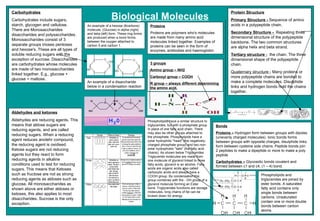
  • 1. Biological Molecules Carbohydrates Carbohydrates include sugars, starch, glycogen and cellulose. There are Monosaccharides disaccharides and polysaccharides. Monosaccharides consist of 3 separate groups trioses pentoses and hexose's. These are all types of soluble reducing sugars with the exception of sucrose. Disaccharides are carbohydrates whose molecules are made of two monosaccharides linked together. E.g., glucose + glucose = maltose. An example of a hexose (6carbons) molecule, (Glucose) in alpha (right) and beta (left) form. These ring forms are produced when a bond forms between the oxygen attached to carbon 5 and carbon 1. An example of a disaccharide below in a condensation reaction Aldehydes and ketones Aldehydes are reducing agents. This means that aldose sugars are reducing agents, and are called reducing sugars. When a reducing agent reduces anotehr compound, the reducing agent is oxidised. Ketose sugars are not reducing agents but they react to form reducing agents in alkaline conditions used to test for reducing sugars. This means that Ketoses such as fructose are not as strong reducing agents as aldoses such as glucose. All monosaccharides as shown above are either aldoses or ketoses, this also applies to most disaccharides. Sucrose is the only exception. Proteins Proteins are polymers who's molecules are made from many amino acid molecules linked together. Examples of proteins can be seen in the form of enzymes, antibodies and haemoglobin. 3 groups Amino group – NH2 Carboxyl group – COOH R group – always different determining the amino acid. Protein Structure Primary Structure - Sequence of amino acids in a polypeptide chain. Secondary Structure - Repeating three dimensional structure of the polypeptide backbone. The two common structures are alpha helix and beta strand. Tertiary structure - the chain. The three dimensional shape of the polypeptide chain. Quaternary structure - Many proteins or more polypeptide chains are bonded to make a complete molecules. Disulphide links and hydrogen bonds hold the chains together. Phospholipids have a similar structure to triglycerides, but with a phosphate group in place of one fatty acid chain. There may also be other groups attached to the phosphate. Phospholipids have a polar hydrophilic "head" (the negatively-charged phosphate group) and two non-polar hydrophobic "tails" (the fatty acid chains). As shown below Triglycerides Triglyceride molecules are made from one molecule of glycerol linked to three fatty acids, glycerol is an alcohol. Fatty acids are organic acids also called carboxylic acids and always have a COOH group. By condensation this group combines with the - OH group of a glycerol molecule forming an Ester bond. Triglycerides functions are storage molecules, long chains of fat can be broken down for energy. Bonds Proteins – Hydrogen form between groups with dipoles (unevenly charged molecules). Ionic bonds forms between groups with opposite charges, disulphide links form between cysteine side chains. Peptide bonds join 2 peptides to make a dipeptide or more to make a poly peptide Carbohydrates – Glycosidic bonds covalent and formed between c1 and c4, (1 – 4) bond. Phospholipids and triglycerides are joined by ester bonds. A saturated fatty acid contains only single bonds between carbons. Unsaturated contain one or more double bonds between carbon atoms.
  • 2.
  • 3.
  • 4.
  • 5. Introduction Organisms are made of a huge variety of compounds. The main organic compounds are carbohydrates, proteins, lipids, and nucleic acids. I will write about these first three groups. The molecules of these groups are mainly made of Carbon, Oxygen and Hydrogen. There are many different lipids, proteins and carbohydrates, they are all made of a small number of simpler molecules joined together. The joining of these smaller molecules happens in a condensation reaction. In which H 2 O is produced. And the opposite can happen by adding water, this is called hydrolysis and is used to break large molecules into smaller ones. E.g. a disaccharide into two monosaccharides. But the first subject topic is lipids. Lipids Lipids are fats, oils and waxes. They are insoluble in water, this is because they have no dipoles and so no charges to attract water molecules. Lipids are made of Carbon oxygen and hydrogen. Most lipids are composed of compounds called triglycerides . Triglycerides are a glycerol bonded to three fatty acid chains. (see fig. 1) They are made from a special type of condensation reaction, called esterfication. Which forms ester bonds between the glycerol and fatty acid. (see fig. 2) Fig. 1 Carbohydrates Carbohydrates are separated into three categories. Monosaccharides - one, single sugar Disaccharides - two, double sugar Polysaccharides - many, multiple sugars. And sugars are separated into reducing sugars, and non-reducing sugars. Reducing sugars can donate electrons to other molecules, all monosaccharides, and some disaccharides are reducing sugars. The Most common monosaccharide is glucose. There are two types of glucose, alpha (  and beta (  .  Glucose is a hexose sugar, because it has a structure like a hexagon (see fig.3). The difference between Alpha and Beta glucose is the 1 st and 4 th carbons. Fig. 3 Chromatography Chromatography is used to separate, then identify the individual chemicals in a mixture. It works by drawing a line on a piece of chromatography paper (in pencil, because ink would run) called the origin. Then dropping a very small amount of liquid onto the origin. You then allow this spot to dry, then repeat the process about 4times, until you have a concentrated spot on the origin line. (see fig. 7) You then put the piece of paper into a solvent, but ensure the line of origin is above the solvent line. Fig. 7 Biological Molecules Holly Stockham Biochemical tests There are tests that you can do to find out what a substance contains. These tests are below: Starch. Adding Iodine to the solution, if it turns blue/black it contains starch. Protein. By adding biurets solution and heating up the solution gently you can see if it contains protein. A positive result would turn the solution lilac. Lipids. You do the emulsion test, which is adding ethanol, shaking, then adding the solution to water. It is called the emulsion test because if the solution contains lipids a white emulsion will be formed. Reducing sugars. Add Benedicts solution, then heat up to near boiling pt. the colour will go from Blue to green to yellow to orange to red . Depending how much reducing sugar it contains. (red containing the most reducing sugar.) Non-reducing sugars. First you must add it to dilute HCL, which hydrolyses the non-reducing sugars to reducing sugars. Then it must be neutralised with NaHCO 3 , then there should be a positive result from the benedicts test (as above) References Advanced Biology by Jones and Jones. CGP AS-level Biology Collins AQA AS Biology Biology toolbox CD Proteins Proteins are made from long chains of amino acids. (see fig. 5) And like lipids and carbohydrates, they are formed by condensation reactions, and broken down by hydrolysis. But the bonds between the amino acids, making the proteins are called peptide bonds (see Fig. 6). Fig.5 An amino acid Fig. 6 Solvent front origin Spot of pigment The chemicals can then be identified comparing Rf values to a table of already known Rf values. The Rf value is calculated by this formula: Rf value = Distance from origin to top of spot Distance from origin to solvent front (Note: The Rf Value will be between 0 and 1.) But sometime the solvent doesn’t completely separate out all the chemicals properly, so you need to do two way chromatography . Where you turn the paper 90 o and place it into another solvent. Which should completely separate out the chemicals. glycerol Fatty acid chain Fig. 2 Ester bonds triglyceride Fatty acids can be saturated or unsaturated. The difference is that saturated fatty acids are completely “covered” in hydrogen, and they have no double bonds. Whereas unsaturated fatty acids have double bonds, and the capability to bond with more hydrogen. If an unsaturated lipid has more than one double bond it is called polyunsaturated. Saturated lipids are normally found in animals, and unsaturated, in plants. There is another type of lipid, a phospholipid . Phospholipids are used in cell membranes because they have a hydrophobic tail and hydrophilic head. So they control what can come in and out. The structure of a phospholipid varies from that of a triglyceride, but not by much. A phospholipid still has the same glycerol backbone as a triglyceride, only instead of having three fatty acid chains, it has two, and a phosphate group, which is ionised to attract water (that’s why the head is hydrophilic). To break up a lipid you need to add water, hydrolysis. Which is normally done by adding a dilute acid, and heating it up. Then you get glycerol and chains of fatty acids again. The main use of triglycerides in the body is as an energy source. They contain much more energy per gram than either carbohydrates or proteins. Triglycerides can be broken down and oxidised in respiration and the energy from them is used to make ATP. Alpha glucose Beta Glucose To form a Disaccharide you must join two monosaccharides together in a condensation reaction (see fig.4) . The monosaccharides bond at carbon 1 and 4, creating a 1-4 glycosidic bond. And produce water. That creates a disaccharide, some key examples of disaccharides are maltose, sucrose, and lactose. Maltose is formed from two alpha glucose molecules, and is the sugar produced when starch is broken down. To break a disaccharide into a monosaccharide/ break down a polysaccharide to a disaccharide you use hydrolysis, it basically means adding water. It reverses the condensation reaction, and is usually done by adding a warm dilute acid. Polysaccharides are formed when 2 or more disaccharides join together. The three main polysaccharides are: Starch (amylose and amylopectin) Amylose is a long unbranched chain, it’s compact, and is therefore good for storage. Amylopectin is branched, which means it is easy to break down the side branches to get at the glucose. Glycogen It’s similar to amylopectin, only with a lot more side branches, so it looks a bit like a brush. This is good for quick energy release, as glycogen is stored in animals. Cellulose Is a straight chain, (a bit like a brick wall) with hydrogen bonds linking the chains together. This is good because it makes the structure strong, as it is used for cell wall. Also enzymes can’t break it, because they can’t reach the glycosidic bonds. Fig. 4 Amine group Functional group Carboxyl group The formation of a protein. Showing the peptide bond The functional group is shown as “R” because it changes in different amino acids. For instance: The Primary structure of a protein is the order of the amino acids. The Secondary structure is the shape e.g. beta pleated sheet or a helix. The Tertiary structure is a globular protein. It’s made up of lots of helixes and beta pleated sheets. It's 3D, enzymes are globular proteins. Globular proteins are therefore affected by all the things you associate with enzymes, like temperature and pH. It is held together by many different bonds, like disulphide, hydrogen bonds, van der waals forces, and ionic bonds. The Quaternary structure is 2 or more polypeptides, so a big group of globular proteins. Beta pleated sheet helix C 4 H 9 leucine C 4 H 9 isoleucine C 3 H 7 valine CH 3 alanine H glycine Functional Group Amino Acid
  • 6. Introduction- A cell is the basic unit from which all living organisms are made. Some cells are big enough to be seen with the naked eye, however most cells are microscopic and can only be seen with microscopes. Bacteria tend to have the smallest cells, around 0.5um across, whereas human cells are between 10um and 30um in diameter. Despite the variations in size, cells have certain features in common. For further information References – www.biologymad.com http://library.thinkquest.org Collins AQA AS Biology Module 1: Molecules, cells and systems Mary Jones and Geoff Jones Advanced Biology Letts Biology Revision guide Organelles Organelles are small structures within cells that perform dedicated functions. As the name implies, you can think of organelles as small organs. Organelles were historically identified through the use of microscopy, and were also identified through the use of cell fractionation. There are many different types of organelles: Eukaryotic cells contain: Cell wall – A layer of tough cellulose fibrils (made of a variety of polysaccharides) that surrounds plant cells to give support and protection. The cell wall allows the cell to become turgid without bursting and maintains the shape of the cell. Cell membrane – A thin boundary around all cells and most organelles that controls what passes in and out of the cell. Proteins are embedded in the cell membrane and are also involved in cell recognition in the immune system. Nucleus – Large, usually round, bounded by double membrane with many pores. The most important part of the cell, controls growth, contains DNA, responsible for cell division, regulates metabolism and protein synthesis. Quite simply, the total control of the cell. Nucleolus – The darkly stained region in the nucleus that is involved with the synthesis of ribosomes. Mitochondrion – Usually round and elongated with a smooth outer membrane and a folded inner membrane. The mitochondria are contained in the cytoplasm of cells and are the site of aerobic respiration and where most of the cells ATP is produced. Chloroplast – A biconcave disc (the perfect compromise between a sphere and a disc) with many internal membranes. Chloroplasts are the site of photosynthesis as they contain the green pigment chlorophyll. Vacuole – A large central organelle in most plant cells, surrounded by a membrane called the tonoplast, full of fluid. Vacuoles store compounds such as sucrose and the difference in water potential compared to the surrounding fluids enables the cell to produce turgor pressure. Rough endoplasmic reticulum (rough ER) – The extensive membrane system that has many cavities and tubes with ribosomes attached. Rough ER is found throughout the cytoplasm connected to nuclear membrane and acts as the transport system in the cytoplasm, collecting, storing, packaging and transporting the proteins made in the ribosomes. Smooth endoplasmic reticulum (smooth ER) – The membrane system with small cavities and no ribosomes, found in small patches in the cytoplasm. The smooth ER is involved in the synthesis of lipids and some steroids. Golgi body – A stack of flattened membrane discs found free in the cytoplasm near to the rough endoplasmic reticulum. The golgi body receives packages (vesicules) of protein from the rough endoplasmic reticulum and is involved in the synthesis and modifying of chemicals before their secretion from the cell. Ribosome – A small and dense structure that acts as an enzyme. The ribosome is attached to the rough endoplasmic reticulum or free in the cytoplasm and is where the genetic code is used to build protein (translation). Prokaryotic cells only contain a cell membrane, cell wall, ribosomes, DNA and a cilia or a flagella (as shown previously). Cell fractionation – Cell fractionation is a process which allows scientists to extract pure samples of a particular organelle. Cell fractionation is a combination of various methods used to separate a cell’s organelles and components. There are two phases of cell fractionation: homogenization and centrifugation. Homogenization is the process of breaking open the cells. Cells are broken apart by chemicals, enzymes, or sound waves. Some scientists even force the cells through small spaces at high pressure to break them apart. Centrifugation is the isolation of the cell organelles. The tissue is chopped up and when the cells are broken apart substances that would normally be kept separate by membranes mix together and can begin to react, so a fluid is added to prevent this which is ice cold, contains a buffer and isotonic. The mixture is then put in a homogeniser (blender) to break up the cells. The resulting mixture is then centrifuged at high speed. The spinning greatly increases the gravitational field, and the organelles separate out according to density and shape. The first most heaviest organelle to sediment out is the nuclei. The remaining fluid, the supernatant, is poured off the centrifuged again to collect other organelles. The order that organelles sediment is: nuclei, mitochondria, chloroplasts (if using plant preparations), rough endoplasmic reticulum, plasma membranes, smooth endoplasmic reticulum and finally ribosomes. Prokaryotic and eukaryotic cells, organelles and microscopy. Sarah Townsend South Bromsgrove High School Microscopy There are two main concepts in microscopy and these are resolution and magnification. Resolution is the ability to distinguish between two clear dots. Magnification is how much bigger a sample appears to be than it is in real life. Light microscope - This microscope uses light to see and magnify objects. The light rays are focused on to a transparent specimen by condenser rays. The rays pass through the specimen and are focused Prokaryotic Cell – (From the Greek meaning ‘before nuclei’ – the DNA is not enclosed by a membrane). All bacteria and cyanobacteria (blue –green algae) are prokaryotes as they lack a membrane – bound nucleus. They are usually smaller than eukaryotic cells and are similar in size to a mitochondrion or a chloroplast. In fact, they probably were prokaryotic cells which came to live inside the larger eukaryotic cells millions of years ago. They have few internal organs that are distinguishable. All prokaryotic cells are surrounded by a cell wall. However this is different from Figure 1 . A prokaryotic cell taken from an electron micrograph a plant cell wall because it contains different polysaccharides. Instead they contain peptidoglycans which are made up of molecules in which peptides and sugars are combined. The cell walls are very strong and important because they prevent the cell from bursting when the cell absorbs water and help to protect against invasion by viruses. Many prokaryotic cells have a thick layer of jelly surrounding them called a capsule which is made of polysaccharides and protects from viruses and antibodies. Beneath the cell wall is a cell surface membrane that is made up of a phospholipid bilayer and the cytoplasm that contains ribosomes that are approximately 20 nm in diameter. The DNA is not chromosomal but in a circular loop called a plasmid. Some contain a flagellum which is used for movement. They divide by binary fission and have three major shapes: rod, spherical and spiral. Figure 2. A eukaryotic cell taken from an electron micrograph. Eukaryotic cell – (From the Greek meaning ‘truly nuclear’ – the larger nucleus contains the DNA inside a membrane). Organisms with complex cells are eukaryotes and are found in plants, animals and fungi. They can be easily distinguished through a membrane bound nucleus (nuclear envelope) and contain membrane bound structures (organelles). The organelles such as the mitrochondrion or chloroplast are there to perform metabollic functions and energy conversion. Other organelles provide structural support and cellular motility. They are generally much larger than prokaryotic cells. Not all eukaryotic cells contain cell walls, only plant cells, not animals cells. The plant cell wall contains pectin, hemicellulose and cellulose (different from a prokaryotic cell). The DNA is linear (chromosomal), complex, organised into chromosomes and combined with histone proteins. They divide by replication (making copies of themselves). The eukaryotic cell is clearly developed from a prokaryotic cell. This is because eukaryotes formed through the merger of prokaryotes, which predate them in the fossil record by some 2 billion years. again by two more lenses – the objective lens and the eyepiece lens. These two lenses produce a magnified image. As many specimens are colourless and nearly transparent, stains are used to make different parts of the specimen show up clearly. Some stains can be added to living cells, but others must be ‘fixed’ by adding a chemical known as a fixative. These chemicals react with substances such as proteins in the cell, making them insoluble and so anchoring them in position. The cells are killed when the fixative is added. Stains may be added either before or after the fixing process. So long as the specimen is thin enough to allow light to pass through, Figure 3. A light microscope they can be used to see major organelles such as the nucleus or mitochondrion. The fluorescent light microscope is the most common and widely used light microscope. This microscope is used to visualise stained cells with fluorescent dyes. However, limitations occur as the microscope can not see beyond 200 nm resolution and x1500 magnification due to the long wavelength of light dissipating if the light is refracted too far. They are relatively cheap. Electron microscope - This microscope uses electrons to see instead of light. They are focused using electromagnets rather than glass lenses. As electrons are easily stopped by air molecules, the space inside must be a vacuum. As our eyes do not respond to electrons, the electrons are allowed to hit a fluorescent screen which emits visible light where the electrons hit. Electrons cannot penetrate materials as well as light, so specimens must be much thinner. This places great limitations on what can be viewed, in particular it is not possible to look at living material, all specimens musty be dead. Specimens are stained using heavy metal ions such as lead or osmium and are taken up by particular parts of the cells. Atoms of these ions have large, positively charged nuclei which scatter electrons rather than letting them pass straight through. These Figure 4. An electron microscope electrons therefore do not arrive on the screen, so leaving a dark area in the image. The structures in a cell which have taken up these heavy metal stains therefore appear dark. Scanning electron microscopes work in a very different way. The electrons do not pass through the specimen but are reflected off its surface. Scanning electron microscopes are used to provide three dimensional images of surfaces. Because the wavelength of electrons are shorter than light, they have a resolution of 1nm and a magnification of x500000. However, they are very expensive. Figure 5. cell fractionation
  • 7. Introduction There are two main types of cells, Eukaryotic and Prokaryotic. They are very different and contain different organelles which help them to do their specific jobs. An Organelle is a discrete structure of a cell with specialized functions. They are similar to the organs in a body because they are vital in the survival of the cell. There are many ways to identify organelles such as microscopy which can be done using different microscopes to give different resolutions and better images. Cell fractionation can also be used as it is a method of splitting up the cell so that all the different organelles are separate. There are two different types of microscopes, light and electron with there being two more different types of electron microscopes. Transition and Scanning which both do different jobs and see different things along with the light microscope. Microscopy There are two different types of Microscopes, Light and Electron microscopes they or both very different with there also being two different types of electron microscopes. Light Microscopes are is a very simple microscope with not a very high resolution or magnification. Resolution being the ability to distinguish between two points on an image and Magnification being how much bigger a sample appears to under the microscope. The resolving power of a microscope is limited by the wavelength of light which is 400 to 600 mm for visible light. To improve the resolving power of a light microscope a shorter wavelength of light is need that is why some microscopes have blue filters as blue has the shortest wavelength of visible light. Light microscopes cost between £100 and £500 compared to the cost of an electron microscope which is over £1,000,000. They are cheep to operate as well as being small and portable. The samples are simple and easy to prepare with little damage done to the samples during this process. A vacuum is not required unlike in the electron microscopes meaning that the specimen has to be dead. The natural colour is maintained in a light microscope even though stains are often needed to make the cells visible but the maximum magnification is only up to 2000 times. Electron microscopes are much more advanced as they have a magnification of over 500,000 times. They use an electron beam which is very expensive and can damage the specimens which also must be stained with an electron dense chemical. Electron microscopes can also be split into Scanning electron microscopes and Transmission electron microscopes. Scanning electron microscopes pas the scanning beam over the surface of the specimen and electrons reflect of it as it has been coated in heavy metals. The reflected electron beams are focused on a florescent screen to make up the image which is linked to a computer to make the image clearer. It is similar to echo location. Transmission electro microscopes pas a beam of electrons through the thin specimen and focusing the image on a screen or film. The TEM is very big, expensive and not very clear. Cells and Microscopy By Abby Pask Organelles There are many different Organelles with many different functions Eukaryotic cells Nucleus- Where DNA is kept and where RNA is transcribed Ribosome's- The site of protein synthesis, where RNA is translated into proteins Mitochondria- The site or aerobic respiration Chloroplasts- the site of photosynthesis Endoplasmic reticulum- There are two types the rough ER and the smooth ER. The ER is the transport network for molecules targeted for certain modifications and specific final destinations. Golgi Apparatus - The ‘post box’ of the cell as it modifies molecules and packages them into small membrane bound sacs called vesicles. Prokaryotic Cells Vacuole- transport and storing nutrients, metabolites, and waste products. Cytoplasm- contains all the enzymes needed for metabolic reactions Nucleoid- The region of the cytoplasm that contains DNA Meosome- A tightly folded region of the cell membrane containing proteins required for respiration and photosynthesis. Capsule- A thick polysaccharide layer outside of the cell wall used for protection and a food reserve. Flagellum- A rigid rotating helical shaped tail used for propulsion. Both types of Cells Cell Membrane- Made of phospholipids and proteins which is used for protection and transport. Cell Wall- Keeps a cell rigid and gives protection. Eukaryotic Cells Eukaryotic cells are the larger of the two types of cell and can be more than 10µm in diameter. They have a true nucleus and contain many organelles that can either be double or single. This makes them very complex cells which have a number or different jobs. The genetics of a Eukaryotic cell is made up of linear DNA and is associated with proteins to form chromatin. It has a true nucleus which means it has a nuclear envelope and is one of the main difference between a eukaryotic cell and a Prokaryotic cell. Another main difference is the number of ribosome's in the cell with the eukaryotic cell having a large number (80S) The cells walls are made up of either chitin in fungi or cellulose in plants but animal cells do not have a cell wall Figure 1. This is a basic diagram of a eukaryotic cell with all the main organelles that it contains including the nucleus and the endoplasmic reticulum Figure 2 . These two photos are different eukaryotic cells that have been magnified using a microscope. The main recognisable organelles have been labels Prokaryotic Cells Prokaryotic cells are much smaller than Eukaryotic cells being less than 5 µm in diameter. They have smaller ribosome's (70s) and less organelles which are all not membrane bound. The genetics of a prokaryotic are also very different as there is circular DNA and no proteins. There is no true nucleus which means that the DNA is free to move within the cytoplasm. The cell walls are made from Glycoprotein's or other polysaccharides, but not cellulose or chitin as these are found in Eukaryotic cells. Prokaryotic cells include bacteria and blue-green algae as they are very small and not very complex Figure 3. This is a basic diagram of a Prokaryotic cell with all the main organelles that it contains including the ribosome's and the Nucleoid Figure 4. This is a prokaryotic cell seen through a microscope it shows the cell wall and cell membrane which outline the cell.
  • 8. Phospholipids have a similar structure to triglycerides, but with a phosphate group in place of one fatty acid chain. There may also be other groups attached to the phosphate. Phospholipids have a polar hydrophilic "head" (the negatively-charged phosphate group) and two non-polar hydrophobic "tails" (the fatty acid chains). This mixture of properties is fundamental to biology, for phospholipids are the main components of cell membranes. RECAP OF LIPIDS What's the difference between a phospholipids and triglyceride Molecule?  Triglycerides are the familiar fats and oils. The molecules are made from one molecule of glycerol linked to three fatty acids. Glycerol is an alcohol. Fatty acids are organic acids (also called carboxylic acids) and also have a -COOH group. BY condensation, this group combines with the -OH group of a glycerol molecule, forming an ester bond. Because triglycerides are fat, they acts as a storage molecule.   Phospholipids have two fatty acids and a phosphate molecule attaches where as a triglyceride molecule only has three fatty acid chains. There function is to form membranes. Biochemical tests These five tests identify the main biologically important chemical compounds. For each test take a small amount of the substance to test, and shake it in water in a test tube. If the sample is a piece of food, then grind it with some water in a pestle and mortar to break up the cells and release the cell contents. Many of these compounds are insoluble, but the tests work just as well on a fine suspension. Starch (iodine test). To approximately 2 cm³ of test solution add two drops of iodine/potassium iodide solution. A blue-black colour indicates the presence of starch as a starch-polyiodide complex is formed. Reducing Sugars (Benedict's test). To approximately 2 cm³ of test solution add an equal quantity of Benedict’s reagent. Shake, and heat for a few minutes at 95°C in a water bath. A precipitate indicates reducing sugar. The colour and density of the precipitate gives an indication of the amount of reducing sugar present, so this test is semi- quantitative. A blue colour means no reducing sugar, a green precipitate means relatively little sugar; a brown or red precipitate means progressively more sugar is present.   Non-reducing Sugars (Benedict's test). Using a separate sample, boil the test solution with dilute hydrochloric acid for a few minutes to hydrolyse the glycosidic bond. Neutralise the solution by gently adding small amounts of solid sodium hydrogen carbonate until it stops fizzing, then test as before for reducing sugars. Biological molecules Chris Kelly 12WH South Bromsgrove High School, A Specialist Technology College, Bromsgrove, Worcestershire, B60 3NL . Lipids (emulsion test). Lipids do not dissolve in water, but do dissolve in ethanol. This characteristic is used in the emulsion test. Do not start by dissolving the sample in water, but instead shake some of the test sample with about 4 cm³ of ethanol. Decant the liquid into a test tube of water, leaving any undissolved substances behind. If there are lipids dissolved in the ethanol, they will precipitate in the water, forming a cloudy white emulsion. The test can be improved by adding the dye Sudan III, which stains lipids red. Protein (biuret test). To about 2 cm³ of test solution add an equal volume of biuret solution, down the side of the test tube. A blue ring forms at the surface of the solution, which disappears on shaking, and the solution turns lilac-purple, indicating protein. Conclusion Biochemistry is a particularly difficult area in biology as it is split up into different sections. For example you have to remember the types, function and importance carbohydrates-monosaccharides, disaccharides and polysaccharides and proteins, Lipids and biochemical tests. Fatty acids are long molecules with a polar, hydrophilic end and a non- polar, hydrophobic "tail". The hydrocarbon chain can be from 14 to 22 CH2 units long, but it is always an even number because of the way fatty acids are made. The hydrocarbon chain is sometimes called an R group, so the formula of a fatty acid can be written as R-COO-. Saturated or unsaturated If there are no C=C double bonds in the hydrocarbon chain, then it is a saturated fatty acid (i.e. saturated with hydrogen). These fatty acids form straight chains, and have a high melting point. If there are C=C double bonds in the hydrocarbon chain, then it is an unsaturated fatty acid (i.e. unsaturated with hydrogen). These fatty acids form bent chains, and have a low melting point. Fatty acids with more than one double bond are called poly-unsaturated fatty acids (PUFAs). These fatty acids are joined by a condensation reaction however in this reaction it is known as ESTERIFICATION and an ESTER bond is formed. With three branches three ester bonds can form so three fatty acids can bond so therefore it is called a triglyceride. Is first molecule of a fatty acid   Acknowledgments Literature cited For further information
  • 9.
  • 10.
  • 11. Jonathan Stevens Module One: Biological Molecules Lipids, Proteins and Carbohydrates Introduction: In the following poster I will go into detail of many things to do with biological molecules, the main aspects I will go into detail are carbohydrates, lipids and proteins. I will show the structures of the molecules and the types of bonding and how they join and get broken down. I will state the chemical tests for the certain types of molecules and what too look for to see If the substance contains the specific type of molecule. I will the state and explains the functions of proteins and etc. Carbohydrates: There are 3 main types of carbohydrates and they are polysaccharides, monosaccharide and disaccharides. All carbohydrates contain 3 elements these elements are carbon, hydrogen and oxygen . Monosaccharide: A monosaccharide is a single sugar this is a molecule of sugar in its smallest form, an example of this is glucose, glucose is a sugar which our body uses, there are two types of glucose there is alpha and beta, the structures of these molecules differ, they are as follows: Alpha Beta Glucose can join together to form long chains which are polysaccharides, the first stage of the joining is when two glucoses join to become a disaccharide the glucoses can join up in different orders and this makes different disaccharides for example alpha plus alpha. The process of joining is between carbon one and carbon four, they join by a glycosilic bond, reaction gives out water and it is called a condensation reaction. The glycosilic bond can also be broken they are broken, they are broken down by adding it with acid, this reaction adds water to the molecule which rejoins the two glucoses, this reaction is a called hydrolosis. The test for monosaccharide is to add benedict's solution to the substance which is at near boiling, if the substance contains monosaccharide the benedict's solution will change to a green – red solution. Disaccharides: Disaccharides are two monosaccharide joined together, they are joined by condensation reaction. The test for disaccharides is as follows, add hydrochloric acid to the solution which is at near boiling, then add benedict's solution to the solution and if the benedict's solution changes to a green to red colour. Polysaccharides: These are massive chains of monosaccharide and disaccharides an example of this is starch which make up bigger molecules. Lipids: Lipids all contain a glycerol backbone which looks as follows: Lipids contain a fattys acid and this has a carboxyl this is shown below, the zig zaged line represents a long hydrocarbon chain, the chain can be saturated or unsaturated. A fatty acid joins to a glycerol backbone they join by esterfication this is a condensation reaction and releases water. A glycerol backbone can join up to three things on its 3 branches, one example it can make is a triglyceride, this is when a glycerol backbone joins to three fatty acids. Esterfication A fatty acid is hydrophobic this means it doesn’t like to react with water this is because the water will breaks the bonds between the glycerol and the fatty acid because if you add water you are adding back hydrogen's, and why do they share hydrogen's if they could have one each. The glycerol head is hydrophilic which means it is water loving. Proteins: Proteins are a key part of the body they have many functions these functions are: ~for enzymes ~anti-bodies ~actin & myosin which make muscles contract ~used to make tissues ~blood clotting involve many proteins ~gives strength to hair and nails Proteins are made up of amino acids and each amino acid has three groups these groups are amino group, functional group and the carboxyl group, a diagram of this is below: The functional group can join to different molecules these molecules make up different proteins and amino acids which have different jobs, examples of these are below: Proteins and amino acids have many structures and these structures are: Primary structure this is the order of the amino acids Secondary structure: this structure is when the long strand of amino acids either curve into a helix or the create a beta pleated sheets, the helix and sheets are joined together by hydrogen bonds. Tertiary structure: the third structure is when the helix’s and sheets join together to form a globular protein this big structure is also known as a enzyme, the big structure is joined together by many bonds these bonds are: di-sulphide, hydrogen, Ionic, Hydrophobic, Van der Waals Quartary structure: this structure is when gobular proteins join together to make bigger structures this structure can join together to make a well known structure which is a red blood cell.
  • 12.