
Download to read offline


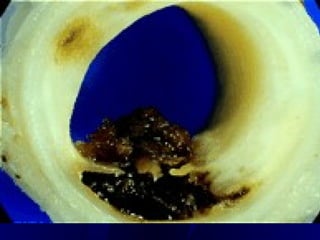

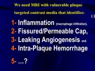
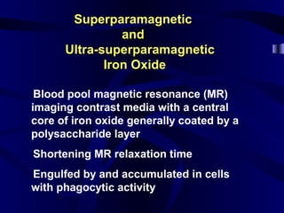
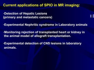
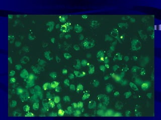
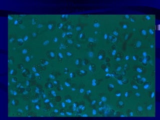
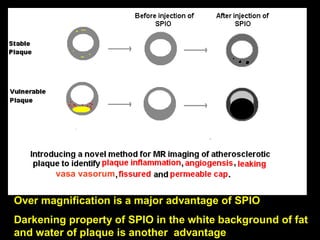
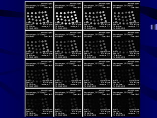

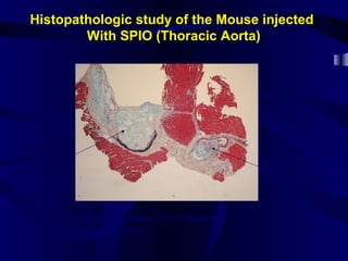
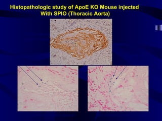
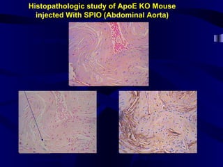
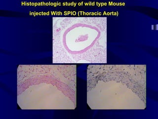
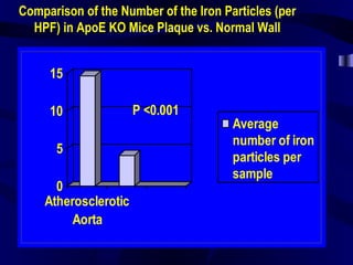
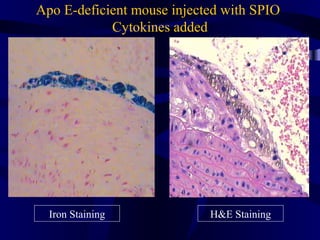
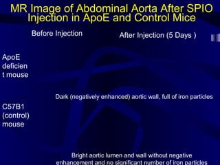
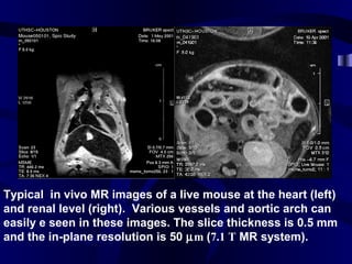

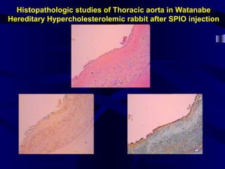
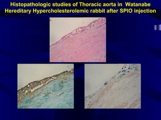
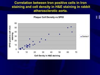
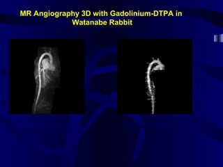
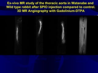
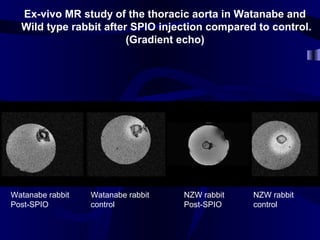

This document discusses research on using superparamagnetic iron oxide (SPIO) nanoparticles and ultra-small superparamagnetic iron oxide (USPIO) as MRI contrast agents to detect vulnerable plaque. In vitro and animal studies show that macrophages engulf SPIOs, which then accumulate in inflamed plaques and decrease the MRI signal. Histological analysis confirms iron particle presence correlates with macrophage density. MRI of atherosclerotic rabbits after SPIO injection shows darker aortic walls with many iron particles compared to controls. This non-invasive method may help identify vulnerable plaque.


