Git j club VCE( GIE Journal + net presentations& references)
•Download as PPT, PDF•
1 like•963 views


Recommended
More Related Content
What's hot
What's hot (20)
Similar to Git j club VCE( GIE Journal + net presentations& references)
Similar to Git j club VCE( GIE Journal + net presentations& references) (20)
More from Shaikhani.
More from Shaikhani. (20)
Recently uploaded
Recently uploaded (20)
Git j club VCE( GIE Journal + net presentations& references)
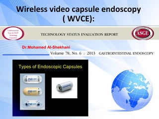
- 1. Wireless video capsule endoscopy ( WVCE): Dr.Mohamed Al-Shekhani
- 2. Definition: • Visualization of the GI tract by transmitting images wirelessly from a disposable capsule to a data recorder worn by the patient. 2
- 3. History: • The first model for the small intestine approved by FDA in 2001. • Over subsequent years, VCE developed with superior resolution, increased battery life& capabilities view different parts of the GI tract. 3
- 5. Companies producing WVCE: • • • • • 5 3 companies that manufacture smallbowel WCE systems approved by FDA: PillCam SB2, Given Imaging, Ltd, Israel; Endocapsule, Olympus America, Inc, Center Valley, Pennsylvania; MiroCam, IntroMedic Company Ltd, Seoul, Korea Capsules for esophageal imaging /colon imaging also are available from Given Imaging.
- 6. 6
- 7. Components: WCE system: 3 components: (1) Capsule endoscope with a light source, camera & battery. (2) A sensing system with sensing pads or a sensing belt to attach to the patient, a data Recorder, a battery pack (3) A personal computer with proprietary software. + handheld viewers allow realtime review of images during WCE examinations 7 Battery life 8-12 hours.
- 9. SI Imagings: OP;Fasting or clear liquids for 10 -24 hs. A full or partial bowel prep improve visualization. Capsule is activated by removal from a magnetic holder. After ingestion, patients are instructed to keep a diary of symptoms and monitor the lights on the data recorder to confirm that the signal is being received. Avoid exercise that may cause the sensors to detach. 9
- 10. SI imaging: A diet of clear liquids is allowed after 2 hours& a light meal after 4 hours. The reusable data-recording system can be disconnected from the patient after the lifespan of the battery has expired. The capsule is disposable& designed to be excreted. The data recorder is subsequently connected to a workstation for transfer of the acquired images. 10
- 12. Esophageal VCE: • Fasting for 2 hours. • the patient drinks 100 mL of water while standing ,ingests the activated capsule in supine position with a 10-mL sip of water. • A 2-minute recording with the patient supine, 2 minutes raised to 30 , additional minute at 60 , followed by an upright position for 15 minutes to maximize time for the capsule to capture images as it traverses the esophagus. 12
- 13. Colon VCE: • Not approved in US but approved in Europe. • Sensitivity less than colonoscopy. 13
- 15. Agile patency system: • A radiopaque non-video, dissolvable capsule . • For those with high risk for retention. • Delivery devices: for those who can not swallow the capsule. 15
- 22. Indications: • (1) (OGIB), overt & occult, including IDA • (2) Suspected Crohn’ s Disease • (3) Surveillance in polyposis syndromes. • (4) Suspected small-intestine tumors • (5) Suspected or refractory malabsorptive syndromes (eg, celiac disease). • FDA approved VCE for the esophagus evaluation for suspected Barrett’ s esophagus, esophagitis, or esophageal varices. 22
- 23. Contraindications: • Relative : • (1) Known or suspected GI obstruction, strictures, or fistulas based on the clinical picture or preprocedure testing • (2) Cardiac pacemakers or other implanted electromedical devices • (3) Swallowing disorders • (4) Pregnant. 23
- 24. Interpreter: • ASGE guidelines:readers should have either undergone formal capsule training during fellowship or have completed a formal GI or surgical society– endorsed training course with proctoring of the first 10 capsule readings. • The average reading time 30-120 mins, dependant on SI transit time&experience. • For VCE esophagus, the average reading time 5-15 minutes. 24
- 25. OGIB: Detection rate 35-77% dependent on various factor: Earlier WCE (within 1 wk of bleeding), Inpatient status Overt tranfusion – requir GI bleeding Male sex. Increasing age. Use of warfarin Liver co-morbidity 25
- 26. Indications: Benefits: OGIB Impacts on patient management (surgery, medical therapy, NSAIDs withdrawal). Better than barium/most imaging studies With the exception of therapeutic potentional both VCE & DBE have comparable results. 26 •. CD: Valuable adjunctive diagnostic test after conventional endoscopy& colonoscopy with ileoscopy. Calculate activity score.
- 27. ... SI tumors: Feasible/safe in known or suspected polyposis syndromes as FAP or PJS. WCE was effective in detecting additional polyps in the jejunum May not be able to adequately visualize the ampulla of Vater. Better than Barium and MRI eneterography sp for small polyps. 27 SI tumors: WCE may have a role in the evaluation of smallbowel tumors, but a negative exam should not preclude further work-up if a lesion is highly suspected.
- 28. ... CELIAC DISEASE: Possible advantage over endoscopy in “ patchy” disease. An additional role of WCE specifically in complicated celiac disease(ulcer,stricture, tumor). 28 Eso diseases WCE is inferior to OGD for the diagnosis of esophagitis&Barrett’. WCE is inferior to endoscopy for the diagnosis/grading of EV for screening. ? MAGNET.
- 36. Safety Generally safe: only concern is retention;1.3% Retention .Remaining for 2 wks or required directed therapy. 36 Risk strictur:e: CD, SI tumors, radiation,NSA ID,Surgical. Zenker div, duodenal div, umbil hernia, Meckel). Diagnosis/trt Abd imaging after 2 weeks if retention is suspected&if confirmed, surgery or endoscopic intervention. perforations • 2 cases in CD. • Tracheal aspiration .
- 37. Interference with cardiac devices: Generally safe, but not used until more data available. MRI should not be done until the VCE passed. 37
- 38. Summary: A valuable test for imaging the SI. It is a safe &easy to perform that can provide valuable information in the diagnosis of SI conditions. Its applications still remain limited within the esophagus& colon. Future developments: improving visualization of esophagus &improved manipulation of the capsule within the GI tract & biopsy capabilities. 38