SlideShare a Scribd company logo
1 of 12
Download to read offline
10/18/2020
1
Ultra Violet Spectroscopy
Dr. Satish Dhirendra Mitragotri
(M.Sc., Ph.D., SET, NET, GATE and M.B.A. (Finance))
Department of Chemistry
Walchand College of Arts and Science, Solapur
Spectroscopy - Terminology
 Spectroscopy - The study of interaction between
electromagnetic radiation and matter is spectroscopy.
Electromagnetic radiation – The oscillating electric
and magnetic fields which are perpendicular to each
other travel through medium and have definite energy
is known as electromagnetic radiation.
Different types of electromagnetic radiations are
o Sun light
oU V light
oVisible light
oX-ray
oMicrowave
oRadio frequency wave etc.
Spectroscopy - Terminology
 Electromagnetic spectrum – Entire range of wave-
length, energy, frequency over which electromagnetic
radiation present is known as electromagnetic
spectrum.
Different terms related to wave motion
1) Wavelength
2) Frequency
3) Wave number
4) Velocity
5) Energy
6) Particle properties of radiation.
Spectroscopy – Electromagnetic wave
Spectroscopy – Electromagnetic wave Spectroscopy - Terminology
1) Wavelength (λ) – The distance between two
particles which are in same phase.
•Wavelength is expressed by Lambda (λ)
•Measured in units of length starting from Km to
nm, more commonly used unit is Ao
1m = 1 X 102 cm = 1 X 106 µm = 1 X 10⁹ nm = 1 X 10¹⁰ Ao
Crest
Trough
10/18/2020
2
Spectroscopy - Terminology
2) Frequency (ν) – The number of waves passing
through a particular point in unit time (one second)
is known as frequency.
Unit of frequency is cycles per second or Hz
 1 Hz = 1 cycle per second
It is direct measure of energy.
 1 MHz = 1 X 10³ KHz = 1 X 10⁶ Hz
A fixed point
Spectroscopy - Terminology
3)Wave number (ν-) – The number of waves per
centimeter is known as wave number.
 Wave number is reciprocal of frequency, ν- = 1/ λ
Unit is kaiser or reciprocal centimeter = cm-1
A fixed Distance = 1 cm
Spectroscopy - Terminology
4)Velocity (C) – The distance travelled by the wave in
one second is called as velocity of radiation.
•Velocity of radiation is fixed and it is
•3 X 10⁸ m/s or 3 X 1010 cm/s
•Energy is directly proportional to velovity.
5)Energy (E) - Energy associated with radiation.
E = h X ν
Longer the wavelength lesser the energy
Shorter the wavelength more is the energy
6)Particle properties of radiation.
Spectroscopy – Electromagnetic Spectrum
Radiation Cosmic
Ray
Gamma
Rays
X-
Rays
UV Visible IR Microwave Radio
freq.
waves
Wavelength
cm
10-9 10-7 10-5 10-4 10-3 10-1 102
Energy
Kcal/mol
109 106 106 10 10-2 10-4 10-6
Frequency
Hz
Function Ioniza
tion
Electr
onic
transi
tion
Mole
cular
Vibra
tion
Rotational
motion
Nuclear
spin
Spectroscopy – Energy of molecule
Energy associated with any molecule can be resolved in four
different components
E Total = E Electronic + E Vibrational + E Rotational + E Spin
When radiation falls on any molecule one or more energy
parameters from molecule are changed and it is called
excitation.
Different type of energy type require different radiation for
this excitation process
Electronic- Electronic transition
Vibrational – IR Radiation
Rotational - Microwave
Spin- Radiofrequency wave
10/18/2020
3
Spectroscopy – Energy of molecule
Energy associated with any molecule can be resolved in
four different components
E Total = E Electronic + E Vibrational + E Rotational + E Spin
Electronic- Electronic transition
A B A+ B-
Vibrational – IR Radiation
A B A B
Rotational – Microwave
A B A
Spin- Radiofrequency wave
U V -Spectroscopy
Ultraviolet absorption spectrum arises from
transition of electrons from lower electronic
energy level to higher electronic energy level.
It is absorption spectroscopy where the amount
of energy absorbed by the molecule from
ultraviolet region is measured.
The wavelength of UV radiation is in between
200nm to 400 nm = 2000 A0 to 4000 A0
= 200 to 400 X 10-8 cm
The region from 100-200 nm is called as Vacuum
ultraviolet region
U V -Spectroscopy
The region from 200-400 nm is called as near or
quartz ultraviolet region
It comes prior to violet region of visible radiation
so it is called as ultra violet.
When there are two energy levels for e.g.
E2 and E1 where E2  E1
E2
E1
E2 – E1 = E
U V -Spectroscopy
When UV light falls on any molecule some part
of it may be absorbed, absorption depends on
two parameters
1. Energy of incident UV radiation
2. Structure of molecule
If the energy of radiation is kept same then
different molecules give different absorption
pattern i.e. UV spectrum.
Thus UV spectrum is a plot of absorbance (A)
verses wavelength.
What is absorbance ?
U V -Spectroscopy
When UV light falls on any sample it can interact
in three different ways
1. Reflection
2. Absorption
3. Transmission
Reflection
Absorption
Transmission
Incident
U V -Spectroscopy
What is absorbance ?
Io = Intensity of incident radiation
Ir = Intensity of reflected radiation
Ia = Intensity of absorbed radiation
It = Intensity of transmitted radiation
Transmittance (T)= It / Io
Opacity (O)= Io /It
Absorbance = log (Io /It)
10/18/2020
4
U V -Spectroscopy
Lambert’s Law - When a beam of
monochromatic radiation passes through the
absorbing medium the intensity of the light
radiation decreases exponentially as the length
of absorbing medium increases.
Beer’s Law – When a beam of monochromatic
radiation passes thorough the absorbing
medium the intensity of incident radiation
decreases with length of absorbing medium as
well as concentration of the soultion.
U V -Spectroscopy
Different types of electrons and electronic
transitions
1.Sigma (σ) electrons-
If compound contain only sigma electrons i.e.
electrons in present in sigma bond
e.g. CH3-H
2.Pi (π) electrons – Electrons present in double
or triple bond but that to C-C only
H2C=CH2
3.Non bonding (n) electrons – Electrons present
in hetero atoms bonded with carbon
CH3-HC=O, CH3-OH
U V –Spectroscopy- Types of Orbitals
σ
S
P
π
π*
n
σ*
S
P
Energy
U V –Spectroscopy – Absorption of energy by electrons
Energy
Bonding orbital
ΔE
Antibonding/Nonbonding orbital
ΔE
ER = hν = hc /λ
ΔE = ER
U V –Spectroscopy
Types of Electronic Transitions
σ to σ*  n to σ*  π to π*  n to π*
Energy
σ* Antibonding
π* Antibonding
n Nonbonding
σ Bonding
π Bonding
U V –Spectroscopy – Types of electronic transitions
Energy
Bonding orbital
Antibonding orbital
Nonbonding orbital
Antibonding orbital
Bonding orbital
σ
σ*
π*
π
n
σ to σ*
π to π*
n to σ*
n to π*
10/18/2020
5
U V -Spectroscopy
Different types of electrons and electronic transitions
1.σ to σ* Transition – Transition of an electron from
bonding sigma orbital(σ) to antibonding sigma (σ*)
orbital is called as σ to σ* . This requires highest energy
and takes place by radiation of short wavelength ( 150
nm).
Stronger bond require shorter wavelength for transition.
e.g. CH3-H
2. n to σ* Transition - Transition of an electron from non
bonding (n) orbital to higher energy, antibonding σ*
orbital is called as n to σ* transition.
This type of transition occurs in compounds containing
hetero atoms like N,O,S,P and halogens
CH3-OH, CH3-SH, CH3 NH2
wavelength ( 180 nm – 200nm).
E-max is more for more probable transition.(R-I  R-Cl)
U V -Spectroscopy
Different types of electrons and electronic transitions
3. π to π * Transition (K band) - Transition of an electron
from bonding (π) orbital to higher energy, antibonding
(π*) orbital is called as π to π * transition.
This type of transition occurs in compounds containing
groups with multiple bonds.
C=C, C=O, N=N, C=N etc.
wavelength (200nm - 400 nm).
4. n to π * Transition (R band) - Transition of an electron
from nonbonding (n) orbital to higher energy,
antibonding (π*)orbital is called as π to π * transition.
This type of transition occurs in compounds containing
groups with multiple bonds with heteroatons and
saturated carbon framework.
R-C=O, R-N=N, R-C=N etc.
wavelength (200nm - 300 nm).
U V -Spectroscopy
Wavelength of maximum absorption = λmax
Wavelength ( λ )
100 150 200 250 300 350 400
not a λmax
not a λmax
Intensity ε
6
5
4
3
2
1
0
U V -Spectroscopy
Maximum absorption intensity = εmax
Wavelength ( λ )
Intensity ε
100 150 200 250 300 350 400
Low absorption intensity = ε
6
5
4
3
2
1
0
U V -Spectroscopy
1.Bathochromic
shift (Red shift)
2.Hypsochromic
shift (Blue shift)
Wavelength ( λ )
Wavelength of maximum absorption λ max
6
5
4
3
2
1
0
Intensity ε
--------------------------------------------------------------
100 150 200 250 300 350 400
U V -Spectroscopy
Wavelength ( λ )
Intensity ε
Maximum absorption intensity = εmax
100 150 200 250 300 350 400
6
5
4
3
2
1
0
3.Hyperchromic shift
4.Hypochromic shift
10/18/2020
6
U V -Spectroscopy
1.Bathochromic shift (Red shift)
2.Hypsochromic shift (Blue
shift)
Wavelength ( λ )
Intensity
E-max
Wavelength of maximum absorption
4.Hypochromic shift
100 150 200 250 300 350
3.Hyperchromic shift
U V -Spectroscopy
Different terms used in UV Spectroscopy
1.Chromophore – It is an isolated functional group
capable of absorbing visible and or ultraviolet
region.
2.It is unsaturated group responsible for electronic
absorption
Molecule which has chromospheres is called as
chromogen.
e.g. C=C, C=O, -NO2, -C=N-, -CN, -COOH, -CONH2 etc.
2. Auxochrome – It is functional group which do not
absorb color above 200nm but when attached to
chromophore causes shift in wavelength and or
intensity of absorption.
e.g. –OH, -X, -OR, -NH2
U V -Spectroscopy
Different terms used in UV Spectroscopy
1.Bathochromic shift (Red shift) – Shift of
absorption maxima to longer wavelength is
called as bathochromic shift or red shift. Here
λmax increases.
This is due to presence of auxochrome such as –
OH, -NH2, etc.
Decrease in solvent polarity causes red shift.
e.g. from alcohol to benzene
When there is extension in conjugation the red
shift is observed e.g. ethyene absorbs at 170nm
where as 1,3-butadiene absorbs at 217nm
U V -Spectroscopy
2.Hypsochromic shift (Blue shift) – Shift of
absorption maxima to shorter wavelength is called
as hypsochromic shift or Blue shift.
Here λmax decreases
Generally change in pH of medium causes this shift.
Aniline in absence of any solvent absorbs at 280 nm
but when HCl is added to it red shift is observed
and it absorbs at 200nm
Phenol in basic medium due to formation of
phenoxide ion has extension in conjugation and
absorbs at higher wavelength but when medium
terns acidic the conjugation is removed as ion is
converted to undissociated phenol and red shift in
absorption is observed.
3.Hyperchromic shift
4.Hypochromic shift
U V -Spectroscopy
3.Hyperchromic shift – Increase in the intensity
of absorption maxima is called as hyperchromic
shift. E-max increase Generally electron donating
effect causes this shift
4.Hypochromic shift - Decrease in the intensity
of absorption maxima is called as hyporchromic
shift. E-max Decrease. Disturbance in conjugation
causes this shift
U V -Spectroscopy
1.Bathochromic shift (Red shift)
2.Hypsochromic shift (Blue shift)
Wavelength ( λ )
Intensity E-max
3.Hyperchromic shift
4.Hypochromic shift
10/18/2020
7
U V –Spectroscopy – Effect of conjugation
Effect of conjugation on position of UV and
visible absorption bands.
n to π * Transition (R band) is the most easy to
detect in normal UV spectrum
Presence of conjugated chromospheres (C=C)
(alternate double and single bond) causes
bathochromic or red shift.
If two chromophores are separated by more
than two single bonds then conjugation is
disturbed and no red shift is observed.
e.g. CH2=CH2 - λmax is 175 nm
CH2=CH2-CH2-CH2=CH2 λmax is 175 nm
U V –Spectroscopy – Effect of conjugation
Energy
P
P
P
π*
π
P ΔE1
ΔE2
ΔE1  ΔE2
π1
π1*
π2
π2*
π3
π3*
U V –Spectroscopy – Effect of conjugation
a)Ethene CH2=CH2 has - λmax is 175 nm also 1,4-
pentadiene also has CH2=CH2-CH2-CH2=CH2 λmax
is 175 nm as the conjugation is disturbed and
thus there is no effect of presence of two
chromophores.
b)If the chromophores are present in
conjugation the red shift is observed.
Ethene - CH2=CH2 has - λmax is 175 nm
1,3-butadiene
CH2=CH-CH=CH2 - λmax is 217 nm
1,3,5-hexatriene
CH2=CH-CH=CH-CH=CH2 - λmax is 256 nm
U V –Spectroscopy – Effect of conjugation
More number of conjugated double bonds lead
to more overlap of HOMO and LUMO and thus
energy difference between the orbitals
decreases leading to absorption at higher
wevelength. Decreasing in energy leads to
increase in wavelength.
c) It two different chromophores are present
e.g. C=C and C=O, e.g.
CH2=CH2 has - λmax is 175 nm
CH2=O has - λmax is 290 nm
Where as CH2=CH-CH2=O has λmax is 320 nm
Thus conjugation increases λmax to higher value.
Applications of U V -Spectroscopy
1.Determination of structure - UV spectroscopy
provides information mainly about the
extension in conjugation.
Though not useful for complete structure
elucidation it provides key information about
different types of electron present in molecule.
If UV spectrum of molecule is available then it
can be compared with unknown spectrum of
identification.
Applications of U V -Spectroscopy
2.Determination of stereochemistry of
compounds – UV spectrum is useful in
determination of stereochemistry of molecules
specially compounds exhibiting cis-trans
isomerism.
The trans conformation has planar structure and
hence the conjugation is not disturbed. In these
molecules thus λmax – Has higher value. Where
as in case of cis isomer the planar nature is
disturbed and thus orbital overlap is not
completely effective resulting in lowering of λmax
10/18/2020
8
Applications of U V -Spectroscopy
This can be confirmed by two examples where
two isomers cis and trans isomers have different
λmax values.
Cis -stilbene has λmax value at 280 nm and ἐ-max
at 13500 where as trans -stilbene has λmax value
at 295 nm and ἐ-max at 27000.
Cis –cinnamic acid has λmax value at 264 nm and
ἐ-max at 10700 where as trans –cinnamic acid
has λmax value at 273 nm and ἐ-max at 15900.
Applications of U V -Spectroscopy
H H H
H
Cis -stilbene
labda max = 280 nm
e-max = 13500
Trans -stilbene
labda max = 295 nm
e-max = 27000
H H
COOH
H
H
COOH
Cis -Cinnamic acid
labda max = 264 nm
e-max = 10700
Trans -Cinnamic acid
labda max = 273 nm
e-max = 15900
U V –Spectroscopy-Woodward-Fieser Rules for
calculations of λmax
Woodward-Fieser rules for calculation of λ max value(nm)
for dienes and trienes
Sr.No. Name of the
Motif
Skeletal structure λ max
value(nm)
1 a Basic value for
Un-substituted
diene
214
1 b Basic value for
Conjugated diene
214
C C C C
C C
C C
C C
U V –Spectroscopy-Woodward-Fieser Rules for
calculations of λmax
Woodward-Fieser rules for calculation of λ max value(nm)
for dienes and trienes
Sr.No. Name of the
Motif
Skeletal structure λ max
value(nm)
1 c Basic value for
Hetero-
annular diene
214
1 d Basic value for
semi cyclic
diene
214
U V –Spectroscopy-Woodward-Fieser Rules for
calculations of λmax
Woodward-Fieser rules for calculation of λ max value(nm)
for dienes and trienes
Sr.No. Name of the
Motif
Skeletal structure λ max
value(nm)
1 e Basic value for
homo-annular
diene
253
2 a Addition for
extended
conjugation
[for every –C=C-]
+ 30 nm
Diene - C C
U V –Spectroscopy-Woodward-Fieser Rules for
calculations of λmax
Woodward-Fieser rules for calculation of λ max value(nm)
for dienes and trienes
Sr.No. Name of the Motif Skeletal structure λ max
value(nm)
2 b Addition for
exocyclic double
bond
+5
2 c Addition for
exocyclic double for
two ring systems at
the same time
+10
10/18/2020
9
U V –Spectroscopy-Woodward-Fieser Rules for
calculations of λmax
Woodward-Fieser rules for calculation of λ max value(nm)
for dienes and trienes
Sr.No. Name of the Motif Skeletal structure λ max
value(nm)
2 c Addition for
exocyclic double for
two ring systems at
the same time
+10
3 a Addition for
substitution (ring
residue Alkyl group
-R)
+ 5 nm
CH3
, C2
H5
, C3
H7
etc.
R =
C C C C
R
U V –Spectroscopy-Woodward-Fieser Rules for
calculations of λmax
Woodward-Fieser rules for calculation of λ max value(nm)
for dienes and trienes
Sr.No. Name of the Motif Skeletal structure λ max
value(nm)
3 b Addition for
substitution –OR
alkoxy group
+ 5 nm
3 c Addition for
substitution –
acyloxy + O nm
CH3, C2H5, C3H7 etc.
R =
C C C C
OR
CH3, C2H5, C3H7 etc.
R =
OCOR
C C C C
U V –Spectroscopy-Woodward-Fieser Rules for
calculations of λmax
Woodward-Fieser rules for calculation of λ max value(nm)
for dienes and trienes
Sr.No. Name of the Motif Skeletal structure λ max
value(nm)
3 d Addition for
substitution –
Halogens
+ 5 nm
3 e Addition for
substitution – N-R
(N-Alkyl) + 60 nm
X
X = Cl, Br
C C C C
NR2
C C C C
U V –Spectroscopy-Woodward-Fieser Rules for
calculations of λmax
Woodward-Fieser rules for calculation of λ max value(nm)
for dienes and trienes
Sr.No. Name of the Motif Skeletal structure λ max
value(nm)
3 f Addition for
substitution – S-R
(S-Alkyl)
+ 30 nm
SR
C C C C
U V –Spectroscopy-Woodward-Fieser Rules for
calculations of λmax
Woodward-Fieser rules for calculation of λ max value(nm) of α-β
unsaturated ketones and aldehydes
Sr.No. Name of the Motif Skeletal structure λ max
value(nm)
1 a Basic value for
acyclic ketone
215
1 b Basic value for six
member ring
ketone 215
O
C C
C O
U V –Spectroscopy-Woodward-Fieser Rules for
calculations of λmax
Woodward-Fieser rules for calculation of λ max value(nm) of α-β
unsaturated ketones and aldehydes
Sr.No. Name of the Motif Skeletal structure λ max
value(nm)
1 c Basic value for five
member ring
ketone
202
1 d Basic value for α-β
unsaturated
aldehydes 207
O
C C
C O
H
10/18/2020
10
U V –Spectroscopy-Woodward-Fieser Rules for
calculations of λmax
Woodward-Fieser rules for calculation of λ max value(nm) of α-β
unsaturated ketones and aldehydes
Sr.No. Name of the Motif Skeletal structure λ max
value(nm)
2 a Addition for
extended
conjugation
[for every –C=C-]
+ 30 nm
2 b Addition for
exocyclic double
bond
+ 5 nm
Diene - C C
U V –Spectroscopy-Woodward-Fieser Rules for
calculations of λmax
Woodward-Fieser rules for calculation of λ max value(nm) of α-β
unsaturated ketones and aldehydes
Sr.No. Name of the Motif Skeletal structure λ max
value(nm)
2 c Addition for
exocyclic double for
two ring systems at
the same time
+ 10 nm
2 d Addition for
exocyclic double for
two ring systems at
the same time
+ 10 nm
U V –Spectroscopy-Woodward-Fieser Rules for
calculations of λmax
Woodward-Fieser rules for calculation of λ max value(nm) of α-β
unsaturated ketones and aldehydes
Sr.No. Name of the Motif Skeletal structure λ max
value(nm)
2 d Addition for homo
annular component
+ 39 nm
Different positions δ ϒ β α
-C=C-C=C-C=O
OR
U V –Spectroscopy-Woodward-Fieser Rules for
calculations of λmax
Substituent at position α β ϒ δ
3 a Addition for substitution –R,
Alkyl group
10 12 18 18
3 b Addition for substitution –OR,
alkoxy group
35 30 17 31
3 c Addition for substitution –
OCOR, acyloxy group
6 6 - 6
3 d Addition for substitution –-OH,
hydroxyl
35 30
-
50
3 e Addition for substitution –
Halogens -Cl (chloro)
15 12 - -
3 f Addition for substitution –
Halogens -Br (Bromo)
25 30 - -
3 g Addition for substitution – N-R
(N-Alkyl)
- 95 -
-
3 h Addition for substitution – S-R
(S-Alkyl) -
85 - -
Calculations of λmax of Dienes and Enones
Diene
C C C C
H
H
H H
H
H
H R
C C C C
H H
H
R R
R
R
R
C C C C
R R
R
R
R
R
R
R
R
R
Calculations of λmax of Dienes and Enones
Enone
Conjugated aldehyde
C C
C O
R
H
R
H O
R
R
C C
C O
H
R
H
R
O
R
R
10/18/2020
11
Calculations of λmax of Dienes and Enones
Structure Component/s Value
in nm
1 Basic value for acyclic diene = 214 nm
Alkyl substituents (2 X 5) = 10 nm
Total = 224 nm
2 Basic value for acyclic diene = 214 nm
Extended conjugation (1 X 30) = 30 nm
Total = 244 nm
3 Basic value for acyclic diene = 214 nm
Alkyl substituents (3 X 5) = 15 nm
Total = 229 nm
EC
Calculations of λmax of Dienes and Enones
Structure Component/s Value
in nm
4 Basic value for acyclic diene = 214 nm
Extended conjugation (1 X 30) = 30 nm
Alkyl substituents (3 X 5) = 15 nm
Total = 259 nm
5 Basic value for acyclic diene = 214 nm
Alkyl substituents (3 X 5) = 15 nm
Exhocyclic double bond (1 X 5) = 5 nm
Total = 234 nm
EC
EDB
Calculations of λmax of Dienes and Enones
Structure Component/s Value
in nm
6 Basic value for acyclic diene =214 nm
Alkyl substituents (2 X 5) = 10 nm
Alkoxy substituent (1 X 6) = 6 nm
Exhocyclic double bond (1 X 5) = 5 nm
Total =235 nm
7 Basic value for homoammular
diene
=253 nm
Extended conjugation (1 X 30) = 30 nm
Exhocyclic double bond (1 X 5) = 5 nm
Alkyl substituents (3 X 5) = 15 nm
Total =303 nm
OMe Alkoxy
EDB
EDB
EC
Calculations of λmax of Dienes and Enones
Structure Component/s Value
in nm
8 Basic value for homoammular diene = 253 nm
Extended conjugation (1 X 30) = 30 nm
Hylogen substituents (1 X 5) = 5 nm
Alkyl substituents (4 X 5) = 20 nm
Total = 308 nm
9 Basic value for homoammular diene = 253 nm
Extended conjugation (1 X 30) = 30 nm
Exhocyclic double bond (2 X 5) = 10 nm
S-Alkyl substituents (1 X 5) = 30 nm
Alkyl substituents (5 X 5) = 25 nm
Total = 348 nm
Cl
Halogen
EC
SCH3
EC
EDB
EDB
S-Alkyl
Calculations of λmax of Dienes and Enones
Structure Component/s Value
in nm
10 Basic value for homoammular diene = 253 nm
Extended conjugation (2 X 30) = 60 nm
Exhocyclic double bond (1 X 5) = 5 nm
Halogen substituents (1X 5) = 5 nm
Alkyl substituents (3 X 5) = 15 nm
Total = 338 nm
Br
EC
EC
EDB
Halogen
Calculations of λmax of Dienes and aldehydes
Structure Component/s Value
in nm
1 Basic value for acyclic enone = 215 nm
Alkyl substituent on α-C (1 X 10) = 10 nm
Total = 225 nm
2 Basic value for acyclic enone = 215 nm
Extended conjugation (1 X 30) = 30 nm
Alkyl substituent on β-C (1 X 10) = 12 nm
Total = 257 nm
O
O
EC
10/18/2020
12
Calculations of λmax of Dienes and aldehydes
Structure Component/s Value in
nm
3 Basic value for α-β unsaturated
aldehyde
= 207 nm
Alkyl substituent on α-C (1 X 10) = 10 nm
Alkyl substituent on β-C (2 X 12) = 24 nm
Total = 241 nm
4 Basic value for α-β unsaturated
aldehyde
= 207 nm
Extended conjugation (1 X 30) = 30 nm
Alkyl substituent on α-C (1 X 10) = 10 nm
Alkyl substituent on β-C (1 X 12) = 12 nm
Alkyl substituent on ϒ-C (1 X 18) = 18 nm
Cl on ϒ-C (1 X 0) = 0 nm
Total = 270 nm
O
O
Cl
EC
Calculations of λmax of Dienes and aldehydes
Structure Component/s Value
in nm
5 Basic value for acyclic enone = 215 nm
Extended conjugation (1 X 30) = 30 nm
Exhocyclic double bond (1 X 5) = 5 nm
Alkyl substituent on ϒ-C (1 X 18) = 18 nm
Alkyl substituent on δ-C (1 X 18) = 18 nm
Alkoxyl substituent on δ-C (1 X 31) = 18 nm
Total = 304 nm
6 Basic value for five member enone = 202 nm
Alkyl substituent on α-C (1 X 10) = 10 nm
Br - substituent on β-C (1 X 18) = 30 nm
Total = 242 nm
O
OMe
EC
Alkoxy
EDB
O
Br
Calculations of λmax of Dienes and aldehydes
Structure Component/s Value in
nm
7 Basic value for six member enone = 215 nm
Extended conjugation (1 X 30) = 30 nm
Homo annular component (1 X 39) = 39 nm
Alkyl substituent on δ-C (2 X 18) = 36 nm
HO - substituent on β-C (1 X 30) = 30 nm
Total = 350 nm
8 Basic value for six member enone = 215 nm
Extended conjugation (1 X 30) = 30 nm
Homo annular component (1 X 39) = 39 nm
Alkyl substituent on α-C (1 X 10) = 10 nm
Alkyl substituent on ϒ-C (1 X 18) = 18 nm
Alkyl substituent on δ-C (2 X 18) = 36 nm
Total = 349 nm
O
OH
O
EC
HAD
Calculations of λmax of Dienes and aldehydes
Structure Component/s Value in
nm
9 Basic value for five member enone = 202 nm
Extended conjugation (1 X 30) = 30 nm
Homo annular component (1 X 39) = 39 nm
Alkyl substituent on α-C (1 X 10) = 10 nm
Alkyl substituent on β -C (1 X 12) = 12 nm
Alkyl substituent on δ-C (1 X 18) = 18 nm
Total = 311 nm
10 Basic value for six member enone = 215 nm
Extended conjugation (1 X 30) = 30 nm
Exhocyclic double bond (1 X 5) = 5 nm
Alkyl substituent on α-C (1 X 10) = 10 nm
Alkyl substituent on ϒ-C (1 X 18) = 18 nm
Total = 273 nm
O
EC
O
EDB
EC

More Related Content

Similar to 06. UV Spectroscopy of Organic Compounds.pdf

Uv visible spectroscopy by dr. pamod r. padole
Uv visible spectroscopy by dr. pamod r. padoleUv visible spectroscopy by dr. pamod r. padole
Uv visible spectroscopy by dr. pamod r. padolepramod padole
 
Ultraviolet and visible spectrophotometer and Its application in pharmaceutic...
Ultraviolet and visible spectrophotometer and Its application in pharmaceutic...Ultraviolet and visible spectrophotometer and Its application in pharmaceutic...
Ultraviolet and visible spectrophotometer and Its application in pharmaceutic...nikhil kadam
 
Ultraviolet spetroscopy by Dr. Monika Singh part-1 as per PCI syllabus
Ultraviolet spetroscopy by Dr. Monika Singh part-1 as per PCI syllabusUltraviolet spetroscopy by Dr. Monika Singh part-1 as per PCI syllabus
Ultraviolet spetroscopy by Dr. Monika Singh part-1 as per PCI syllabusMonika Singh
 
Fundamentals of spectroscopy
Fundamentals of spectroscopyFundamentals of spectroscopy
Fundamentals of spectroscopydebasishsarmah2
 
Introduction of spectroscopy
Introduction of spectroscopyIntroduction of spectroscopy
Introduction of spectroscopyZainab&Sons
 
UV Visible Spectroscopy PDF.pdf
UV Visible Spectroscopy PDF.pdfUV Visible Spectroscopy PDF.pdf
UV Visible Spectroscopy PDF.pdfPrabhat Kumar
 
UV-Visible Spectroscopy
UV-Visible SpectroscopyUV-Visible Spectroscopy
UV-Visible SpectroscopySudha Reddy
 
M.Sc.Part-II Sem- III (Unit - IV) Nuclear Magnetic Resonance Spectroscopy
M.Sc.Part-II Sem- III (Unit - IV)  Nuclear Magnetic Resonance SpectroscopyM.Sc.Part-II Sem- III (Unit - IV)  Nuclear Magnetic Resonance Spectroscopy
M.Sc.Part-II Sem- III (Unit - IV) Nuclear Magnetic Resonance Spectroscopypramod padole
 
UV-VISIBLE SPECTROSCOPY.pptx
UV-VISIBLE SPECTROSCOPY.pptxUV-VISIBLE SPECTROSCOPY.pptx
UV-VISIBLE SPECTROSCOPY.pptxAbbas Ali
 
Visible and ultraviolet spectroscopy
Visible and ultraviolet spectroscopyVisible and ultraviolet spectroscopy
Visible and ultraviolet spectroscopyRawat DA Greatt
 
9_2020_05_07!07_18_38_AM.pdf
9_2020_05_07!07_18_38_AM.pdf9_2020_05_07!07_18_38_AM.pdf
9_2020_05_07!07_18_38_AM.pdfZuniRehman
 
Uv visible Spectroscopy
Uv visible SpectroscopyUv visible Spectroscopy
Uv visible Spectroscopyknowledge1995
 

Similar to 06. UV Spectroscopy of Organic Compounds.pdf (20)

Spectroscopy module 6
Spectroscopy module 6Spectroscopy module 6
Spectroscopy module 6
 
Uv visible spectroscopy by dr. pamod r. padole
Uv visible spectroscopy by dr. pamod r. padoleUv visible spectroscopy by dr. pamod r. padole
Uv visible spectroscopy by dr. pamod r. padole
 
Ultraviolet and visible spectrophotometer and Its application in pharmaceutic...
Ultraviolet and visible spectrophotometer and Its application in pharmaceutic...Ultraviolet and visible spectrophotometer and Its application in pharmaceutic...
Ultraviolet and visible spectrophotometer and Its application in pharmaceutic...
 
Ultraviolet spetroscopy by Dr. Monika Singh part-1 as per PCI syllabus
Ultraviolet spetroscopy by Dr. Monika Singh part-1 as per PCI syllabusUltraviolet spetroscopy by Dr. Monika Singh part-1 as per PCI syllabus
Ultraviolet spetroscopy by Dr. Monika Singh part-1 as per PCI syllabus
 
Fundamentals of spectroscopy
Fundamentals of spectroscopyFundamentals of spectroscopy
Fundamentals of spectroscopy
 
Introduction of spectroscopy
Introduction of spectroscopyIntroduction of spectroscopy
Introduction of spectroscopy
 
UV-VIS.pdf
UV-VIS.pdfUV-VIS.pdf
UV-VIS.pdf
 
Spectroscpoic techniques
Spectroscpoic techniquesSpectroscpoic techniques
Spectroscpoic techniques
 
Lect. 11 energy level diagram degree of freedom
Lect. 11 energy level diagram degree of freedomLect. 11 energy level diagram degree of freedom
Lect. 11 energy level diagram degree of freedom
 
Basic uv spectroscopy
Basic uv spectroscopyBasic uv spectroscopy
Basic uv spectroscopy
 
UV Visible Spectroscopy PDF.pdf
UV Visible Spectroscopy PDF.pdfUV Visible Spectroscopy PDF.pdf
UV Visible Spectroscopy PDF.pdf
 
Spectroscopy of uv visible
Spectroscopy of uv visibleSpectroscopy of uv visible
Spectroscopy of uv visible
 
UV-Visible Spectroscopy
UV-Visible SpectroscopyUV-Visible Spectroscopy
UV-Visible Spectroscopy
 
M.Sc.Part-II Sem- III (Unit - IV) Nuclear Magnetic Resonance Spectroscopy
M.Sc.Part-II Sem- III (Unit - IV)  Nuclear Magnetic Resonance SpectroscopyM.Sc.Part-II Sem- III (Unit - IV)  Nuclear Magnetic Resonance Spectroscopy
M.Sc.Part-II Sem- III (Unit - IV) Nuclear Magnetic Resonance Spectroscopy
 
Molecular Spectroscopy.pdf
Molecular Spectroscopy.pdfMolecular Spectroscopy.pdf
Molecular Spectroscopy.pdf
 
UV-VISIBLE SPECTROSCOPY.pptx
UV-VISIBLE SPECTROSCOPY.pptxUV-VISIBLE SPECTROSCOPY.pptx
UV-VISIBLE SPECTROSCOPY.pptx
 
Visible and ultraviolet spectroscopy
Visible and ultraviolet spectroscopyVisible and ultraviolet spectroscopy
Visible and ultraviolet spectroscopy
 
Basic uv,visible
Basic uv,visibleBasic uv,visible
Basic uv,visible
 
9_2020_05_07!07_18_38_AM.pdf
9_2020_05_07!07_18_38_AM.pdf9_2020_05_07!07_18_38_AM.pdf
9_2020_05_07!07_18_38_AM.pdf
 
Uv visible Spectroscopy
Uv visible SpectroscopyUv visible Spectroscopy
Uv visible Spectroscopy
 

Recently uploaded

TOPIC 8 Temperature and Heat.pdf physics
TOPIC 8 Temperature and Heat.pdf physicsTOPIC 8 Temperature and Heat.pdf physics
TOPIC 8 Temperature and Heat.pdf physicsssuserddc89b
 
Call Girls in Munirka Delhi 💯Call Us 🔝9953322196🔝 💯Escort.
Call Girls in Munirka Delhi 💯Call Us 🔝9953322196🔝 💯Escort.Call Girls in Munirka Delhi 💯Call Us 🔝9953322196🔝 💯Escort.
Call Girls in Munirka Delhi 💯Call Us 🔝9953322196🔝 💯Escort.aasikanpl
 
Behavioral Disorder: Schizophrenia & it's Case Study.pdf
Behavioral Disorder: Schizophrenia & it's Case Study.pdfBehavioral Disorder: Schizophrenia & it's Case Study.pdf
Behavioral Disorder: Schizophrenia & it's Case Study.pdfSELF-EXPLANATORY
 
Recombination DNA Technology (Microinjection)
Recombination DNA Technology (Microinjection)Recombination DNA Technology (Microinjection)
Recombination DNA Technology (Microinjection)Jshifa
 
Call Girls in Mayapuri Delhi 💯Call Us 🔝9953322196🔝 💯Escort.
Call Girls in Mayapuri Delhi 💯Call Us 🔝9953322196🔝 💯Escort.Call Girls in Mayapuri Delhi 💯Call Us 🔝9953322196🔝 💯Escort.
Call Girls in Mayapuri Delhi 💯Call Us 🔝9953322196🔝 💯Escort.aasikanpl
 
SOLUBLE PATTERN RECOGNITION RECEPTORS.pptx
SOLUBLE PATTERN RECOGNITION RECEPTORS.pptxSOLUBLE PATTERN RECOGNITION RECEPTORS.pptx
SOLUBLE PATTERN RECOGNITION RECEPTORS.pptxkessiyaTpeter
 
Biopesticide (2).pptx .This slides helps to know the different types of biop...
Biopesticide (2).pptx  .This slides helps to know the different types of biop...Biopesticide (2).pptx  .This slides helps to know the different types of biop...
Biopesticide (2).pptx .This slides helps to know the different types of biop...RohitNehra6
 
Genomic DNA And Complementary DNA Libraries construction.
Genomic DNA And Complementary DNA Libraries construction.Genomic DNA And Complementary DNA Libraries construction.
Genomic DNA And Complementary DNA Libraries construction.k64182334
 
Call Us ≽ 9953322196 ≼ Call Girls In Mukherjee Nagar(Delhi) |
Call Us ≽ 9953322196 ≼ Call Girls In Mukherjee Nagar(Delhi) |Call Us ≽ 9953322196 ≼ Call Girls In Mukherjee Nagar(Delhi) |
Call Us ≽ 9953322196 ≼ Call Girls In Mukherjee Nagar(Delhi) |aasikanpl
 
STERILITY TESTING OF PHARMACEUTICALS ppt by DR.C.P.PRINCE
STERILITY TESTING OF PHARMACEUTICALS ppt by DR.C.P.PRINCESTERILITY TESTING OF PHARMACEUTICALS ppt by DR.C.P.PRINCE
STERILITY TESTING OF PHARMACEUTICALS ppt by DR.C.P.PRINCEPRINCE C P
 
Recombinant DNA technology( Transgenic plant and animal)
Recombinant DNA technology( Transgenic plant and animal)Recombinant DNA technology( Transgenic plant and animal)
Recombinant DNA technology( Transgenic plant and animal)DHURKADEVIBASKAR
 
Grafana in space: Monitoring Japan's SLIM moon lander in real time
Grafana in space: Monitoring Japan's SLIM moon lander  in real timeGrafana in space: Monitoring Japan's SLIM moon lander  in real time
Grafana in space: Monitoring Japan's SLIM moon lander in real timeSatoshi NAKAHIRA
 
PossibleEoarcheanRecordsoftheGeomagneticFieldPreservedintheIsuaSupracrustalBe...
PossibleEoarcheanRecordsoftheGeomagneticFieldPreservedintheIsuaSupracrustalBe...PossibleEoarcheanRecordsoftheGeomagneticFieldPreservedintheIsuaSupracrustalBe...
PossibleEoarcheanRecordsoftheGeomagneticFieldPreservedintheIsuaSupracrustalBe...Sérgio Sacani
 
Module 4: Mendelian Genetics and Punnett Square
Module 4:  Mendelian Genetics and Punnett SquareModule 4:  Mendelian Genetics and Punnett Square
Module 4: Mendelian Genetics and Punnett SquareIsiahStephanRadaza
 
Luciferase in rDNA technology (biotechnology).pptx
Luciferase in rDNA technology (biotechnology).pptxLuciferase in rDNA technology (biotechnology).pptx
Luciferase in rDNA technology (biotechnology).pptxAleenaTreesaSaji
 
Orientation, design and principles of polyhouse
Orientation, design and principles of polyhouseOrientation, design and principles of polyhouse
Orientation, design and principles of polyhousejana861314
 
The Black hole shadow in Modified Gravity
The Black hole shadow in Modified GravityThe Black hole shadow in Modified Gravity
The Black hole shadow in Modified GravitySubhadipsau21168
 
Analytical Profile of Coleus Forskohlii | Forskolin .pdf
Analytical Profile of Coleus Forskohlii | Forskolin .pdfAnalytical Profile of Coleus Forskohlii | Forskolin .pdf
Analytical Profile of Coleus Forskohlii | Forskolin .pdfSwapnil Therkar
 

Recently uploaded (20)

TOPIC 8 Temperature and Heat.pdf physics
TOPIC 8 Temperature and Heat.pdf physicsTOPIC 8 Temperature and Heat.pdf physics
TOPIC 8 Temperature and Heat.pdf physics
 
Call Girls in Munirka Delhi 💯Call Us 🔝9953322196🔝 💯Escort.
Call Girls in Munirka Delhi 💯Call Us 🔝9953322196🔝 💯Escort.Call Girls in Munirka Delhi 💯Call Us 🔝9953322196🔝 💯Escort.
Call Girls in Munirka Delhi 💯Call Us 🔝9953322196🔝 💯Escort.
 
Behavioral Disorder: Schizophrenia & it's Case Study.pdf
Behavioral Disorder: Schizophrenia & it's Case Study.pdfBehavioral Disorder: Schizophrenia & it's Case Study.pdf
Behavioral Disorder: Schizophrenia & it's Case Study.pdf
 
Recombination DNA Technology (Microinjection)
Recombination DNA Technology (Microinjection)Recombination DNA Technology (Microinjection)
Recombination DNA Technology (Microinjection)
 
Call Girls in Mayapuri Delhi 💯Call Us 🔝9953322196🔝 💯Escort.
Call Girls in Mayapuri Delhi 💯Call Us 🔝9953322196🔝 💯Escort.Call Girls in Mayapuri Delhi 💯Call Us 🔝9953322196🔝 💯Escort.
Call Girls in Mayapuri Delhi 💯Call Us 🔝9953322196🔝 💯Escort.
 
The Philosophy of Science
The Philosophy of ScienceThe Philosophy of Science
The Philosophy of Science
 
SOLUBLE PATTERN RECOGNITION RECEPTORS.pptx
SOLUBLE PATTERN RECOGNITION RECEPTORS.pptxSOLUBLE PATTERN RECOGNITION RECEPTORS.pptx
SOLUBLE PATTERN RECOGNITION RECEPTORS.pptx
 
Biopesticide (2).pptx .This slides helps to know the different types of biop...
Biopesticide (2).pptx  .This slides helps to know the different types of biop...Biopesticide (2).pptx  .This slides helps to know the different types of biop...
Biopesticide (2).pptx .This slides helps to know the different types of biop...
 
Genomic DNA And Complementary DNA Libraries construction.
Genomic DNA And Complementary DNA Libraries construction.Genomic DNA And Complementary DNA Libraries construction.
Genomic DNA And Complementary DNA Libraries construction.
 
Call Us ≽ 9953322196 ≼ Call Girls In Mukherjee Nagar(Delhi) |
Call Us ≽ 9953322196 ≼ Call Girls In Mukherjee Nagar(Delhi) |Call Us ≽ 9953322196 ≼ Call Girls In Mukherjee Nagar(Delhi) |
Call Us ≽ 9953322196 ≼ Call Girls In Mukherjee Nagar(Delhi) |
 
STERILITY TESTING OF PHARMACEUTICALS ppt by DR.C.P.PRINCE
STERILITY TESTING OF PHARMACEUTICALS ppt by DR.C.P.PRINCESTERILITY TESTING OF PHARMACEUTICALS ppt by DR.C.P.PRINCE
STERILITY TESTING OF PHARMACEUTICALS ppt by DR.C.P.PRINCE
 
Recombinant DNA technology( Transgenic plant and animal)
Recombinant DNA technology( Transgenic plant and animal)Recombinant DNA technology( Transgenic plant and animal)
Recombinant DNA technology( Transgenic plant and animal)
 
Grafana in space: Monitoring Japan's SLIM moon lander in real time
Grafana in space: Monitoring Japan's SLIM moon lander  in real timeGrafana in space: Monitoring Japan's SLIM moon lander  in real time
Grafana in space: Monitoring Japan's SLIM moon lander in real time
 
Engler and Prantl system of classification in plant taxonomy
Engler and Prantl system of classification in plant taxonomyEngler and Prantl system of classification in plant taxonomy
Engler and Prantl system of classification in plant taxonomy
 
PossibleEoarcheanRecordsoftheGeomagneticFieldPreservedintheIsuaSupracrustalBe...
PossibleEoarcheanRecordsoftheGeomagneticFieldPreservedintheIsuaSupracrustalBe...PossibleEoarcheanRecordsoftheGeomagneticFieldPreservedintheIsuaSupracrustalBe...
PossibleEoarcheanRecordsoftheGeomagneticFieldPreservedintheIsuaSupracrustalBe...
 
Module 4: Mendelian Genetics and Punnett Square
Module 4:  Mendelian Genetics and Punnett SquareModule 4:  Mendelian Genetics and Punnett Square
Module 4: Mendelian Genetics and Punnett Square
 
Luciferase in rDNA technology (biotechnology).pptx
Luciferase in rDNA technology (biotechnology).pptxLuciferase in rDNA technology (biotechnology).pptx
Luciferase in rDNA technology (biotechnology).pptx
 
Orientation, design and principles of polyhouse
Orientation, design and principles of polyhouseOrientation, design and principles of polyhouse
Orientation, design and principles of polyhouse
 
The Black hole shadow in Modified Gravity
The Black hole shadow in Modified GravityThe Black hole shadow in Modified Gravity
The Black hole shadow in Modified Gravity
 
Analytical Profile of Coleus Forskohlii | Forskolin .pdf
Analytical Profile of Coleus Forskohlii | Forskolin .pdfAnalytical Profile of Coleus Forskohlii | Forskolin .pdf
Analytical Profile of Coleus Forskohlii | Forskolin .pdf
 

06. UV Spectroscopy of Organic Compounds.pdf

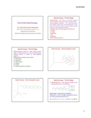
  • 1. 10/18/2020 1 Ultra Violet Spectroscopy Dr. Satish Dhirendra Mitragotri (M.Sc., Ph.D., SET, NET, GATE and M.B.A. (Finance)) Department of Chemistry Walchand College of Arts and Science, Solapur Spectroscopy - Terminology Spectroscopy - The study of interaction between electromagnetic radiation and matter is spectroscopy. Electromagnetic radiation – The oscillating electric and magnetic fields which are perpendicular to each other travel through medium and have definite energy is known as electromagnetic radiation. Different types of electromagnetic radiations are o Sun light oU V light oVisible light oX-ray oMicrowave oRadio frequency wave etc. Spectroscopy - Terminology Electromagnetic spectrum – Entire range of wave- length, energy, frequency over which electromagnetic radiation present is known as electromagnetic spectrum. Different terms related to wave motion 1) Wavelength 2) Frequency 3) Wave number 4) Velocity 5) Energy 6) Particle properties of radiation. Spectroscopy – Electromagnetic wave Spectroscopy – Electromagnetic wave Spectroscopy - Terminology 1) Wavelength (λ) – The distance between two particles which are in same phase. •Wavelength is expressed by Lambda (λ) •Measured in units of length starting from Km to nm, more commonly used unit is Ao 1m = 1 X 102 cm = 1 X 106 µm = 1 X 10⁹ nm = 1 X 10¹⁰ Ao Crest Trough
  • 2. 10/18/2020 2 Spectroscopy - Terminology 2) Frequency (ν) – The number of waves passing through a particular point in unit time (one second) is known as frequency. Unit of frequency is cycles per second or Hz 1 Hz = 1 cycle per second It is direct measure of energy. 1 MHz = 1 X 10³ KHz = 1 X 10⁶ Hz A fixed point Spectroscopy - Terminology 3)Wave number (ν-) – The number of waves per centimeter is known as wave number. Wave number is reciprocal of frequency, ν- = 1/ λ Unit is kaiser or reciprocal centimeter = cm-1 A fixed Distance = 1 cm Spectroscopy - Terminology 4)Velocity (C) – The distance travelled by the wave in one second is called as velocity of radiation. •Velocity of radiation is fixed and it is •3 X 10⁸ m/s or 3 X 1010 cm/s •Energy is directly proportional to velovity. 5)Energy (E) - Energy associated with radiation. E = h X ν Longer the wavelength lesser the energy Shorter the wavelength more is the energy 6)Particle properties of radiation. Spectroscopy – Electromagnetic Spectrum Radiation Cosmic Ray Gamma Rays X- Rays UV Visible IR Microwave Radio freq. waves Wavelength cm 10-9 10-7 10-5 10-4 10-3 10-1 102 Energy Kcal/mol 109 106 106 10 10-2 10-4 10-6 Frequency Hz Function Ioniza tion Electr onic transi tion Mole cular Vibra tion Rotational motion Nuclear spin Spectroscopy – Energy of molecule Energy associated with any molecule can be resolved in four different components E Total = E Electronic + E Vibrational + E Rotational + E Spin When radiation falls on any molecule one or more energy parameters from molecule are changed and it is called excitation. Different type of energy type require different radiation for this excitation process Electronic- Electronic transition Vibrational – IR Radiation Rotational - Microwave Spin- Radiofrequency wave
  • 3. 10/18/2020 3 Spectroscopy – Energy of molecule Energy associated with any molecule can be resolved in four different components E Total = E Electronic + E Vibrational + E Rotational + E Spin Electronic- Electronic transition A B A+ B- Vibrational – IR Radiation A B A B Rotational – Microwave A B A Spin- Radiofrequency wave U V -Spectroscopy Ultraviolet absorption spectrum arises from transition of electrons from lower electronic energy level to higher electronic energy level. It is absorption spectroscopy where the amount of energy absorbed by the molecule from ultraviolet region is measured. The wavelength of UV radiation is in between 200nm to 400 nm = 2000 A0 to 4000 A0 = 200 to 400 X 10-8 cm The region from 100-200 nm is called as Vacuum ultraviolet region U V -Spectroscopy The region from 200-400 nm is called as near or quartz ultraviolet region It comes prior to violet region of visible radiation so it is called as ultra violet. When there are two energy levels for e.g. E2 and E1 where E2 E1 E2 E1 E2 – E1 = E U V -Spectroscopy When UV light falls on any molecule some part of it may be absorbed, absorption depends on two parameters 1. Energy of incident UV radiation 2. Structure of molecule If the energy of radiation is kept same then different molecules give different absorption pattern i.e. UV spectrum. Thus UV spectrum is a plot of absorbance (A) verses wavelength. What is absorbance ? U V -Spectroscopy When UV light falls on any sample it can interact in three different ways 1. Reflection 2. Absorption 3. Transmission Reflection Absorption Transmission Incident U V -Spectroscopy What is absorbance ? Io = Intensity of incident radiation Ir = Intensity of reflected radiation Ia = Intensity of absorbed radiation It = Intensity of transmitted radiation Transmittance (T)= It / Io Opacity (O)= Io /It Absorbance = log (Io /It)
  • 4. 10/18/2020 4 U V -Spectroscopy Lambert’s Law - When a beam of monochromatic radiation passes through the absorbing medium the intensity of the light radiation decreases exponentially as the length of absorbing medium increases. Beer’s Law – When a beam of monochromatic radiation passes thorough the absorbing medium the intensity of incident radiation decreases with length of absorbing medium as well as concentration of the soultion. U V -Spectroscopy Different types of electrons and electronic transitions 1.Sigma (σ) electrons- If compound contain only sigma electrons i.e. electrons in present in sigma bond e.g. CH3-H 2.Pi (π) electrons – Electrons present in double or triple bond but that to C-C only H2C=CH2 3.Non bonding (n) electrons – Electrons present in hetero atoms bonded with carbon CH3-HC=O, CH3-OH U V –Spectroscopy- Types of Orbitals σ S P π π* n σ* S P Energy U V –Spectroscopy – Absorption of energy by electrons Energy Bonding orbital ΔE Antibonding/Nonbonding orbital ΔE ER = hν = hc /λ ΔE = ER U V –Spectroscopy Types of Electronic Transitions σ to σ* n to σ* π to π* n to π* Energy σ* Antibonding π* Antibonding n Nonbonding σ Bonding π Bonding U V –Spectroscopy – Types of electronic transitions Energy Bonding orbital Antibonding orbital Nonbonding orbital Antibonding orbital Bonding orbital σ σ* π* π n σ to σ* π to π* n to σ* n to π*
  • 5. 10/18/2020 5 U V -Spectroscopy Different types of electrons and electronic transitions 1.σ to σ* Transition – Transition of an electron from bonding sigma orbital(σ) to antibonding sigma (σ*) orbital is called as σ to σ* . This requires highest energy and takes place by radiation of short wavelength ( 150 nm). Stronger bond require shorter wavelength for transition. e.g. CH3-H 2. n to σ* Transition - Transition of an electron from non bonding (n) orbital to higher energy, antibonding σ* orbital is called as n to σ* transition. This type of transition occurs in compounds containing hetero atoms like N,O,S,P and halogens CH3-OH, CH3-SH, CH3 NH2 wavelength ( 180 nm – 200nm). E-max is more for more probable transition.(R-I R-Cl) U V -Spectroscopy Different types of electrons and electronic transitions 3. π to π * Transition (K band) - Transition of an electron from bonding (π) orbital to higher energy, antibonding (π*) orbital is called as π to π * transition. This type of transition occurs in compounds containing groups with multiple bonds. C=C, C=O, N=N, C=N etc. wavelength (200nm - 400 nm). 4. n to π * Transition (R band) - Transition of an electron from nonbonding (n) orbital to higher energy, antibonding (π*)orbital is called as π to π * transition. This type of transition occurs in compounds containing groups with multiple bonds with heteroatons and saturated carbon framework. R-C=O, R-N=N, R-C=N etc. wavelength (200nm - 300 nm). U V -Spectroscopy Wavelength of maximum absorption = λmax Wavelength ( λ ) 100 150 200 250 300 350 400 not a λmax not a λmax Intensity ε 6 5 4 3 2 1 0 U V -Spectroscopy Maximum absorption intensity = εmax Wavelength ( λ ) Intensity ε 100 150 200 250 300 350 400 Low absorption intensity = ε 6 5 4 3 2 1 0 U V -Spectroscopy 1.Bathochromic shift (Red shift) 2.Hypsochromic shift (Blue shift) Wavelength ( λ ) Wavelength of maximum absorption λ max 6 5 4 3 2 1 0 Intensity ε -------------------------------------------------------------- 100 150 200 250 300 350 400 U V -Spectroscopy Wavelength ( λ ) Intensity ε Maximum absorption intensity = εmax 100 150 200 250 300 350 400 6 5 4 3 2 1 0 3.Hyperchromic shift 4.Hypochromic shift
  • 6. 10/18/2020 6 U V -Spectroscopy 1.Bathochromic shift (Red shift) 2.Hypsochromic shift (Blue shift) Wavelength ( λ ) Intensity E-max Wavelength of maximum absorption 4.Hypochromic shift 100 150 200 250 300 350 3.Hyperchromic shift U V -Spectroscopy Different terms used in UV Spectroscopy 1.Chromophore – It is an isolated functional group capable of absorbing visible and or ultraviolet region. 2.It is unsaturated group responsible for electronic absorption Molecule which has chromospheres is called as chromogen. e.g. C=C, C=O, -NO2, -C=N-, -CN, -COOH, -CONH2 etc. 2. Auxochrome – It is functional group which do not absorb color above 200nm but when attached to chromophore causes shift in wavelength and or intensity of absorption. e.g. –OH, -X, -OR, -NH2 U V -Spectroscopy Different terms used in UV Spectroscopy 1.Bathochromic shift (Red shift) – Shift of absorption maxima to longer wavelength is called as bathochromic shift or red shift. Here λmax increases. This is due to presence of auxochrome such as – OH, -NH2, etc. Decrease in solvent polarity causes red shift. e.g. from alcohol to benzene When there is extension in conjugation the red shift is observed e.g. ethyene absorbs at 170nm where as 1,3-butadiene absorbs at 217nm U V -Spectroscopy 2.Hypsochromic shift (Blue shift) – Shift of absorption maxima to shorter wavelength is called as hypsochromic shift or Blue shift. Here λmax decreases Generally change in pH of medium causes this shift. Aniline in absence of any solvent absorbs at 280 nm but when HCl is added to it red shift is observed and it absorbs at 200nm Phenol in basic medium due to formation of phenoxide ion has extension in conjugation and absorbs at higher wavelength but when medium terns acidic the conjugation is removed as ion is converted to undissociated phenol and red shift in absorption is observed. 3.Hyperchromic shift 4.Hypochromic shift U V -Spectroscopy 3.Hyperchromic shift – Increase in the intensity of absorption maxima is called as hyperchromic shift. E-max increase Generally electron donating effect causes this shift 4.Hypochromic shift - Decrease in the intensity of absorption maxima is called as hyporchromic shift. E-max Decrease. Disturbance in conjugation causes this shift U V -Spectroscopy 1.Bathochromic shift (Red shift) 2.Hypsochromic shift (Blue shift) Wavelength ( λ ) Intensity E-max 3.Hyperchromic shift 4.Hypochromic shift
  • 7. 10/18/2020 7 U V –Spectroscopy – Effect of conjugation Effect of conjugation on position of UV and visible absorption bands. n to π * Transition (R band) is the most easy to detect in normal UV spectrum Presence of conjugated chromospheres (C=C) (alternate double and single bond) causes bathochromic or red shift. If two chromophores are separated by more than two single bonds then conjugation is disturbed and no red shift is observed. e.g. CH2=CH2 - λmax is 175 nm CH2=CH2-CH2-CH2=CH2 λmax is 175 nm U V –Spectroscopy – Effect of conjugation Energy P P P π* π P ΔE1 ΔE2 ΔE1 ΔE2 π1 π1* π2 π2* π3 π3* U V –Spectroscopy – Effect of conjugation a)Ethene CH2=CH2 has - λmax is 175 nm also 1,4- pentadiene also has CH2=CH2-CH2-CH2=CH2 λmax is 175 nm as the conjugation is disturbed and thus there is no effect of presence of two chromophores. b)If the chromophores are present in conjugation the red shift is observed. Ethene - CH2=CH2 has - λmax is 175 nm 1,3-butadiene CH2=CH-CH=CH2 - λmax is 217 nm 1,3,5-hexatriene CH2=CH-CH=CH-CH=CH2 - λmax is 256 nm U V –Spectroscopy – Effect of conjugation More number of conjugated double bonds lead to more overlap of HOMO and LUMO and thus energy difference between the orbitals decreases leading to absorption at higher wevelength. Decreasing in energy leads to increase in wavelength. c) It two different chromophores are present e.g. C=C and C=O, e.g. CH2=CH2 has - λmax is 175 nm CH2=O has - λmax is 290 nm Where as CH2=CH-CH2=O has λmax is 320 nm Thus conjugation increases λmax to higher value. Applications of U V -Spectroscopy 1.Determination of structure - UV spectroscopy provides information mainly about the extension in conjugation. Though not useful for complete structure elucidation it provides key information about different types of electron present in molecule. If UV spectrum of molecule is available then it can be compared with unknown spectrum of identification. Applications of U V -Spectroscopy 2.Determination of stereochemistry of compounds – UV spectrum is useful in determination of stereochemistry of molecules specially compounds exhibiting cis-trans isomerism. The trans conformation has planar structure and hence the conjugation is not disturbed. In these molecules thus λmax – Has higher value. Where as in case of cis isomer the planar nature is disturbed and thus orbital overlap is not completely effective resulting in lowering of λmax
  • 8. 10/18/2020 8 Applications of U V -Spectroscopy This can be confirmed by two examples where two isomers cis and trans isomers have different λmax values. Cis -stilbene has λmax value at 280 nm and ἐ-max at 13500 where as trans -stilbene has λmax value at 295 nm and ἐ-max at 27000. Cis –cinnamic acid has λmax value at 264 nm and ἐ-max at 10700 where as trans –cinnamic acid has λmax value at 273 nm and ἐ-max at 15900. Applications of U V -Spectroscopy H H H H Cis -stilbene labda max = 280 nm e-max = 13500 Trans -stilbene labda max = 295 nm e-max = 27000 H H COOH H H COOH Cis -Cinnamic acid labda max = 264 nm e-max = 10700 Trans -Cinnamic acid labda max = 273 nm e-max = 15900 U V –Spectroscopy-Woodward-Fieser Rules for calculations of λmax Woodward-Fieser rules for calculation of λ max value(nm) for dienes and trienes Sr.No. Name of the Motif Skeletal structure λ max value(nm) 1 a Basic value for Un-substituted diene 214 1 b Basic value for Conjugated diene 214 C C C C C C C C C C U V –Spectroscopy-Woodward-Fieser Rules for calculations of λmax Woodward-Fieser rules for calculation of λ max value(nm) for dienes and trienes Sr.No. Name of the Motif Skeletal structure λ max value(nm) 1 c Basic value for Hetero- annular diene 214 1 d Basic value for semi cyclic diene 214 U V –Spectroscopy-Woodward-Fieser Rules for calculations of λmax Woodward-Fieser rules for calculation of λ max value(nm) for dienes and trienes Sr.No. Name of the Motif Skeletal structure λ max value(nm) 1 e Basic value for homo-annular diene 253 2 a Addition for extended conjugation [for every –C=C-] + 30 nm Diene - C C U V –Spectroscopy-Woodward-Fieser Rules for calculations of λmax Woodward-Fieser rules for calculation of λ max value(nm) for dienes and trienes Sr.No. Name of the Motif Skeletal structure λ max value(nm) 2 b Addition for exocyclic double bond +5 2 c Addition for exocyclic double for two ring systems at the same time +10
  • 9. 10/18/2020 9 U V –Spectroscopy-Woodward-Fieser Rules for calculations of λmax Woodward-Fieser rules for calculation of λ max value(nm) for dienes and trienes Sr.No. Name of the Motif Skeletal structure λ max value(nm) 2 c Addition for exocyclic double for two ring systems at the same time +10 3 a Addition for substitution (ring residue Alkyl group -R) + 5 nm CH3 , C2 H5 , C3 H7 etc. R = C C C C R U V –Spectroscopy-Woodward-Fieser Rules for calculations of λmax Woodward-Fieser rules for calculation of λ max value(nm) for dienes and trienes Sr.No. Name of the Motif Skeletal structure λ max value(nm) 3 b Addition for substitution –OR alkoxy group + 5 nm 3 c Addition for substitution – acyloxy + O nm CH3, C2H5, C3H7 etc. R = C C C C OR CH3, C2H5, C3H7 etc. R = OCOR C C C C U V –Spectroscopy-Woodward-Fieser Rules for calculations of λmax Woodward-Fieser rules for calculation of λ max value(nm) for dienes and trienes Sr.No. Name of the Motif Skeletal structure λ max value(nm) 3 d Addition for substitution – Halogens + 5 nm 3 e Addition for substitution – N-R (N-Alkyl) + 60 nm X X = Cl, Br C C C C NR2 C C C C U V –Spectroscopy-Woodward-Fieser Rules for calculations of λmax Woodward-Fieser rules for calculation of λ max value(nm) for dienes and trienes Sr.No. Name of the Motif Skeletal structure λ max value(nm) 3 f Addition for substitution – S-R (S-Alkyl) + 30 nm SR C C C C U V –Spectroscopy-Woodward-Fieser Rules for calculations of λmax Woodward-Fieser rules for calculation of λ max value(nm) of α-β unsaturated ketones and aldehydes Sr.No. Name of the Motif Skeletal structure λ max value(nm) 1 a Basic value for acyclic ketone 215 1 b Basic value for six member ring ketone 215 O C C C O U V –Spectroscopy-Woodward-Fieser Rules for calculations of λmax Woodward-Fieser rules for calculation of λ max value(nm) of α-β unsaturated ketones and aldehydes Sr.No. Name of the Motif Skeletal structure λ max value(nm) 1 c Basic value for five member ring ketone 202 1 d Basic value for α-β unsaturated aldehydes 207 O C C C O H
  • 10. 10/18/2020 10 U V –Spectroscopy-Woodward-Fieser Rules for calculations of λmax Woodward-Fieser rules for calculation of λ max value(nm) of α-β unsaturated ketones and aldehydes Sr.No. Name of the Motif Skeletal structure λ max value(nm) 2 a Addition for extended conjugation [for every –C=C-] + 30 nm 2 b Addition for exocyclic double bond + 5 nm Diene - C C U V –Spectroscopy-Woodward-Fieser Rules for calculations of λmax Woodward-Fieser rules for calculation of λ max value(nm) of α-β unsaturated ketones and aldehydes Sr.No. Name of the Motif Skeletal structure λ max value(nm) 2 c Addition for exocyclic double for two ring systems at the same time + 10 nm 2 d Addition for exocyclic double for two ring systems at the same time + 10 nm U V –Spectroscopy-Woodward-Fieser Rules for calculations of λmax Woodward-Fieser rules for calculation of λ max value(nm) of α-β unsaturated ketones and aldehydes Sr.No. Name of the Motif Skeletal structure λ max value(nm) 2 d Addition for homo annular component + 39 nm Different positions δ ϒ β α -C=C-C=C-C=O OR U V –Spectroscopy-Woodward-Fieser Rules for calculations of λmax Substituent at position α β ϒ δ 3 a Addition for substitution –R, Alkyl group 10 12 18 18 3 b Addition for substitution –OR, alkoxy group 35 30 17 31 3 c Addition for substitution – OCOR, acyloxy group 6 6 - 6 3 d Addition for substitution –-OH, hydroxyl 35 30 - 50 3 e Addition for substitution – Halogens -Cl (chloro) 15 12 - - 3 f Addition for substitution – Halogens -Br (Bromo) 25 30 - - 3 g Addition for substitution – N-R (N-Alkyl) - 95 - - 3 h Addition for substitution – S-R (S-Alkyl) - 85 - - Calculations of λmax of Dienes and Enones Diene C C C C H H H H H H H R C C C C H H H R R R R R C C C C R R R R R R R R R R Calculations of λmax of Dienes and Enones Enone Conjugated aldehyde C C C O R H R H O R R C C C O H R H R O R R
  • 11. 10/18/2020 11 Calculations of λmax of Dienes and Enones Structure Component/s Value in nm 1 Basic value for acyclic diene = 214 nm Alkyl substituents (2 X 5) = 10 nm Total = 224 nm 2 Basic value for acyclic diene = 214 nm Extended conjugation (1 X 30) = 30 nm Total = 244 nm 3 Basic value for acyclic diene = 214 nm Alkyl substituents (3 X 5) = 15 nm Total = 229 nm EC Calculations of λmax of Dienes and Enones Structure Component/s Value in nm 4 Basic value for acyclic diene = 214 nm Extended conjugation (1 X 30) = 30 nm Alkyl substituents (3 X 5) = 15 nm Total = 259 nm 5 Basic value for acyclic diene = 214 nm Alkyl substituents (3 X 5) = 15 nm Exhocyclic double bond (1 X 5) = 5 nm Total = 234 nm EC EDB Calculations of λmax of Dienes and Enones Structure Component/s Value in nm 6 Basic value for acyclic diene =214 nm Alkyl substituents (2 X 5) = 10 nm Alkoxy substituent (1 X 6) = 6 nm Exhocyclic double bond (1 X 5) = 5 nm Total =235 nm 7 Basic value for homoammular diene =253 nm Extended conjugation (1 X 30) = 30 nm Exhocyclic double bond (1 X 5) = 5 nm Alkyl substituents (3 X 5) = 15 nm Total =303 nm OMe Alkoxy EDB EDB EC Calculations of λmax of Dienes and Enones Structure Component/s Value in nm 8 Basic value for homoammular diene = 253 nm Extended conjugation (1 X 30) = 30 nm Hylogen substituents (1 X 5) = 5 nm Alkyl substituents (4 X 5) = 20 nm Total = 308 nm 9 Basic value for homoammular diene = 253 nm Extended conjugation (1 X 30) = 30 nm Exhocyclic double bond (2 X 5) = 10 nm S-Alkyl substituents (1 X 5) = 30 nm Alkyl substituents (5 X 5) = 25 nm Total = 348 nm Cl Halogen EC SCH3 EC EDB EDB S-Alkyl Calculations of λmax of Dienes and Enones Structure Component/s Value in nm 10 Basic value for homoammular diene = 253 nm Extended conjugation (2 X 30) = 60 nm Exhocyclic double bond (1 X 5) = 5 nm Halogen substituents (1X 5) = 5 nm Alkyl substituents (3 X 5) = 15 nm Total = 338 nm Br EC EC EDB Halogen Calculations of λmax of Dienes and aldehydes Structure Component/s Value in nm 1 Basic value for acyclic enone = 215 nm Alkyl substituent on α-C (1 X 10) = 10 nm Total = 225 nm 2 Basic value for acyclic enone = 215 nm Extended conjugation (1 X 30) = 30 nm Alkyl substituent on β-C (1 X 10) = 12 nm Total = 257 nm O O EC
  • 12. 10/18/2020 12 Calculations of λmax of Dienes and aldehydes Structure Component/s Value in nm 3 Basic value for α-β unsaturated aldehyde = 207 nm Alkyl substituent on α-C (1 X 10) = 10 nm Alkyl substituent on β-C (2 X 12) = 24 nm Total = 241 nm 4 Basic value for α-β unsaturated aldehyde = 207 nm Extended conjugation (1 X 30) = 30 nm Alkyl substituent on α-C (1 X 10) = 10 nm Alkyl substituent on β-C (1 X 12) = 12 nm Alkyl substituent on ϒ-C (1 X 18) = 18 nm Cl on ϒ-C (1 X 0) = 0 nm Total = 270 nm O O Cl EC Calculations of λmax of Dienes and aldehydes Structure Component/s Value in nm 5 Basic value for acyclic enone = 215 nm Extended conjugation (1 X 30) = 30 nm Exhocyclic double bond (1 X 5) = 5 nm Alkyl substituent on ϒ-C (1 X 18) = 18 nm Alkyl substituent on δ-C (1 X 18) = 18 nm Alkoxyl substituent on δ-C (1 X 31) = 18 nm Total = 304 nm 6 Basic value for five member enone = 202 nm Alkyl substituent on α-C (1 X 10) = 10 nm Br - substituent on β-C (1 X 18) = 30 nm Total = 242 nm O OMe EC Alkoxy EDB O Br Calculations of λmax of Dienes and aldehydes Structure Component/s Value in nm 7 Basic value for six member enone = 215 nm Extended conjugation (1 X 30) = 30 nm Homo annular component (1 X 39) = 39 nm Alkyl substituent on δ-C (2 X 18) = 36 nm HO - substituent on β-C (1 X 30) = 30 nm Total = 350 nm 8 Basic value for six member enone = 215 nm Extended conjugation (1 X 30) = 30 nm Homo annular component (1 X 39) = 39 nm Alkyl substituent on α-C (1 X 10) = 10 nm Alkyl substituent on ϒ-C (1 X 18) = 18 nm Alkyl substituent on δ-C (2 X 18) = 36 nm Total = 349 nm O OH O EC HAD Calculations of λmax of Dienes and aldehydes Structure Component/s Value in nm 9 Basic value for five member enone = 202 nm Extended conjugation (1 X 30) = 30 nm Homo annular component (1 X 39) = 39 nm Alkyl substituent on α-C (1 X 10) = 10 nm Alkyl substituent on β -C (1 X 12) = 12 nm Alkyl substituent on δ-C (1 X 18) = 18 nm Total = 311 nm 10 Basic value for six member enone = 215 nm Extended conjugation (1 X 30) = 30 nm Exhocyclic double bond (1 X 5) = 5 nm Alkyl substituent on α-C (1 X 10) = 10 nm Alkyl substituent on ϒ-C (1 X 18) = 18 nm Total = 273 nm O EC O EDB EC