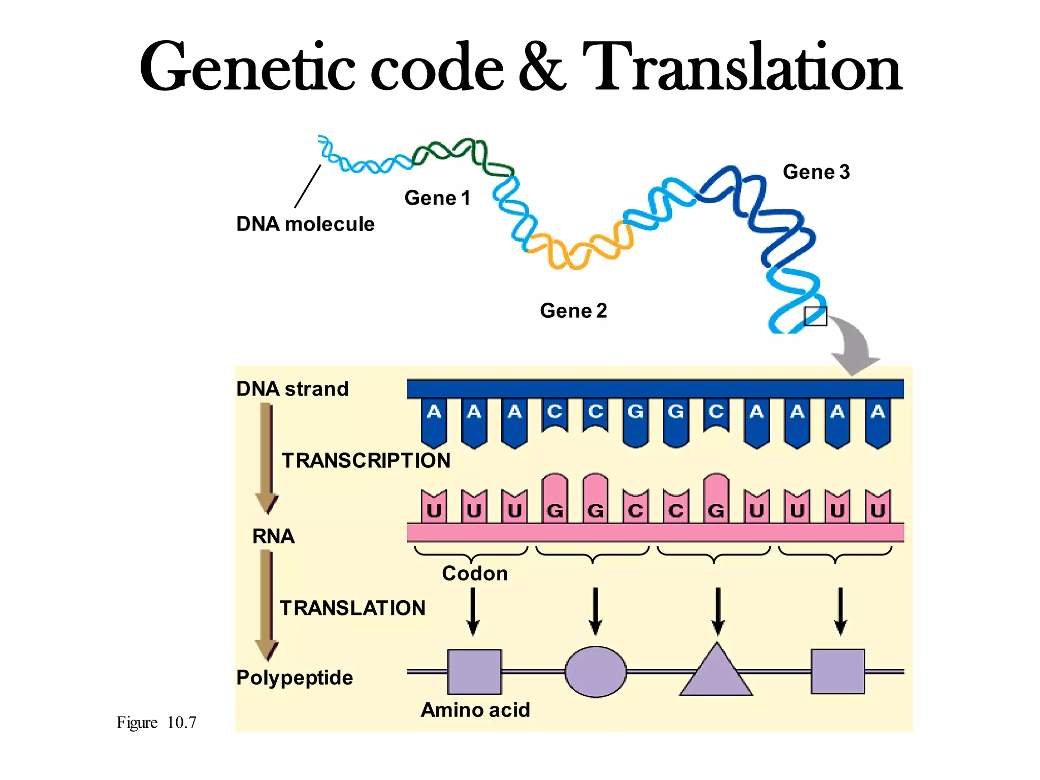
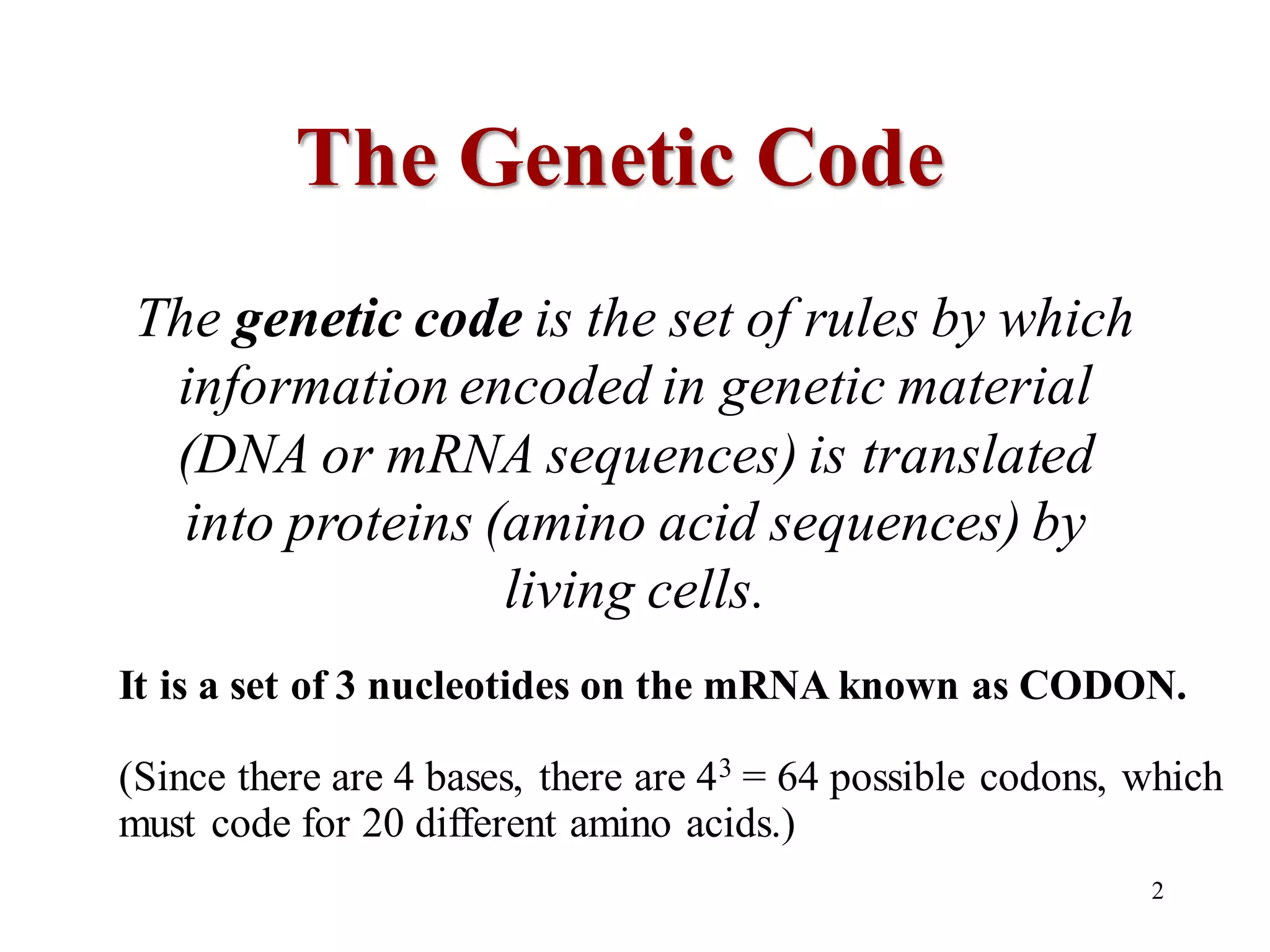
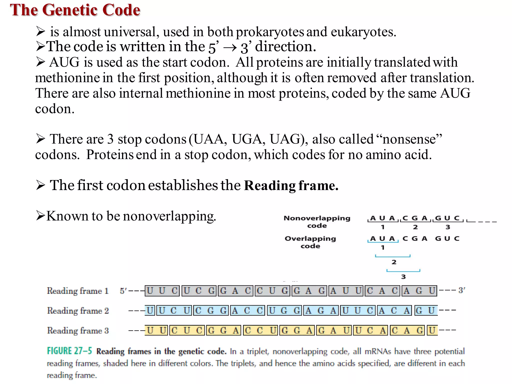
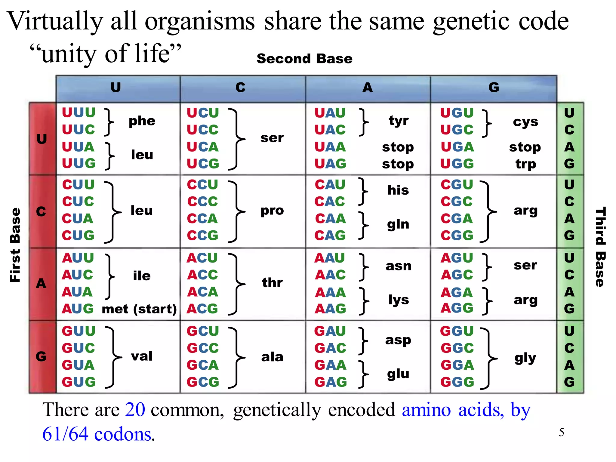
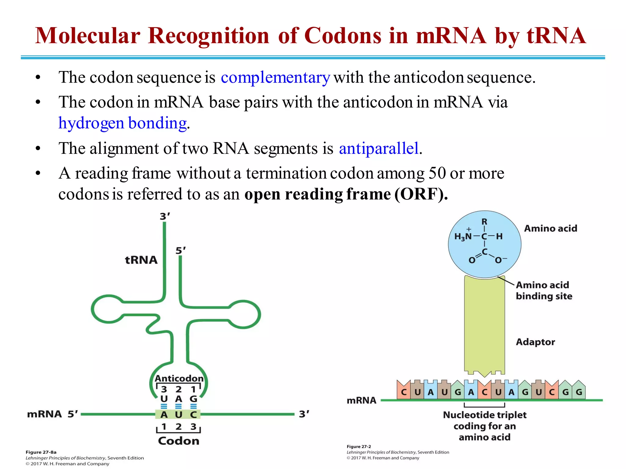
2. The genetic code is the set of rules by which
information encoded in genetic material
(DNA or mRNA sequences) is translated
into proteins (amino acid sequences) by
living cells.
The Genetic Code
It is a set of 3 nucleotides on the mRNA known as CODON.
(Since there are 4 bases, there are 43 = 64 possible codons, which
must code for 20 different amino acids.)
2
3. The Genetic Code
➢ is almost universal, used in both prokaryotesand eukaryotes.
➢The code is written in the 5’ → 3’ direction.
➢ AUG is used as the start codon. All proteins are initially translatedwith
methioninein the first position, although it is often removed after translation.
There are also internal methionine in most proteins, coded by the same AUG
codon.
➢ There are 3 stop codons(UAA, UGA, UAG), also called “nonsense”
codons. Proteinsend in a stop codon, which codes for no amino acid.
➢ The first codonestablishes the Reading frame.
➢Known to be nonoverlapping.
4. Breaking the Genetic Code
• One of the greatest scientific developments of the
twentieth century
• First clue from Nirenberg and Matthaei:
– poly(U) and 20 radiolabeled amino acids fed to E.coli
→only *Phe produced
• Hence, UUU codes for Phe.
• Another step forward from Khorana:
– used defined mRNAs in planned patterns
• that is, (AC)n (alternating ACA and CAC codons → His and Thr)
5. U C A G
U
C
A
G
G
A
C
U
G
A
C
U
G
A
C
U
G
A
C
U
UUU
UUC
UUA
UUG
CUU
CUC
CUA
CUG
AUU
AUC
AUA
AUG
GUU
GUC
GUA
GUG
phe
leu
leu
ile
met (start)
val
UCU
UCC
UCA
UCG
CCU
CCC
CCA
CCG
ACU
ACC
ACA
ACG
GCU
GCC
GCA
GCG
ser
pro
thr
ala
UAU
UAC
UAA
UAG
CAU
CAC
CAA
CAG
AAU
AAC
AAG
AAA
GAU
GAC
GAA
GAG
tyr
stop
stop
his
gln
asn
lys
asp
glu
UGU
UGC
UGA
UGG
CGU
CGC
CGA
CGG
AGU
AGC
AGA
AGG
GGU
GGC
GGA
GGG
cys
stop
trp
arg
ser
arg
gly
First
Base
Third
Base
Second Base
Virtually all organisms share the same genetic code
“unity of life”
5
There are 20 common, genetically encoded amino acids, by
61/64 codons.
6. Most Amino Acids Have More
Than One Codon
• Some codons are less subject to causing a mutation
in an amino acid sequence because of degeneracy or
because of the abundance of such tRNAs.
• There are 20 amino acids with 61 possible codons.
• Only Met and Trp have a single codon.
7. Codon Correlation with Amino Acids
TABLE 27-3 Degeneracy of the Genetic Code
Amino acid Number of codons Amino acid
Number of
codons
Met 1 Tyr 2
Trp 1 Ile 3
Asn 2 Ala 4
Asp 2 Gly 4
Cys 2 Pro 4
Gln 2 Thr 4
Glu 2 Val 4
His 2 Arg 6
Lys 2 Leu 6
Phe 2 Ser 6
8. The Genetic Code Is Resistant to Mutations
• Degenerate code allows certain mutations to
still code for the same amino acid.
– “silent” mutations―different nucleotide in DNA
but same amino acid in protein
• Mutation in the first base of a codon usually
produces a conservative substitution.
– Example: GUU → Val but AUU →Leu
9. The Genetic Code Is Universal,
With a Few Exceptions
• It is used by prokaryotes and eukaryotes, across
species.
• Mitochondria contain DNA and use a slightly
different code.
– UGA encodes Trp in vertebrate mtDNA (instead of STOP).
– AGA/AGG encodes STOP in vertebrate mtNDA (instead
of Arg).
• Mitochondria encode their own tRNAs, using 22
instead of 32.
10. Molecular Recognition of Codons in mRNA by tRNA
• The codon sequenceis complementarywith the anticodonsequence.
• The codon in mRNA base pairs with the anticodon in mRNA via
hydrogen bonding.
• The alignment of two RNA segments is antiparallel.
• A reading frame without a termination codon among 50 or more
codonsis referred to as an open reading frame (ORF).
11. “Wobble” Pairings in tRNA with
mRNA Can Occur in the Third Base
• The third base of a codon can form noncanonical base
pairs with its complement (anticodon) in tRNA.
• Some tRNAs contain Inosinate (I), which can H-bond
with U,C, and A.
– These H-bonds are weaker and were named by Crick as
“wobble” base pairs.
– Example: In yeast, CGA, GCU, and CGC all bind to
tRNAArg, which has the anticodon 3’-GCI-5’.
• Although sequences are usually written 5’→3’, the anticodon here is
written 3’→5’ to illustrateits bondingto the mRNA codons.
12. a. When the first base of the anticodon is C
or A, base pairing is specific and only one
codon is recognized.
b. When the first base is U or G, binding is
less specific and two different codons may
be read.
c. When inosine (I) is the first (wobble)
nucleotide of an anticodon, three different
codons can be recognized-the maximum
number for any tRNA.
Wobble Hypothesis
1. The first two bases of codon always form strong base pairs with the
corresponding bases anticodon and confer the coding specificity.
2. When an amino acid is specifled by several different codons, the codons
that differ in either of the first two bases require different tRNAs.
3. A minimum of 32 tRNAs are required to translate all 61 codons (31 to
encode the amino acids and 1 for initiation).
4. The first base of the anticodon (reading in the 5'-3' direction; this pairs with
the third base of the codon) determines the number of codons recognized
by the tRNA.
12
13. Some mRNAs Are Edited Before Protein
Synthesis
• Editing involves alteration, addition, or deletion of nucleotides
in mRNA.
• Editing uses guide RNAs (gRNAs) that temporarily hybridize
with the mRNA and act as templates for editing.
14. Deamination Reactions Yield
• RNA editing by
alteration of
nucleotides most
commonly involves
the enzymatic
deamination of
adenosine or
cytidine residues.
15. Five Stages of Protein Synthesis
1. Activation of amino acids
– tRNA aminoacylated
2. Initiation of translation
– mRNA and aminoacylated tRNA bind to ribosome
3. Elongation
– cycles of aminoacyl-tRNA binding and peptide bond
formation…until a STOP codon is reached
4. Termination and ribosome recycling
– mRNA and protein dissociate, ribosome recycled
5. Folding and posttranslational processing
– catalyzed by a variety of enzymes
17. TABLE 27-5
Components Required for the Five Major Stages of Protein Synthesis in E.
Coli
Stage Essential components
1. Activation of amino acids 20 amino acids
20 aminoacyl-tRNA syntheses
32 or more tRNAs
ATP
Mg2+
2. Initiation mRNA
N-Formylmethionyl-tRNAfMet
Initiation codon in mRNA (AUG)
30S ribosomal subunit
50S ribosomal subunit
Initiation factors (IF1, IF2, IF3)
GTP
Mg2+
3. Elongation Functional 70S ribosomes (initiation complex)
Aminoacyl-tRNAs specified by codons
Elongation factors (EF-Tu, EF-Ts, EF-G)
GTP
Mg2+
4. Termination and ribosome recycling Termination codon in mRNA
Release factors (RF1, RF2, RF3, RRF)
EF-G
IF3
5. Folding and posttranslational
processing
Chaperones and folding enzymes (PPI, PDI); specific enzymes, cofactors, and other
components for removal of initiating residues and signal sequences, additional proteolytic
processing, modification of terminal residues, and attachment of acetyl, phosporyl, methyl,
carboxyl, carbohydrate, or prosthetic groups
18. The Ribosome Is a Key Player in Protein Synthesis
• Make up ~25% of dry weight of bacteria
• ~65% rRNA, 35% protein
– rRNA forms the core.
– RNA does the catalysis of peptidebond formation.
• Made of two subunits bound together (30S and 50S) in
bacteria, with mRNA running through them
TABLE 27-6 RNA and Protein Components of the E. Coli Ribosome
Subunit
Number of different
proteins
Total number of
proteins Protein designations Number and type of rRNAs
30S 21 21 S1–S21 1 (16S rRNA)
50S 33 36 L1–L36a 2 (5S and 23S rRNAs)
aThe L1 to L36 protein designations do not correspond to 36 different proteins. The protein originally designated L7 is a modified form of L12, and L8 is
a complex of three other proteins. Also, L26 proved to be the same protein as S20 (and not part of the 50S subunit). This gives 33 different proteins in
the large subunit. There are four copies of the L7/L12 protein, with the three extra copies bringing the total protein count to 36.
19. • Overall, very similar
• Still two subunits with mRNA
running between
• In eukaryotes, larger (80S), more
complex, contain > 80 proteins
• Chloroplasts and mitochondria have
ribosomes simpler than those in
bacteria.
Ribosomes in Bacteria and Eukaryotes
20. rRNAs Have Complex
Secondary Structures
• The three ss rRNAs having
specific 3-D structure with
extensive intrachain base
pairs.
• Shape of rRNAs are highly
conserved.
• The red, yellow, and purple indicate
areas where the structures of the
rRNAs from bacteria, archaea, and
eukaryotes have diverged.
• Conserved regions are shown in
green.
21. Characteristics of tRNAs
• ssRNA of 73–93 nucleotides in both bacteria and eukaryotes
• Cloverleaf structure in 2-D, “Twisted L” shape in 3-D
• Serve as interpreters during translation
• Most have G at 5’-end; all have CAA at 3’-end
• Have modified bases
– methylated bases, and so on
3-D Structure of Yeast tRNAPhe
22. • Amino acid arm
– has amino acid esterified via
carboxyl group to the 2’-OH or 3’-
OH of the A of the terminal CAA
codon
• Anticodon arm
• D arm
– contains dihydrouridine (D)
– contributes to folding
• TC arm
– contains pseudouridine ()―has
bonding between base and ribose
– helps in folding
Characteristics of tRNAs
23. • Each tRNA molecule has a triplet anticodonon one end and an amino acid
attachment site on the other
• Blackdots are same in all tRNAs
• Green & Blue Dots are the Recognitionpoints (orange for one ) or (green
for more) aminoacyl-tRNA synthetases.
23
Recognition points of aminoacyl-tRNA synthetases
23
24. Aminoacyl-tRNA synthetases esterify the 20 amino acids to
their corresponding tRNAs. Each enzyme is specific for one amino
acid attachment to one or more corresponding tRNAs.
•Two classesi.e. Class I and Class II: Based on substantial differencesin reaction
mechanism
Amino acid + tRNA + ATP → aminoacyl-tRNA + AMP + Ppi
Stage 1: Activation of Amino Acids
(1) Activation: it activates an amino acid
for peptide bond formation
(2) Proof reading: it ensures appropriate
placement of the amino acid in a growing
polypeptide.
24
27. The Second Genetic Code
• Aminoacyl-tRNA synthetases must be specific for both
amino acid and tRNA.
– Matching each amino acid with correct tRNA can be viewed
as the “second genetic code.”
– The “code” is in molecular recognition of a specific tRNA
molecule by a specific synthetase.
• Only a few nucleotides in tRNA confer the binding
specificity.
– anticodon region
– The primary determinant in Ala-tRNA
is a single G=U in the amino acid arm.
28. Shine-Dalgarno sequence:
Consensus Sequence of 4-9 purine residue provide the initiation
signals and binds to 16S rRNA
(named for Australian researchers John Shine and Lyrrn Dalgarno, who identified it)
28
29. Stage 2: Initiation
Bacterial Ribosomes have three sites that bind
tRNAs,
Aminoacyl (A) site, the peptidyl (P) site, and
exit (E) site
Step 1: The 30S ribosomal subunit binds two
initiation factors, IF-1 and IF-3.
Factor IF-3 prevents the 30S and 50S subunits
from combining prematurely.
Factor IF-1 binds at the A site and prevents tRNA
binding at this site during initiation.
The mRNA then binds to the 30S subunit. The
initiating (5)AUG is guided to its correct position
by the ShineDalgarno sequence.
Step 2: The complex consisting of the 30S
ribosomal subunit, IF-3, and mRNA is joined by
both GTP-bound IF-2 and the initiating fMet-
tRNAfMet. The anticodon of this tRNA now pairs
correctly with the mRNA’s initiation codon.
Step 3: This large complex combines with the 50S
ribosomal subunit; simultaneously, the GTP bound
to IF-2 is hydrolyzed to GDP and Pi , which are
released from the complex. All three initiation
factors depart from the ribosome at this point.
29
30. 30
Eukaryotic mRNAs are bound to the ribosome as a
complex with a number of specific binding proteins.
1. At the 3 end, the mRNA is bound by the poly(A)
binding Proteins (PAB).
2. The protein eIF4A has an RNA helicase activity
removes secondary structure within the mRNA
seq.
3. eIF4F complex (eIF4A, E, & G) and eIF4B scan
mRNA from the 5 end until the first AUG is
encountered.
Initiation in Eukaryotes
31. Stage 3: Elongation
Elongationrequires
1. the initiation complex described above,
2. aminoacyl-tRNAs,
3. a set of three solublecytosolicproteins called
elongation factors (EF-Tu, EF-Ts, and EF-G in
bacteria).
4. GTP.
Elongation Step 1: Binding of an
Incoming Aminoacyl-tRNA
a) The appropriate incoming aminoacyl-tRNA
binds to a complex of GTP-bound EF-Tu.
b) The resulting aminoacyltRNA–EF-Tu–GTP
complex binds to the A site of the 70S initiation
complex.
c) The GTP is hydrolyzed and an EF-Tu–GDP
complex is released from the 70S ribosome.
d) The EF-Tu–GTP complex is regenerated in a
process involving EF-Ts and GTP.
32. ElongationStep 2: PeptideBond Formation
a) A peptide bond is formed by the transfer of
the initiating N-formylmethionyl group from
its tRNA to the amino group of the second
amino acid, now in the A site.
b) The amino group of the amino acid in the A
site acts as a nucleophile, displacing the
tRNA in the P site to form the peptide bond.
c) This reaction produces a dipeptidyl tRNA in
the A site, and the now “uncharged”
(deacylated) tRNAfMet remains bound to the
P site.
d) The enzymatic activity that catalyzes peptide
bond formation has historically been referred
to as peptidyl transferase (assumed to be
intrinsic to one or more of the proteins in the
large ribosomal subunit). We now know that
this reaction is catalyzed by the 23S rRNA
adding to the known catalytic property of
ribozymes.
33. Elongation Step 3: Translocation
a) The ribosome moves one codon toward the 3
end of the mRNA. This movement shifts the
anticodon of the dipeptidyl tRNA, which is still
attached to the second codon of the mRNA,
from the A site to the P site, and shifts the
deacylated tRNA from the P site to the E site,
from where the tRNA is released into the
cytosol.
b) The third codon of the mRNA now lies in the A
site and the second codon in the P site.
c) Movement of the ribosome along the mRNA
requires EF-G (also known as translocase) and
the energy provided by hydrolysis of another
molecule of GTP.
d) A change in the three-dimensional
conformation of the entire ribosome results in
its movement along the mRNA. Because the
structure of EF-G (bind to A site and displace
the peptidyl-tRNA) mimics the structure of the
EF-Tu–tRNA complex.
34. Stage 3: Elongation (Eukaryotic)
• Steps are similar to bacteria
• Elongation factors – eEF1α (EF-Tu), eEF1βγ
(EF-Ts), eEF2 (EF-G)
• Difference: Eukaryotic ribosomes do NOT have
an E site; the uncharged tRNAs are released
from the P site.
35. Stage 4:Termination
• Termination occurs in responseTo a termination
codon In theA site.
• First, a release factor, ( RF-1orRF-2,depending
on which termination Codon is present) binds to
theA site.
• This leads to hydrolysis of the ester linkage
between the nascent polypeptideand the tRNA in
the P site and release of the completed
polypeptide.
• Finally the mRNA, deacylatedtRNA, and release
factor leave the ribosome which dissociates in to
its 30S and 50S subunitsaided by ribosome
recycling factor (RRF). IF-3, and energy.
provided by EF-G-mediated GTP hydrolysis.
• The 30S subunit complex with IF-3 is ready to
begin anothercycle of translation.
35
36. Features of Protein Synthesis
• Large energy cost (122 kJ/mol of
phosphodiesterbond energy, to generate a
peptidebond, which has a standard free
energy of hydrolysis of about -21 kJ/mol)
• Can be rapid when
accomplished on clusters of
ribosomes called a polysome
• In bacteria, tightly coupled to
transcription
– Translation can begin before
transcription is finished.
37. Newly Synthesize polypeptide chains Undergo Folding and Processing
1. Amino-Terminal and Carboxy-Terminal Modifications
➢ Removal of Formyl-Methionine and Methionine
➢ Removal of Additional Amine Terminal
➢ Removal of Additional Carboxy Terminal
➢ Acetylation at N-terminal (50% of eukaryotic proteins)
2. Removal of Signal Sequence (Direct the movement of proteinsin ER)
3. Phosphorylation: ex: Casien
4. Attachment of Carbohydrate Side Chains
5. Addition of Isoprenyl Groups
6. Addition of Prosthetic Groups (Biotin in carboxylases, heme in
hemoglobin and cytochromes)
7. Proteolytic Processing
8. Formation of Disulfide Cross-Links 37
Posttranslational Modifications
38. • Modifying amino acids with
additional phosphates, carboxylic
acid groups, and so on
Posttranslational Modifications
39. More Modified Amino Acids
Addition of isoprenyl groups (such as farnesyl pyrophosphate)
from intermediates of cholesterol synthesis pathway
Isoprene group helps anchor proteins in membranes for
carcinogenic activity.
Product of ras
oncogene
40. Directing Peptides to the ER and Beyond by
signal sequence
signal sequence is bound
by signal recognition
particle (SRP).
delivered to the ER lumen.
modification takes place
in ER (i.e. glycosylation)
Transport vesicles then
take proteins to Golgi
apparatus for final
dispatch.
41. Glycosylation of Proteins
• Glycoproteins form by linking an oligosaccharide to a side group
on the peptide.
• N-linked (nitrogen of Asn or Arg)
– begins in the RER
• O-linked (oxygen of Ser, Thr, Tyr)
typically occurs in the golgi or cytosol
for proteinsnot enteringthe ER
• Proteins are sorted in the
Golgi and sent on to their final
destinations.