1. VIRUSES
Definition
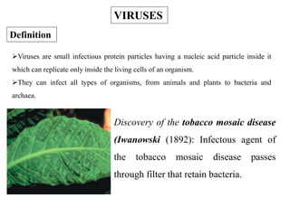
Viruses are small infectious protein particles having a nucleic acid particle inside it
which can replicate only inside the living cells of an organism.
They can infect all types of organisms, from animals and plants to bacteria and
archaea.
Discovery of the tobacco mosaic disease
(Iwanowski (1892): Infectous agent of
the tobacco mosaic disease passes
through filter that retain bacteria.
2. Size of virus particle (virion) varies between 20-300 nm
SIZE OF VIRUS PARTICLE
3. GENERAL PROPERTIES OF VIRUSES
Viruses are obligate parasites
They can not perform processes for energy conservation or for biosynthesis
Replication occurs within a host (intracellular)
Occur as nucleic acid in the host (intracelluar) or as virion (extracellular)
Virions consist of a genome (DNA or RNA), capsid (protein coat), and
often virus-specific enzymes
Enveloped viruses are enclosed in a membrane (lipid bilayer)
5. CLASSIFICATION OF VIRUSES
Viruses are mainly classified by phenotypic characteristics, such as
morphology, nucleic acid type, mode of replication, host organisms, and the type
of disease they cause. Currently, two main schemes are used for the
classification of viruses: the International Committee on Taxonomy of Viruses
(ICTV) system and Baltimore classification system, which places viruses into
one of seven groups.
Baltimore classification (first defined in 1971) is a classification system
that places viruses into one of seven groups depending on a combination of their
nucleic acid (DNA or RNA), strandedness (singlestranded or doublestranded),
Sense, and method of replication. Named after David Baltimore, a Nobel
Prizewinning biologist, these groups are designated by Roman numerals.
6. Viral classification starts at the level of order and continues as follows,
with the taxon suffixes given in italics:
Order (virales)
Family (viridae)
Subfamily (virinae)
Genus (virus)
Species
Species names generally take the form of [Disease] virus.
Currently (2012), seven orders, 96 families, 22 subfamilies, 420
genera, and 2,618 species of viruses have been defined by the ICTV.
7. Caudovirales are tailed dsDNA (group I) bacteriophages.
Herpesvirales contain large eukaryotic dsDNA viruses.
Ligamenvirales contains linear, dsDNA (group I) archaean viruses.
Mononegavirales include nonsegmented (-)strand ssRNA (Group V)
plant and animal viruses.
Nidovirales are composed of (+) strand ssRNA (Group IV) viruses
with vertebrate hosts.
Picornavirales contains small (+) strand ssRNA viruses that infect a
variety of plant, insect and animal hosts.
Tymovirales contain monopartite (+) ssRNA viruses that infect
plants.
ICTV GROUPS
8. I: dsDNA viruses (e.g. Adenoviruses, Herpesviruses, Poxviruses)
II: ssDNA viruses (+ strand or "sense") DNA (e.g. Parvoviruses)
III: dsRNA viruses (e.g. Reoviruses)
IV: (+)ssRNA viruses (+ strand or sense) RNA (e.g. Picornaviruses,
Togaviruses)
V: (−)ssRNA viruses (− strand or antisense) RNA (e.g. Orthomyxoviruses,
Rhabdoviruses)
VI: ssRNA RT viruses (+ strand or sense) RNA with DNA intermediate in
lifecycle (e.g. Retroviruses)
VII: dsDNA RT viruses (e.g. Hepadnaviruses)
Baltimore classification
12. Endocytosis: Many viruses enter cells via receptor mediated
endocytosis . In this pathway, viruses bind to receptors at coated pits. The
coated pits pinch off to form coated vesicles, which are uncoated and then
fuse with endocytic vesicles, and eventually with lysosomes. As they go
through this process, the endosomes become more acidic (remember
lysosomes are a very acidic environment where the breakdown of cellular
macromolecules occurs). Viral genomes must therefore escape the endosome
before they are destroyed by proteases, nucleases, etc.
Direct Membrane Fusion: Some enveloped viruses directly fuse
with the plasma membrane. In these cases the activity of a fusion protein is
not dependent on pH change, but rather is induced in response to receptor
binding.
13. However, most plant viruses can get inside the cell through wound or
minute passage of the cell. Bacteriophages are evolved by special mechanism to
inject the viral genome in to the bacterial cell while the capsid remains outside.
III: Uncoating and Targeting
With some viruses, the genome is completely released from the capsid during or
after penetration. This is known as "uncoating". In others, such as retroviruses and
reoviruses, the first stages of the viral replication cycle (transcription, replication)
actually occur inside the capsid. These capsids have undergone some
conformational changes during infection that allow viral gene expression and/or
replication to begin, and the resulting structures are sometimes known as partially
uncoated particles. Since almost all DNA viruses replicate in the nucleus of
infected cells, they must be targeted there. In many cases the entire nucleocapsid
enters the nucleus, where uncoating then takes place.
14. IV. Replication of viral genome:
In order for new virus to be assembled, both new viral
genomes and other virion components (proteins) must be produced.
Exactly how this occurs varies greatly depending on the family (and
Baltimore Class) of virus. Summaries of common schemes are given
below ……
15. DNA Viruses
A) Viruses with Small, Double Stranded DNA genomes
1. Early gene expression. The first stage in the viral replication cycle is
expression of the viral early genes. Transcription of these genes
occurs using cellular RNA polymerase II and cellular
transcription factors. These proteins bind to the viral DNA in
regions called early promoters/enhancers, and promote synthesis of
the early pre mRNAs. The early RNAs are then transported to the
cytoplasm where they are translated, giving rise to the early proteins.
16. 2. Viral DNA replication. Once the viral early genes have been expressed,
and the cells have been induced to enter S phase, viral DNA is replicated.
This occurs in the nucleus of infected cells, and gives rise to new viral
genomes. Many hundreds or thousands of new viral genomes can be
produced in the nucleus of a lytically infected cell.
3. Late gene expression. After viral DNA replication has begun, the late
genes are transcribed and translated to give rise to late proteins. Late
genes encode the structural proteins of the virus, including capsid
proteins and, for enveloped viruses, the matrix and envelope proteins.
Both late and early viral proteins are synthesized in the cytoplasm, but
are often transported back to the nucleus where both viral replication and
nucleocapsid assembly occurs.
17. B. Viruses with Single Stranded DNA Genomes.
The sole family in this class is the Parvoviridae, which includes
a number of animal pathogens including feline panleukopenia virus and
canine parvovirus. The genomes of these viruses are approximately 5 kb
in length, and only the negative strand is encapsidated. Before any viral
genes can be expressed, the single stranded genome must first be
converted to double stranded DNA, which is then transcribed to give rise
to the viral mRNAs. Replication takes place in the nucleus, and requires
host S phase functions.
18. Viruses with RNA Genomes
A) Replication and Gene Expression of + Strand Viruses
These viruses share the common feature that the genome itself
can serve as mRNA and be directly translated to give rise to proteins.
The naked RNA ( with no protein attached) of these viruses is infectious
because the viral replicase, which is not present in uninfected cells, can
be translated directly from the viral RNA.
19. The general replication scheme used by + strand viruses is the
following.
1) Synthesis of viral proteins (including replicase) from the
viral genomic RNA.
2) Synthesis of viral RNA (strand, then new + strand) using
the viral replicase.
3) Use of the new progeny + strand for 2 purposes
a) production of more viral proteins (including
structural proteins)
b) to be packaged into virions as new virus
genomes.
20. B) Replication and Gene Expression Strategies of (-) Strand RNA Viruses:
By definition, the genome of these viruses cannot be used as a
template for protein synthesis, so an RNA replicase (RNA dependent RNA
polymerase) cannot be synthesized directly as it is with the + sense viruses.
Since cells do not contain such an activity, (-) strand viruses must contain a
replicase/polymerase as part of the virion. The overall replication/gene
expression strategy for strand viruses is outlined below.
21. 1) Synthesis of + strands using the virion associated polymerase.
These + strands can either be full length or subgenomic mRNAs.
2) Translation of viral mRNAs (+ strand) to make viral proteins,
including additional polymerase.
3) Production of new strands from the intact + strands, which are
then used either as templates for additional + strands, or packaged into
virions as new genomes.
22. C) Replication and Expression of Viruses with Double Stranded RNA
Genomes
Members of the Reoviridae and Birnaviridae families contain
segmented, double stranded RNA genomes. This gives them the ability to
undergo antigenic shift, as discussed for Orthomyxoviruses. The strategy for
Reovirus gene expression is very interesting. Early transcription occurs in
partially uncoated virions within the cytoplasm, using enzymes that are
brought in with the virus. This givesrise to early mRNAs. These mRNAs then
translated into protein. The final stages of viral replication and gene expression
actually occur inside newly formed virions within the infected cell.
23. V: Virus Assembly and Release
Once new viral genomes and proteins have been produced, they
are assembled into new virions. This usually occurs in a very specific
order. For example, for many viruses, the viral capsid is partially
assembled (ie, the newly synthesized capsid proteins associate together
into a capsidlike structure). The viral genome is then inserted into the
capsid to form a nucleocapsid, which then undergoes some type of
maturation that can include proteolytic cleavage of capsid proteins. In the
case of nonenveloped viruses, these newly formed virions accumulate in
the cell and are released by cell lysis.
25. LIFE CYCLE OF BACTERIOPHAGE
The first step in the infection process is the adsorption of the
phage to the bacterial cell. This step is mediated by the tail fibers or by
some analogous structure on those phages that lack tail fibers. Phages
attach to specific receptors on the bacterial cell such as proteins on the
outer surface of the bacterium, LPS, pili, and lipoprotein. This process is
reversible. One or more of the components of the base plate mediates
irreversible binding of phage to a bacterium.
Adsorption
26. The irreversible binding of the phage to the bacterium results
in the contraction of the sheath (for those phages which have a sheath)
and the hollow tail fiber is pushed through the bacterial envelope.
Some phages have enzymes that digest various components of the
bacterial envelope. Nucleic acid from the head passes through the
hollow tail and enters the bacterial cell. The remainder of the phage
remains on the outside of the bacterium as “ghost”. Even a non-
susceptible bacterium can be artificially infected by injecting phage
DNA by a process known as transfection.
Penetration
29. PRIONS AND VIROIDS
PRIONS-
Structure
Prions are infectious agents composed exclusively of a single sialoglycoprotein
called PrP 27-30. They contain no nucleic acid. PrP 27-30 has a mass of 27,000
30,000 daltons and is composed of 145 amino acids with glycosylation at or
near amino acids 181 and 197. The carboxy terminus contains a
phosphatidylinositol glycolipid whose components are ethanolamine,
phosphate, myoinositol and stearic acid. This protein polymerizes into rods
possessing the ultrastructural and histochemical characteristics of amyloid.
Amyloid is a generic term referring to any optically homogenous, waxy,
translucent glycoprotein; it is deposited intercellularly and/or intracellularly in
many human diseases such as:
30. •Alzheimer's disease
•CreutzfeldtJakob disease
•Down's syndrome
•Fatal familial insomnia
•Gerstmann Straussler syndrome
•Kuru Leprosy
Replication
The prion is a product of a human gene, termed the PrP gene, found on
chromosome 20. This gene contains two exons separated by a single intron.
Exon I and Exon II are transcribed and the two RNAs ligated into a single
mRNA. This mRNA contains an open reading frame (ORF) or protein coding
region which is translated into the PrP protein. The PrP protein is a precursor of
the prion protein. It is termed PrP 33-35.
31.
32. The PrP 33-35 undergoes several post-translational events to become the
prion protein (PrP 27-30):
1. Glycosylation - at two sites.
2. Formation of a disulfide bond between two cysteine residues.
3. Removal of the N-terminal signal peptide.
4. Removal of the C-terminal hydrophobic segment.
5. Addition of a phosphatidylinositol glycolipid at the C-terminal.
6. Removal of the N-terminal first 57 amino acids.
33. In normal cells only the PrP 33-35 protein is synthesized. It is found in
the neural cell membrane where it's function is to sequester Cu++ ions. In
abnormal ("infected") cells, the PrP 27-30 is produced from the PrP 33-35
protein. The PrP 27-30 triggers a series of reactions that produce more PrP 27-
30 proteins, i.e., PrP 27-30 induces its own synthesis. In addition to the post
translational modifications, the PrP 27-30 protein differs from the PrP 33-35
protein in a single amino acid residue. Residue 178 in the PrP 27-30 contains
an asparagine residue whereas the PrP 33-35 protein has an aspartate residue at
this position. This causes a conformational change in the PrP 27-30 protein
from an a-helix to a b-sheet. This conformational change in the PrP 27-30
protein has three effects:
34. 1. It imparts to the PrP 27-30 protein the ability to induce the same a-helix to
b-sheet conformation in the PrP 33-35 protein. This is a permanent
conformational change. It thus induces its own "replication.“
2. The b-sheet-forming peptides aggregate to form amyloid fibrils.
3. The amyloid fibrils kill thalamus neurons through apoptosis, a programmed
series of events that leads to cell death.
35.
36. Transmission
Spread of the disease is via horizontal transmission, i.e., transmission
from one person to another, either directly or by fomites or by ingestion of
contaminated meat.
Diagnosis
In the past, diagnosis of prion disease was made through examination of
brain biopsies taken from patients in advanced stages of the disease or, more
commonly, after they had died. In January of 1999 it was found that the prion
protein accumulated in the tonsils and could be detected by an immuno
fluorescence test on tonsilar biopsies. A second test was simultaneously developed
which was based on a Western blot. Later that year a third test was developed that
had the high sensitivity necessary to detect the prion protein in blood. This test is
based on capillary electrophoresis with laser-induced fluorescence. It detects as
little as 10-18 mole.
37. Viroids
Viroids are infectious agents composed exclusively of a single piece of
circular single stranded RNA which has some double-stranded regions.
Because of their simplified structures both prions and viroids are
sometimes called subviral particles. Viroids mainly cause plant diseases but
have recently been reported to cause a human disease.
Catalytic RNAs are those that have the intrinsic ability to break and
form covalent bonds; Viroids are catalytic RNA's (ribozymes) that cleave RNA
to produce fragments containing a 5'-hydroxyl and a 2', 3'-cyclic phosphate.
38. This is a nonhydrolytic reaction in which the same number of
phosphodiester bonds are maintained and the transesterification reaction is
theoretically reversible. This reaction is considered to play an essential role
in the replication of these RNAs in vivo. Such reactions are all
intramolecular and hence quasi-catalytic with single turnover. These RNAs
can be manipulated, however, to provide true catalytic cleavage in trans-
reactions.
39. Replication
Circular, pathogenic RNAs are replicated by a rolling circle mechanism in
vivo. There are two variations of this rolling circle mechanism:
In the first variation (A), the circular plus strand is copied by viroid RNA-
dependent RNA polymerase to form a concatameric minus strand (step 2). Site-specific
cleavage (arrows) of this strand produces a monomer that is circularized by a host RNA
ligase (step 3) and then copied by the RNA polymerase to produce a concatameric plus
strand. Cleavage of this strand (step 5) produces monomers which, on circularization,
produces the progeny circular, plus RNA, the dominant form in vivo.
In the other variation (B), the concatameric minus strand of step 1 is not
cleaved but is copied directly to give a concatameric plus strand (step 3), which is cleared
specifically to monomers for ligation to the circular progeny. Those RNAs that self-
cleave only in the plus strand in vitro are considered to follow this route.