
Recommended
More Related Content
What's hot
What's hot (20)
Similar to Reproduction In Sphenopsida
Similar to Reproduction In Sphenopsida (20)
More from rasikapatil26
More from rasikapatil26 (16)
Recently uploaded
Recently uploaded (20)
Reproduction In Sphenopsida
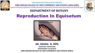
- 1. Reproduction In Equisetum DR. RASIKA N. PATIL ASSISTANT PROFESSOR DEPARTMENT OF BOTANY SHRI SHIVAJI COLLEGE OF ARTS COMMERCE AND SCIENCE AKOLA
- 2. Spore-Producing Organs in Equisetum
- 3. Cone: Fertile aerial, unbranched shoots bear at their apices (Fig. 243) the spore-bearing compact organs known as strobili (sing, strobilus) or cones. In some rare cases branched fertile axis is also present (Fig. 244). Each cone or strobilus has a thick central axix known as strobilus axis or cone axis. On the strobilus axis are attached many umbrella like sporangiophores in whorl. Each sporangiophore is a stalked structure, the free end of which becomes flattened to form a peltate disc (Fig. 245). The disc is a hexagonal structure and present at right angle to the stalk. On the undersurface of disc are present many sporangia, horizontally towards the axis of strobilus. Each sporangium is elongated and pendant, and contains a rounded apex . The sporangium is surrounded by jacket and tapetal layers which enclose many spores. Equisetum is homosporous . A ring-like outgrowth is present below each strobilus. This is called annulus. Morphological nature of annulus is controversial. According to some it is the uppermost whorl of sterile leaves while others are of the opinion that it is the lowermost sterile sporangiophore .
- 4. Reproduction in Equisetum: • Equisetum reproduces vegetatively and by means of spores. Vegetative Reproduction: The subterranean rhizomes of some species (e.g., E. telmateia, E. arvense) form tubers (Fig. 7.83) which, on separation from the parent plant, germinate to produce new sporophytic plants. The tubers develop due to irregular growth of some buds at the nodes of the rhizomes. Reproduction by Spores: • Spores are produced within the sporangia. The sporangia are borne on the sporangiophores which are aggregated into a compact structure termed strobilus or cone or sporangiferous spike. • Strobilus: • The strobilus are terminal in position and generally are borne terminally on the chlorophyllous vegetative shoot . However, they may be borne terminally on a strictly non- chlorophyllous axis . • The strobilus is composed of an axis with whorls of sporangiophores. Each sporangiophore is a stalked structure bearing a hexagonal peltate disc at its distal end. under surface of the sporangiophore disc 5-10 elongate, cylindrical hanging sporangia are borne near the periphery in a ring. • The flattened tips of the sporangiophores fit closely together which provide protection to the developing sporangia. The axis bears a ring-like outgrowth, the so-called annulus immediately below the whorls of sporangiophores which provide additional protection during early development. • The annulus has been interpreted as a rudimentary leaf sheath by some botanists, whereas others consider it to be sporangiophoric in nature as occasionally it bears small sporangia.
- 5. Development of Sporangium: The mode of development of sporangium is eusporangiate, as it is not entirely formed from a single initial. Superficial cells adjacent to the original initial may also take part in the development of sporangium. Sporangia are initiated in single superficial cell around the rim of the young sporangiophore. The periclvnal division of sporangium initial forms an inner and an outer cell. The inner cell, by further divisions in various planes, gives rise to sporogenous tissue. The outer cell, by periclinal and anticlinal divisions, gives rise to irregular tiers of cells, the inner tiers of which may transform into sporogenous tissue and the outer tiers become the future sporangial wall cells. The innermost layer of the sporangial wall differentiates as the tapetum. The sporogenous cells separate from each other, round off and eventually transform into spore mother cell. All but the two outermost wall layers disorganise to form periplasmodial fluid. However, not all of the sporogenous cells function as spore mother cells. Many of them degenerate to form a multinucleate nourishing substance for the spore mother cells. Each spore mother cell undergoes meiotic division (reductional division) and produces spore tetrad. All spores in a sporangium are of same size and shape i.e., homosporous.
- 6. Dehiscence of Sporangium: At maturity, the strobilar axis elongates, as a result the sporangiophores become separated and exposed. Then the sporangium splits open by a longitudinal line due to the differential hygroscopic response of the wall cells. Spores: The spores are spherical and filled with densely packed chloroplasts. The spore wall is laminated and shows four concentrate layers. The innermost is the delicate intine, followed by thick exine, the middle cuticular layer and the outermost epispore or perispore. The intine (endospore) and exine (exospore) are the true walls of the spore. The outer two layers i.e., cuticular layer and epispore are derived due to the disintegration of the nonfunctional spore mother cells and tapetal cells. At maturity, the epispore (the outermost layer) splits to produce four ribbon like bands or strips with flat spoon-like tips. These bands are free from the spore wall except for a common point of attachment and remain tightly coiled around the spore wall until the sporangium is fully matured. These are called elaters. The elaters are hygroscopic in nature. The spores remain moist atearly stages of development, thus the elaters are spirally coiled round the spore. The spores dry out at maturity and consequently the elaters become uncoiled. These uncoiled elaters become entangled with the elaters of other spores. Through these actions the elaters help in the dehiscence process and also the dispersal of spores in large groups from the sporangium. The elaters of Equisetum are different from those of the bryophytes.
- 7. Gametophyte Generation: • Equisetum is a homosporous pteridophytes. The haploid spores germinate to form gametophyte. The germination takes place immediately if the spores land on a suitable substratum. If the spores do not germinate immediately, their viability decreases significantly. • The spores swell up by absorbing water and shed their exine. The first division of the spore results in two unequal cells: a small and a large cell. The smaller cell elongates and forms the first rhizoid. The larger cell divides irregularly to produce the prothallus. The prevailing environmental conditions determine the size and shape of the prothallus. • If a large number of spores are developed together within a limited space, then theprothalli formed are of thin filamentous type. • But a relatively thick and cushion-shaped prothalli are formed from sparsely germinating spores. Mature gametophytic plants may range in size from a few millimeters up to 3 centimeters e.g., E. debile) in diameter. • They are dorsiventral and consist of a basal non-chlorophyllous cushion-like portion from which a number of erect chlorophyllous muticellular lobes develop upwards. • Unicellular rhizoids are formed from the basal cells of cushion. • The prothallus bears sex organs and reproduces by means of sexual method.
- 8. SexOrgans of Equisetum: • Antheridium: • In monoecious species, antheridia develop later than archegonia. They are of two types - projecting type and embedded type. • Antheridia first appear on the lobes of the gametophyte. The periclinal division of the superficial antheridial initial gives rise to jacket initial and an androgonial cell. • The jacket initial divides anticlinally to form a single-layered jacket. The repeated divisions of androgonial cells form numerous cells which, on metamorphosis, produce spermatids/antherozoids. The antherozoids escape through a pore created by the separation of the apical jacket cell. • The apical part of the antherozoid is spirally coiled, whereas the lower part is, to some extent, expanded. Each antherozoid has about 120 flagella attached to the anterior end. • Many antherozoids pass through the canal of the archegonium but only one of them fuses with the egg. Thus diploid zygote is formed. Generally more than one archegonia are fertilized
- 11. Embryo (The New Sporophyte): • The embryo is the mother cell of the next sporophytic generation. Unlike most pteridophytes, several sporophytes develop on the same prothallus. The first division of the zygote is transverse. This results in an upper epibasal cell and lower hypobasal cell. • The embryo is therefore exoscopic (where the apical cell is duacted outward i.e., towards the neck of the archegonium) in polarity. • No suspensor is formed in Equisetum. The epibasal and hypobasal cells then divide at right angles to the oogonial wall, and as a result a tour-celled quadrant stage is • established. All the four cells of the quadrant are of different size and shape. • The four-celled embryo undergoes subsequent divisions and the future shoot apex originates from the largest cell and leaf initials from the remaining cells of one quadrant of the epibasal hemisphere. • One cell of the epibasal quadrant and a portion of the adjacent quadrant of the hypobasal region contribute to the development of root. The first root develops from one of the epibasal quadrants and a portion of the adjacent hypobasal quadrant. The shoot grows rapidly. • Later the root grows directly downward and penetrates the gametophytic tissue to reach the soil or substratum. A number of such sporophytes may develop from a large mature gametophyte if more than one egg is fertilized.
- 15. THANK YOU