1. Pradip Hamal
BMLT 2nd Year
Chitwan School of Medical
Sciences
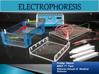
Cathode
( -)
Anode
( +)
2. Introduction:
• Electrophoresis is the popular technique used in
the clinical and research laboratories for the
separation of closely related compounds like
mixture of proteins, amino acid.
• Electrophoresis is the process of moving charged
molecules in solution by applying an electrical field
across the mixture.
3. Definition
The movement of charged particles in an
electrical field resulting in their migration
towards the oppositely charged electrode is
known as electrophoresis.
4. The velocity of migration of each molecule in an electric field
is dependent upon the net charge on the molecule, strength of
the electric field and is inversely proportional to the molecular
weight.
- ve charge(cathode) …….. +ve charge(anode)
+ve charge(anode)………… -ve charge(cathode)
Molecules moved with a speed dependent on their charge, shape and size [charge
and mass].
5. 1. Depending upon the nature of supporting
medium:
a. Cellulose acetate electrophoresis(paper strip
electrophoresis): where cellulose acetate paper serves as the
supporting medium.
b. Polyacrylamide gel electrophoresis(PAGE):
Here the acrylamide and methylene bisacrylamide forms
a polymer.
c. Agar gel ectrophoresis(AGE):
Where agar gel is used as supporting medium.
Types of electrophoresis
6. 2. Depending upon the mode of technique:
a. Slide gel electrophoresis
b. Tube gel electrophoresis
c. Disc electrophoresis
d. Low and high voltage electrophoresis
7. Commonly used electrophoresis for Hb:
1. Starch agarose gel electrophoresis (8.6)
2. Cellulose acetate membrane electrophoresis (PH
8.4-8.6)
3. Citrate agar gel electrophoresis (6.0)
4. Globin chain electrophoresis (PH 8.5)
8. Applications:
1. Haemoglobin separation
2. Separating serum protein for diagnostic
purposes
3. Lipoprotein separation and identification
4. Isoenzyme separation and their analysis
5. Nucleic acid studies
6. Determination of molecular weight of
proteins.
9. SUPPORT MEDIA
The commonly using supporting media are :
• agarose,
• Polyacrylamide &
• Cellulose acetate membrane.
Agarose
• highly purified polysaccharide derived from agar, long
sugar polymers held together by hydrogen and
hydrophobic bonds.
Acrylamide
• (CH2=CH-CO-NH2)
• Polyacrylamide gel structure held together by covalent
cross-links
10.
11. Collection of blood for electrophoresis.
• Anticoagulated blood(EDTA) is used. Lysate is
prepare for electrophoresis.
12. Preparation of lysate
1. Collect 2 ml of blood and mix in EDTA.
2.Wash RBCs three times with normal saline ( to
remove trap plasma) because plasma contain
plasma protein like albumin, globulin,
fibrinogen, Hb also type of protein. Plasma Protein gives band
as Hb As a result, false positive result appears.
1. Lyse RBCs with equal volume of distilled
water.
2. Mix in vertex mixture and add equal volume
of carbon tetrachloride(dissolve cell stroma,
13. 4. Centrifuge at 3000 rpm for 30 min to remove
carbon tetrachloride which settle down at bottom.
5. Transfer clear supernatant to clean test tube and
adjust Hb concentration 7- 10 gm with water.
6. Perform electrophoresis as soon as possible. If
delayed, add 1 drop of 1M/dl potassium cyanide
and store in the refrigerator.
15. • The cellulose acetate membrane is manufactured
by CELLOGEL (Milano, Italy)
• It is simple, reliable and rapid technique.
• It is satisfactory for the detection of most
common clinically important haemoglobin
variants.
• Haemoglobin electrophoresis at pH 8.4-8.6.
16. Principle:
Electrophoresis is the movement of charged
particles in an electrical field.
The rate of movement depends on the
electrical charge of the colloidal particles(Hb)
and on the intensity of the electrical field.
In alkaline Tris EDTA borate buffer, PH 8.5,
most haemoglobins have a negative charge and
migrate towards the positive pole.
17. Electrophoresis is accomplished at
350v or 450v for over 30 min.
The migration of haemoglobins may be
examined unstained or are stained
with 0.5% ponceau S, a protein stain.
A visual comparision of unknown with
a normal control is usually adequate
for the interpretation of the pattern.
19. 2. Protein stain/ fixative:
Ponceau S : 0.5 gm
Trichloroacetic acid : 7.5 gm
Distilled water : 1 Lit
3. Destaining solution/Rinse solution
5% acetic acid : 30 ml
Distilled water upto : 1 lit
20. Equipment Required
1. Electrophoratic tank and power supply
2. Chromatography paper or wicks of filter
3. Blotting paper
4. Applicators
5. Cellulose acetate membrane
6. Staining equipment
21. Procedure:
1. At the first haemolysate is prepared. Its
concentration is maintained between 7-10gm/dl
with distilled water.
2. The compartment of electrophoretic tank is filled
with TEB buffer.
3. Soak wicks and position them .
4. Soak cellulose plates in buffer for at least 5 min.
22. 5. Blot plates between 2 pieces of filter paper
quickly and evenly to remove excess
moisture.
6. Apply 10µl sample to cellulose acetate side of
plate at points approximately 5 mm from
cathode using microdispenser.
23. 7. Cover the chamber and apply 350v for 30 minutes
at room temp. Time should be adjusted depending
on migration of Hb bands.
8. Remove cellulose acetate plate from chamber and
stain for 3-5min with Ponceau ‘S’ stain.
9. Remove from stain and wash for 2 min in 5%GAA
three times until background is white.
24. 10. Plate may be fixed in absolute methanol for 3-5
min and cleared in 20% acetic acid in absolute
methanol for 10 min.
11. Plate is placed between blotter sheets and kept
pressed under weight until dry.
12. Label the membrane and store in protective
plastic envelope.
25.
26. Migration pattern of Hemoglobin
• Normal hemoglobins present in an adult:
- Hb A migrates the fastest, followed by Hb F.
- Hb A2 moves only slightly from the point of origin near the
cathode.
• Abnormal hemoglobins:
- Hb C migrates with Hb A2 near the cathode.
- Hb E also migrates with A2 and Hb S lies between Hb A2 and
Hb F.
- Hb H and Bart's hemoglobin are unstable and very fast
moving, with Hb H being the faster of the two. They are
located nearer the anode past Hb A .
27. Interpretation:
All haemoglobins move from cathode (-)(application point) towards anode (+) in a
definite pattern and speed. The following diagram of cellulose acetate at pH 8.5 will
provide a rough pattern of normal and abnormal haemoglobins
31. Agarose gels are commercially available as
substitutes for both alkaline and acid
separation system with acid agarose system.
The principle of the test is the same as that of
above electrophoresis at the same ph but
there is significant differences in mobility of
some variant Hb.