
Recommended
More Related Content
What's hot
What's hot (20)
Similar to Separation techniques by NSK
Similar to Separation techniques by NSK (20)
More from Dr. Santhosh Kumar. N
More from Dr. Santhosh Kumar. N (20)
Recently uploaded
Recently uploaded (20)
Separation techniques by NSK
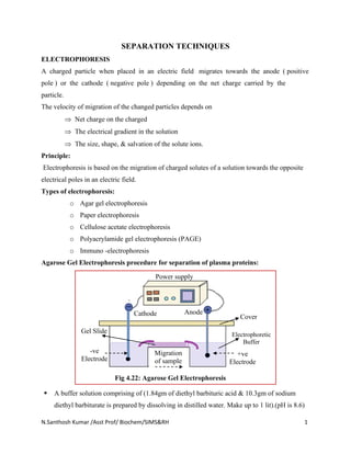
- 1. N.Santhosh Kumar /Asst Prof/ Biochem/SIMS&RH 1 SEPARATION TECHNIQUES ELECTROPHORESIS A charged particle when placed in an electric field migrates towards the anode ( positive pole ) or the cathode ( negative pole ) depending on the net charge carried by the particle. The velocity of migration of the changed particles depends on Net charge on the charged The electrical gradient in the solution The size, shape, & salvation of the solute ions. Principle: Electrophoresis is based on the migration of charged solutes of a solution towards the opposite electrical poles in an electric field. Types of electrophoresis: o Agar gel electrophoresis o Paper electrophoresis o Cellulose acetate electrophoresis o Polyacrylamide gel electrophoresis (PAGE) o Immuno -electrophoresis Agarose Gel Electrophoresis procedure for separation of plasma proteins: ▪ A buffer solution comprising of (1.84gm of diethyl barbituric acid & 10.3gm of sodium diethyl barbiturate is prepared by dissolving in distilled water. Make up to 1 lit).(pH is 8.6) Electrophoretic Buffer Migration of sample Cover Anode Power supply Cathode Gel Slide +ve Electrode -ve Electrode Fig 4.22: Agarose Gel Electrophoresis
- 2. N.Santhosh Kumar /Asst Prof/ Biochem/SIMS&RH 2 ▪ Agarose gel dissolved into 100ml of buffer solution. It is heated over a flame and then poured on clean slides; ▪ The slides are allowed to set, as thin layer. ▪ 2 to 3 μl of the serum sample is applied in the nick, made with a cover slip, at one end of the slide. ▪ Voltage around 120 volts and a current of 7 milli amperes (7 MA) is applied per slide for around 60min. ▪ The slides are now placed in a fixative (fixative is ethanol: water: acetic acid (70 : 25 : 5 v/v) for 30min), and then placed in a staining solution for 30min (Stain solution: bromophenol blue – 1 gm in 100ml of 95% ethanol saturated with mercuric chloride). ▪ The slides are finally rinsed under tap water and dried. ▪ The bands which have separated can be visualized. Electrophoretic patterns of plasma proteins in diagnosis: ▪ In paper or gel electrophoresis, the serum can be separated into a number of fractions- albumin, globulin, but if plasma is used instead of serum, a band of fibrinogen fraction is seen between β and δ globulin regions. Fig 4.23: Normal Electrophoresis pattern of Plasma Proteins Abnormal Electrophoretic patterns of plasma proteins in diagnosis DISEASES ALBUMIN 1- band 2- band - band -band Nephrotic Syndrome --- --- Chronic Hepatitis --- Multiple Myeloma --- --- --- (M-Band) Note : stands for decrease and stands for increase, normal level --- stands for no change In multiple myeloma, Extra M –band will be appears in b/w the β globulin and γ-globulin regions
- 3. N.Santhosh Kumar /Asst Prof/ Biochem/SIMS&RH 3 CLINICAL SIGNIFICANCE OF SERUM ELECTROPHORESIS Point of application ELECTROPHORETIC PATTERNS – NORMAL AND DISEASED CONDITION Electrophoresis helps in the detection of the charges in the indicated protein fractions in the serum or plasma and in detecting abnormal bands in certain disease conditions. ❖ In acute inflammation, albumin is normal or decreased. There is an increase in α1 and α2 globulin, normal or decrease in γ globulin. ❖ In chronic infection, albumin is same or decreased with an increase in α1 and α2 globulin, β globulin may be same or increased and γ globulin is increased. ❖ In cirrhosis of liver, there is marked decrease in albumin, α2 globulin and β globulin but increase in γ globulin. Alb α1 α2 β γ + Normal pattern Nephrotic syndrome Albumin decreased α2 increased Multiple myeloma Hypogammaglobulinemia γ- region decreased Alb α1 α2 β γ Alb α1 α2 β γ M- band
- 4. N.Santhosh Kumar /Asst Prof/ Biochem/SIMS&RH 4 ❖ In nephrotic syndrome, there is marked decrease in albumin, increase in α2 globulin with same or decreased γ globulin. ❖ In hypogammaglobulinemia, there is marked decrease in γ globulins. ❖ In Hypoproteinemia, there is marked decrease in albumin, normal or increase in α1 and α2 globulin, increase in β globulin and normal or increased γ globulin. ❖ In leukemia, there is decrease in albumin and increase in γ globulin. ❖ In multiple myeloma, the characteristic feature is the presence of an abnormal band close to or within γ band & β band (M-Band). Applications of electrophoresis Electrophoresis technique can also be applied in, 1. Separation and identification of isoenzymes. 2. Separation and identification of abnormal hemoglobin. 3. Separation and identification of lipoproteins. 4. Determination of molecular weight of proteins.
- 5. N.Santhosh Kumar /Asst Prof/ Biochem/SIMS&RH 5 CHROMATOGRAPHY Chromatography is a powerful tool for the separation of a wide variety of physiologically important substances. The primary goal of the chromatographic process is to separate and resolve a mixture of substances into individual components which are called ‘solutes’. Chromatography is defined as the technique of separation of mixture of solutes, dissolved in a common solvent, by virtue of their differences in their partition co efficient in two different media. Principle: Chromatography consists of a mobile phase and a stationary phase. ▪ Mobile phase refers to the mixture of substance dissolved in a liquid or gas. ▪ Stationary phase is a porous solid matrix through which the sample contained in the mobile phase percolates. ▪ The interaction between the mobile and stationary phases results in the separation of the compound from the mixture. PAPER CHROMATOGRAPHY: It uses a special paper sheet as the inert solid support whose cellulose fibers hold as immobilized polar liquid in their meshes as a stationary phase. a) Paper Chromatography Descending Paper chromatography Partition Chromatography Ion –Exchange Chromatography Absorption Chromatography TYPES OF CHROMATOGRAPHY Ascending Paper chromatography b) Gas Liquid Chromatography Column Absorption chromatography Thin Layer chromatography Gel Filtration Chromatography Affinity Chromatography High performance liquid Chromatography (HPLC)
- 6. N.Santhosh Kumar /Asst Prof/ Biochem/SIMS&RH 6 It is a type of partition chromatography Procedure: ▪ A whatman paper no 3 is select and cut into a size of around 22cm X 22cm. ▪ The solvent for the chromatography is now prepared butanol: water: acetic acid (12:5:3). ▪ The solvent is powered in chromatography chamber. ▪ A line is drawn on the chromatography paper, about ½’ to 1” above the height of the solvent measured in the chromatography jar. ▪ Two marks are made about 1” from the edge of the paper and the distance between the second points in divided into four equal reagent, for the application of the amino acid solution. ▪ Two strings are attached to the top edge of the paper on both sides. ▪ The amino acid mixtures are prepared, taking around 10mg of amino acid (powder form) and dissolving in 1ml of distilled water. ▪ A capillary tube used for the application of the amino acid solution. ▪ Amino acids are select and a mixture formed by dissolving the amino acids. ▪ The capillary tube is used for the application of amino acid such that, the diameter of each circle is less than 0.5cm. The applied solutions are allowed to dry. ▪ The paper is now dipped in the solvent and the lid of the jar is sealed with grease. ▪ The run is allowed to continue for 4 to 6hr. ▪ Remove the whatman paper, then allowed to dry in air. ▪ Prepare staining solution [Ninhydrin solution: 0.2 % of ninhydrin, 95% of n-butanol, 5% acetic acid). Tank Cover Paper support Spotting lines (Samples) Chromatography Tank Whatman Filter paper with the Stationary phase Solvent front Direction of solvent flow Fig 4.18: Descending Paper Chromatography Direction of solvent flow Trough containing solvent (Mobile phase) Spotting lines (Samples) Fig 4.19: Ascending Paper Chromatography
- 7. N.Santhosh Kumar /Asst Prof/ Biochem/SIMS&RH 7 ▪ The staining solution is sprayed on the chromatography paper and allowed to dry. ▪ The spots formed by the migration of the amino acids can now be identified and then Rf values are calculated. The migration of a substance is expressed as Rf value The Rf is constant for a particular amino acid for a given solvent mixture. And also estimated by either densitometer & scintillation spectrometry. Applications: ▪ It separates sugars, amino acids, lipids, steroids and athletic doping drugs and also used to identify drugs, contaminants and adulterants. ▪ HPLC has contributed to analytical solutions in diverse fields such as pharmaceuticals, foods, life sciences, environment, forensics, etc. ▪ A fully automated HPLC system is used to separate and determine HbA2, HbF, HbS and glycosylated Hb. ▪ It employed for the detection and estimation of hormones such as epinephrine, nor epinephrine and ACTH, vitamins like vitamin – A, calcitriol, drugs like phenytoin, LSD & AZT and metabolites like metanephrines). Distance travelled by the solute (Amino Acid) Distance travelled by the solvent Rf =