2. INTRODUCTION
• One of the most common causes of cancer related death in the world
and accounts for 8.8% of all cancer-related deaths The age-
standardized mortality rate of stomach cancer worldwide is 8.9 per
100,000 persons. The highest mortality rates are in Eastern Asia
(14.3/100,000) and the lowest rates in Northern America
(2.1/100,000).
• Gastric cancer is xterised by its predominance in males. Men are
affected 2 to 3 times more often than women (12.3 per 100,000 years
vs. 6.0 per 100,000 years).
De Pope
3. Continua…………
• Shows regional variations between and within countries. Incidence of
gastric tumor increases with age. At presentation, most gastric cancer
patients are diagnosed with an advanced disease, with a 5-year
survival rate lower than 30%
• Although incidence of gastric cancer has decreased during last 2
decades, it is still the 4th most common cancer and the second
leading cause of cancer deaths worldwide. More than 950,000 new
gastric cancers and 700,000 deaths were estimated in 2012.
De Pope
4. Continua………
• In Uganda gastric cancer was found to be prevalent in tribes
inhabiting volcanic areas of south western Uganda especially the
Banyakole (25%). With the commonest mode of clinical presentation:
epigastric pain, weight loss, constipation, epigastric tenderness,
palpable epigastric mass and anaemia.
• The most accurate mode of investigation was by endoscopy followed
by barium meal, commonest locality was pyloric antrum (40%)
adenocarcinoma predominated histologically (95.5%) ( Ibingira C. B.
2001 management of stomach cancer at mulago hosp)
De Pope
5. SURGICAL ANATOMY
• The stomach contains four anatomic regions: 1. Fundus; 2. Cardia; 3.
Body; 4. Pyloric part contains pyloric antrum, pyloric canal.
Fundus:
• Projects upwards and lies in contact with the left dome of diaphragm.
It’s usually full of gas.
Significance:
• To identify the side (right or left) of the body in a plain X-ray
abdomen.
• In achalasia cardia, fundic air bubble is absent.
• Fundic ‘Wrap’ is used in hiatus hernia.
De Pope
6. Continua…………
• During mobilisation of the fundus as in splenectomy or other upper
gastric surgery, short gastric arteries need to be divided. If ligatures
are too close to the stomach near the fundus, gastric fistula may
occur due to necrosis of the stomach.
• GISTs (gastrointestinal stromal tumours) are common in fundus.
Body:
• From fundus to incisura angularis. It has a lesser curvature and a
greater curvature.
De Pope
7. Continua……….
Significance:
• Ability to have a large meal is due to receptive relaxation of the body
of the stomach.
• Greater curvature is located at the level of umbilicus.
• Classical gastrojejunostomy (GJ), anterior or posterior, involves using
body of the stomach.
• Posteriorly, it is related to the lesser sac and pancreas. Carcinoma of
the body may infiltrate pancreas— necessitates careful dissection to
separate from pancreas (sometimes not resectable).
De Pope
8. Continua………..
Pyloric Antrum:
• From incisura till pylorus. Pylorus is thicker than the rest of the
stomach. It is a sphincter of circular muscle fibres. Its canal is usually
closed.
Significance:
• Common site for gastritis, ulcer and carcinoma.
• Incompetence of pyloric sphincter results in severe duodenogastric
reflux.
De Pope
9. Continua……….
• It is in close contact with the head of pancreas. During gastrectomy,
extreme care has to be taken to mobilise the antrum to avoid
bleeding in the pancreatic head region.
Greater Omentum:
It lies in contact with transverse colon and gastrocolic omentum. This
has to be divided from transverse colon during gastrectomy which is
done for carcinoma or ulcer.
De Pope
10. Continua…………
Lesser Omentum:
• Double-layered structure.
• It is suspended between the lesser curvature of the stomach and the
proximal 0.5 inch (2 cm) of the first part of the duodenum inferiorly
and the porta hepatis and the fissure of the ligamentum venosum
superiorly.
De Pope
11. Continua……….
• The lesser omentum is divided into two ligaments:
1. Hepatogastric
2. Hepatoduodenal
Located within the lesser omentum are the hepatic triad, branches of
the anterior vagus nerve, some lymph nodes, and the right and left
gastric arteries.
De Pope
12. Continua………..
Vagus Nerves:
• The left and right vagus nerves descend parallel to the oesophagus
and form oesophageal vagal plexus between the level of the tracheal
bifurcation and level of the diaphragm.
• From this plexus, two vagal trunks, anterior and posterior, form and
pass through the oesophageal hiatus of the diaphragm (mnemonic
LARP: Left trunk—anterior gastric wall; right trunk—posterior gastric
wall).
De Pope
15. Blood Supply of the Stomach
• Mainly supplied by coeliac trunk and its branches:
1. Left gastric artery: direct branch of coeliac trunk. Ascends up to
oesophageal hiatus and turns to the right along the lesser curvature of
stomach. Branches and anastomoses with branches of right gastric
artery and supplies anterior and posterior wall of the stomach. There
is true anastomosis between branches of left gastric artery and
branches from other arteries.
2. Right gastric artery: branch of hepatic artery, comes from coeliac
trunk. Also supplies lesser curvature and body of stomach, along with
left gastric artery
De Pope
16. Continua……….
3. Left gastroepiploic artery: arises from splenic artery and supplies
greater curvature of stomach and anastomoses with right
gastroepiploic artery.
4. Right gastroepiploic artery: branch of gastroduodenal artery, which
is a branch of hepatic artery.
5. Short gastric arteries: branches of splenic artery. They supply the
fundus of the stomach. They are also called vasa braevia.
De Pope
19. Surgical Importance
• Because of extensive anastomoses of blood vessels (extramural and
intramural collateral vessels), stomach can survive with right gastric
and right gastroepiploic arteries only. Thus stomach can be used to
replace the entire oesophagus after oesophagectomy—gastric pull
up.
• The order of ligation of blood vessels in gastrectomy is as follows: Left
gastroepiploic, right gastroepiploic, right gastric (then stomach is
divided) and lastly, left gastric arteries.
De Pope
20. Continua…………
• Gastroduodenal artery, branch of hepatic artery runs behind first part
of duodenum and divides into right gastroepiploic artery and superior
pancreaticoduodenal artery. It is this artery which bleeds when a
posterior duodenal ulcer erodes into it.
De Pope
21. LYMPHATIC DRAINAGE OF STOMACH
• It is an important pathway for spread of carcinoma of the stomach.
The spread occurs both by emboli and permeation. (intrinsic and
extrinsic network)
• Right gastric nodes/suprapyloric nodes mainly drain the pyloric
antrum.
• Subpyloric nodes/gastroepiploic nodes (right) drain the greater
curvature of stomach and pyloric antrum.
• Left gastroepiploic nodes (splenic nodes) drain the upper portion of
stomach, mainly the fundus (carcinoma of fundus).
De Pope
22. Continua…………
• Left gastric (superior gastric) nodes drain the lesser curvature and
body of the stomach (anterior and posterior wall).
• Coeliac nodes receive lymph from the entire foregut (including
stomach) and drain directly into the cisterna chyli and the thoracic
duct. Later, mediastinal nodes and left supraclavicular nodes (Virchow
nodes) are involved.
De Pope
25. NERVE SUPPLY OF STOMACH
• Intrinsic innervation occurs through myenteric plexus of Auerbach
and submucous plexus of Meissner
• Right vagus is posterior and left vagus is anterior.
• Posterior vagus gives criminal nerve of Grassi which supplies lower
oesophagus and fund us of stomach, which, if not cut properly during
vagotomy, may lead to recurrent ulcer.
• Vagus also gives splanchnic branches (hepatic and coeliac branches),
ends as nerve of Latarjet which supplies the antrum and maintains
the antral pump
De Pope
26. Continua………
• Parietal branches help in HCI secretion, which is an important concept
in vagotomy that is done as a treatment in duodenal ulcer
De Pope
28. CARCINOMA STOMACH
• More common (2 times) in men compared to women.
• Rare below 40, Average age is 63 years.
• Worldwide, it is the fourth most common cancer and second leading
cancer of death.
• Incidence of proximal gastric carcinoma is increased— may be due to
obesity and in rich socioeconomic status patients.
• 3 most common primary malignant gastric neoplasms are
adenocarcinoma (95 percent), lymphoma (4 percent), and malignant
gastrointestinal stromal tumor (GIST) (1 percent).
De Pope
29. Continua……………
• Carcinoma distal stomach is more commonly associated with H.
pylori.
• Proximal carcinomas are more advanced at the time of presentation
than distal carcinomas.
• Overall 5-year survival after the diagnosis of gastric cancer is 10 to
20%.
• Those who undergo potentially curative resection (R–0) have a 5-year
survival rate of 25–50%.
De Pope
30. Risk Factors for Carcinoma Stomach
Environmental and Dietary Factors:
• Increased in persons who consume red meat, cabbage, spices, spirits,
salt-fish.
• Smoked salmon fish was responsible for increased incidence of
carcinoma stomach in Japanese population. Theory: release of
polycyclic hydrocarbons and aromatic amino acids. Smoking, spicy
food and alcohol consumed over a period of many years produce
chronic gastritis which may change into carcinoma of stomach.
De Pope
31. Continua………
Precancerous Conditions:
• Atrophic gastritis: This may be due to smoking, spicy food, continuous
ingestion of drugs, reflux of bile into the stomach, etc.
• Pernicious anaemia: Pts have increased risk (4 to x6) of development of
carcinoma when compared to general population. It causes atrophic
gastritis and precipitates carcinoma of fundus of the stomach. Lesions
are polypoidal and multicentric.
• Patients with hypogammaglobulinaemia (50-fold increase) are at high-
risk.
De Pope
32. Continua…………
• H. pylori infection results in atrophic gastritis, followed by the
intestinal type of gastric mucosa, metaplasia and then dysplasia.
Eventually, it leads to intestinal type of gastric cancer. H. pylori can
also cause proliferation of gastric cancer cells and decrease secretion
of vitamin C. Cytotoxin associated gene A (Cag A) is associated with
increased risk.
• Also both type A and type B gastritis can predispose to carcinoma
stomach. Type A—proximal stomach, type B—distal stomach. It is an
important modifiable risk factor.
De Pope
33. Continua………..
• Adenomatous polyps which occur in the antrum have highest risk of
malignant transformation (larger polyps, i.e. more than 2 cm—10 to
20% malignant transformation).
• Polyp more than 2 cm, pedunculated polyp can be removed by
endoscopically. Higher chances of malignancy is seen in sessile
polyps. 5 types of gastric epithelial polyps: inflammatory,
hamartomatous, heterotopic, hyperplastic, and adenoma.
De Pope
34. Continua……………
• Menetrier’s disease, a protein-losing enteropathy, along with giant
hypertrophy of gastric mucosal folds. It is a precancerous condition.
• Gastric ulcer (benign): Incidence of malignancy is 2% (0.5 to 5%).
Carcinoma arising in a gastric ulcer is called “Ulcer Cancer of the
Stomach”.
• Previous GJ or gastric resection predisposes to development of
carcinoma of the stomach after a period of 15–20 years. Stump
carcinoma. Pathogenesis is related to dvlpmnt of atrophic gastritis,
achlorhydria and duodenogastric bile reflux.
De Pope
35. Continua…………
Genetic and Familial Factors:
• Carcinoma stomach can run in families. However, only 10% of
patients give family hx of carcinoma stomach.
• Carcinoma stomach is more common in patients with blood group A.
These pts have different mucopolysaccharide secretion in the
stomach and greater susceptibility to ingested carcinogens. They dvlp
diffuse type of carcinoma. (In Ug its pipo with blood grp O Rh+)
• Genetics: activation of oncogenes, inactivation of tumour suppressor
genes p53 and p16, reduction or loss in the cell adhesion molecule E-
cadherin (met protooncogene).
De Pope
36. Continua…………
• Mutation of E-cadherin gene causes hereditary diffuse gastric cancer.
Defective DNA mismatch repair (MLH1 or MSH2 mutation) causes
Lynch syndrome. They have increased risk of gastric and colon cancer
De Pope
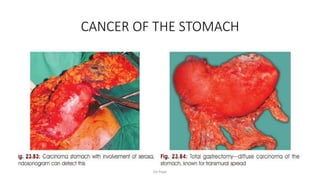
38. Gross Types
1. Cauliflower-like growth with friable tissue. This variety can give rise to
melaena or bleeding causing anaemia.
2. Infiltrative type of lesion (diffuse) with dense submucosal fibrosis
which converts the stomach into a small contracted, functionless
stomach—linitis plastica or leather bottle stomach. Mucosa may
appear normal.
3. Ulcerative variety, with classical everted edges with central slough.
4. Ulcer cancer: carcinoma arising in a preexisting gastric ulcer. In this
variety, complete destruction of the muscle coat is present.
De Pope
39. Continua…………..
5. Colloid carcinoma: In this condition, malignant cells are separated by
colloid material. Common in women and gives rise to Krukenberg’s
tumour—bilateral, bulky ovarian metastasis common in
premenopausal women (signet ring carcinoma produces this).
De Pope
40. Pathology
• 95% of all malignant gastric neoplasms are adenocarcinomas.
• The Lauren system separates gastric adenocarcinoma into intestinal
or diffuse types based on histology.
• Other histologic types include squamous cell carcinoma,
adenoacanthoma, carcinoid tumours, GI stromal tumours, and
lymphoma.
De Pope
42. EARLY GASTRIC CANCER
• Defined as adenocarcinoma limited to the mucosa and submucosa of
the stomach, regardless of lymph node status.
• The entity is common in Japan, where gastric cancer is the number
one cause of cancer death, and where aggressive surveillance
programs have been established.
• Approx 10% of pts with early gastric cancer will have lymph node
metastasis.
• Approx 70% of early gastric cancers are well differentiated and 30%
are poorly differentiated.
De Pope
43. Continua………….
• The overall cure rate with adequate gastric resection and
lymphadenectomy is 95%.
• In some Japanese centres, 50% of the gastric cancers treated are early
gastric cancers.
• In the US, less than 20% of resected gastric adenocarcinomas are
early gastric cancers.
De Pope
45. Criticism for Early Gastric Cancer
• Five-year survival in node negative early gastric cancer is more than
95%. However, it falls to 70% if nodes are positive. Hence, the
suggestion is that node-positive cases should not be included under
early gastric cancer.
De Pope
46. ADVANCED GASTRIC CANCER
• Refers to involvement of muscularis mucosa and/or serosa with or
without involvement of lymph nodes.
• The Borrmann classification system dvlped in 1926 remains useful
today for the description of endoscopic findings.
• The Borrmann system divides gastric carcinoma into five types
depending on the lesion’s macroscopic appearance.
De Pope
47. Continua………..
• Type 1 represents polypoid or fungating lesions
• Type 2 ulcerating lesions surrounded by elevated borders
• Type 3 ulcerating lesions with infiltration into the gastric wall
• Type 4 diffusely infiltrating lesions
• Type 5 lesions that do not fit into any of the other categories.
De Pope
51. WHO CLASSIFICATION: Five Main Categories
1. Adenocarcinoma 95%
• Papillary, tubular
• Mucinous
• Signet ring
2. Adenosquamous cell carcinoma
3. Squamous cell carcinoma
4. Undifferentiated carcinoma
5. Unclassified carcinoma
De Pope
52. Clinical Features of Carcinoma Stomach
(mnemonic: SOLID)
• Very often pts would have vague symptoms— early satiety, flatulence,
discomfort, pain in the upper abdomen.
• Early satiety is due to decreased distensibility of the stomach.
• Anaemia is due to many factors
De Pope
53. Continua…………
• S Silent: Growth is silent but manifests as secondaries in the liver, ascites,
Virchow’s node, rectovesical deposits, (Blumer’s shelf), umbilical nodule
(Sister Mary Joseph’s nodule), left axillary nodes (Irish nodes), palpable
ovarian mass (Krukenberg tumour).
• O Obstruction at pylorus (pyloric antrum) producing pyloric obstruction
with features of vomiting with/ without blood. Visible gastric peristalsis can
also be present. Obstruction at cardio-oesophageal junction produces
dysphagia.
• L Lump in the abdomen which is hard and irregular. Clinically, stomach
mass is differentiated from liver mass by features mentioned below.
• I Insidious in onset: Anaemia, anorexia and asthenia of short duration.
• D Dyspepsia in a man over the age of 40: Carcinoma stomach should be
ruled out. Early gastric cancer presents as dyspepsia.
De Pope
55. Features of Stomach Mass
• Stomach moves with respiration.
• Upper border of the stomach mass can be made out.
• Anatomical location of the mass: Right hypochondrium in a pyloric
mass, epigastrium and left hypochondrium in a mass arising from
body of the stomach.
• Knee elbow position: The mass falls forwards, unless fixed.
• The mass may have intrinsic mobility.
De Pope
56. Spread
1. Penetration of gastric serosa: most important prognostic indicator.
When serosa is NOT penetrated, 50% survive for 5 years after
resection. When serosa is penetrated, this figure drops to 20%.
Once serosa is involved, adjacent organs such as liver, pancreas,
spleen, omentum, transverse colon get involved.
2. Lymphatic spread: 420 lymph nodes have been identified
• Lymph node involvement is a poor prognostic indicator.
• Involvement of 4 or more nodes is less favourable.
De Pope
57. Continua………..
3. Blood spread: Most common sites are liver and lungs. It produces
extensive secondaries. They are signs of inoperability.
4. Transcoelomic spread results in ascites, Krukenberg tumour—
bilateral bulky ovarian deposits and rectovesical deposits (Blumer’s
shelf).
De Pope
62. Investigations
1. CBC: 20% of early gastric cancer patients have iron deficiency
(microcytic, hypochromic) anaemia. Preoperative blood transfusion
may be necessary. Hb-haematocrit.
2. Routine examination, fasting and postprandial sugars, ECG, renal
function for fitness before surgery.
3. Flexible oesophagogastroduodenoscopy: To know the extent of the
lesion, To confirm the diagnosis, To take multiple biopsy—6 pieces,
Also to aid brush cytology.
De Pope
63. Continua………
4. Ultrasound of the abdomen:
• To rule out secondaries in the liver.
• To look for enlarged coeliac nodes.
• Can detect ascites—guided fluid tap and cell cytology.
• To detect Krukenberg tumour (pelvic CT).
• Useful in detecting metastatic disease.
5. CECT of the abdomen, pelvis and chest should be done
• It is superior to ultrasound
• Resectability—specially infiltration into pancreas, retroperitoneal structures
are better appreciated.
• However, detection of peritoneal disease sensibility is only around 30–35%.
De Pope
64. Continua………
6. Endoscopic ultrasonography can detect advanced tumours in 80% of
patients. Overall staging accuracy is about 75%, however, it has
significant limitations for staging mucosal disease. Hence, routinely not
done.
7. Laparoscopy: CT cannot detect liver or peritoneal metastasis
(small<5mm) and small lymph nodes.
Laparoscopy is an ideal investigation. Almost 20 to 30% of so-called
operable cases become inoperable. Laparoscopic peritoneal lavage for
cytology is best test.
De Pope
67. Continua…………..
8. Role of PET scan
• Not routinely used in staging gastric cancer.
• PET scan is done to rule out metastatic disease.
9. CEA: Carcinoembryonic antigen is elevated in about 60–70% pts. It
indicates extensive spread of the disease. Also CA 19-9 (carbohydrate
antigen) and a-FP can be done.
10. Barium meal may show intrinsic, persistent, irregular, filling defect.
Double contrast air-barium study is used for mass screening in Japan to
detect early cases.
De Pope
68. Continua…….....
• Barium meal study useful in cases of linitis plastica wherein mucosa
may appear to be normal in early cases.
• Today use of barium has become almost nil with the availability of
endoscopy.
De Pope
70. Histopathology
• Adenocarcinoma of the stomach. Basically two types of gastric
carcinomas as per Lauren’s classification.
1. Diffuse is more common in young, females and carries poor
prognosis. The leather-bottle stomach or linitis plastica is poorly
differentiated with anaplastic cells.
2. Intestinal is more common in elderly males. It shows areas of
intestinal metaplasia. It has better prognosis.
De Pope
72. Treatment of Carcinoma Stomach
• Surgery is the main modality of the treatment. Adjuvant
chemotherapy only beneficial in a few patient.
• Resectable means the growth can be removed.
• Inoperable means there are no chances of cure but growth may be
resectable.
• Operable means cure is possible.
De Pope
73. Signs of inoperability
• Growth fixed to pancreas or posterior abdominal wall
• Secondaries in the liver, hard nodular liver
• Rectovesical deposits, due to peritoneal seedings which are felt
during per rectal examination
• Enlarged, fixed coeliac nodes, para-aortic nodes and left
supraclavicular nodes
• Krukenberg tumour, malignant ascites
• Sister Mary Joseph’s nodule
De Pope
74. Aims of Surgery
1. Curative resection (R0) should be done whenever possible.
2. Stage IA: Regionally confined disease should undergo primary
surgical R-0 resection. Stage II and III: Neoadjuvant therapy
followed by resection.
3. Stage T1a: Endoscopic resection for tumours ≤2 cm.
4. Bypass procedure (GJ) to relieve vomiting in advanced cases of
pyloric obstruction.
5. Palliative gastrectomy can be done to remove a fungating,
ulcerative, bleeding mass. It gives better palliation.
De Pope
76. Curative Resections
A resection is considered to be curative, if:
• There is no evidence of microscopic or gross residual tumour.
• Serosa is not involved (this means that curative resection is not
possible for T3/T4 tumours).
• There is no evidence of metastatic disease.
• Minimum 5 cm margin is required.
De Pope
78. Extent of Gastrectomy
• Mostly subtotal gastrectomy followed by reconstruction by Bill
Billroth II gastrojejunostomy but if less than 20% of stomach left then
a Roux reconstruction is done.
• Total gastrectomydiscoutraged except when R-0 can be achieved
(proximal gastric adenocarcinoma-jejunal pouch/esophageal
anastosmosis)
• Small tumors <2cm and confined to mucosa with EUS node neg-
endoscopic resection.
De Pope
79. Extent of Lymphadenectomy
• The Japanese have labeled all the lymph node stations which
potentially drain the stomach.
• Generally grouped into level N1 (e.g., stations 1–6), level N2 (e.g.,
stations 7–11), and level N3 (e.g., stations 12–16) nodes.
• The nodal stations level N1, N2, and N3 varies depending on the
location of the tumor.
• General, N1 nodes are within 3 cm of the tumor, N2 nodes are along
the celiac branches and N3 nodes are the most distant from the
tumor (portal triad, retropancreatic, mesenteric root, middle colic,
para-aortic).
De Pope
80. Continua……….
• D1 resection: removes the tumor and the N1 nodes.
• D2 gastrectomy: extensive lymphadenectomy (removal of N1 and N2
nodes). In addition to the tissue removed in a D1 resection, the
standard D2 gastrectomy removes the peritoneal layer over the
pancreas and anterior mesocolon, along with nodes along the hepatic
and splenic arteries, and the crural nodes.
• Note: minimum of 15 LNs should be resected with gastrectomy to
avoid understaging.
De Pope
82. Carcinoma of Pyloric Antrum and Distal Body
of the Stomach
• Radical subtotal gastrectomy which includes the removal of 60–70%
of the stomach, greater omentum along with enlarged lymph nodes
(N1) followed by gastrojejunal anastomosis is the treatment of choice.
De Pope
83. Carcinoma of Proximal Stomach and Diffuse
Carcinoma
• Oesophagogastrectomy: Removal of the entire stomach, lower end of
oesophagus, with regional lymph nodes, followed by
oesophagojejunal anastomosis.
De Pope
84. Palliative Surgery
• Carcinoma pyloric antrum (inoperable): Palliative anterior GJ is done
to relieve vomiting, by anastomosing a jejunal loop to the stomach in
the prepyloric region. If posterior GJ is done, the growth may involve
the GJ stoma early resulting in stomal obstruction. With anterior GJ,
entero-enterostomy can be added to prevent bilious vomiting.
• Palliative gastrectomy to get rid of ulcerated, necrotic or bleeding
lesion.
• Endoscopic palliation: Thermal photodestruction by laser.
De Pope
86. Endoscopic Mucosal Resection
• This is indicated in early gastric cancer confined to mucosa.
• The cancer should be less than 2 cm and there should not be node
enlargement.
• Ideally cancer should be elevated variety and well differentiated.
• Normal saline is injected into submucosal plane and lesion gets
elevated.
• It is excised with 1 cm margin up to muscularis propria at a deeper
plane.
De Pope
87. Endoluminal Gastric Surgery
• Small, high up lesions are ideal.
• Here, laparoscopic instrumentation is done under endoscopic
guidance.
• Stomach is suitable for endoluminal surgery because it can be
distended and contents are sterile.
De Pope
88. Paraneoplastic Syndromes Associated with
Carcinoma Stomach
• Trousseau’s syndrome—Thrombophlebitis
• Acanthosis nigricans—Hyperpigmentation of the axilla and groin.
• Peripheral neuropathy.
De Pope
89. THE ADJUVANT TREATMENT
• Chemotherapy: Injection 5-FU (fluorouracil) 500 mg IV daily for five
days, every 28 days. It can be given by IV infusion or IV bolus over 15
minutes/ combination with adriamycin, mitomycin and cisplatin
• Intraperitoneal mitomycin and mitomycin C— impregnated charcoal
have also been used (target the recurrence site—gastric bed).
• Postoperative chemotherapy
• Chemoradiotherapy
• Immunochemotharapy
De Pope
91. REFERENCES
• Bailey and Love’s short practice of surgery 27th edition
• Manipal Manual of Surgery, 5th edition
• Schwartz principles of surgery 10th edition
Knowledge must be free for all...
De Pope