3. Introduction
• Myocardial infarction (MI) (i.e heart attack) is the irreversible
necrosis of the heart muscle secondary to prolonged ischaemia.
Patients with ischemic heart disease fall into two large groups:
1. patients with chronic coronary artery disease (CAD) who most
commonly present with stable angina and
2. patients with acute coronary syndromes (ACSs).
The latter group, in turn, is composed of patients with acute
myocardial infarction (MI) with ST-segment elevation on their
presenting electrocardiogram (STEMI) and those with unstable angina
and non-ST-segment elevation MI (UA/NSTEMI)
4. Impact
• Every year in the United States, ∼1.3 million patients are admitted to
hospitals with UA/NSTEMI in comparison with ∼300,000 patients with acute
STEMI.
• The relative incidence of UA/NSTEMI in comparison with STEMI appears to
be increasing.
• Almost one-half of patients with UA/NSTEMI are women, while more than
three-fourths of patients with STEMI are men.
• In Ug CVD accounts for 10% of all deaths making it the most common
noncommunicable cause of death in the country.
• The burden has been increasing with the number of deaths attributed to IHD
increasing by 13.4% btn 2007 and 2017. (Ssinabulya et al., 2020)
5. Causes of Acute Coronary
Syndromes
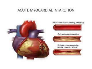
• Atherosclerotic plaque rupture with
superimposed thrombus/95%/
• Vasculitic syndromes
• Coronary embolism (e.g., from endocarditis, artificial
heart valves)
• Congenital anomalies of the coronary arteries
• Coronary trauma or aneurysm
• Severe coronary artery spasm (primary or cocaine-
induced)
• Increased blood viscosity (e.g.,
polycythemia vera, thrombocytosis)
• Spontaneous coronary artery dissection
• Markedly increased myocardial oxygen demand (e.g.,
severe
aortic stenosis)
6. Risk factors
• American Heart Association guide to risk
factors for coronary artery disease.
• Resource:“Cardiology explained”
7. Risk factors
European Society of Cardiology table of lifestyles and characteristics
associated with an increased risk of a future coronary heart disease
event.
Resource: “Cardiology explained”
8. UNSTABLE ANGINA AND NON-ST-ELEVATION
MYOCARDIAL INFARCTION
• Diagnosis of UA: based largely on the clinical presentation. Stable
angina pectoris is xterised by chest or arm discomfort that may not
be described as pain but is reproducibly associated with physical
exertion or stress and is relieved within 5–10 min by rest and/or
sublingual nitroglycerin.
• UA is defined as angina pectoris or equivalent ischemic discomfort
with at least one of three features: (1) it occurs at rest (or with
minimal exertion), usually lasting >10 min; (2) it is severe and of new
onset (i.e., within the prior 4–6 weeks); and/or (3) it occurs with a
crescendo pattern (i.e., distinctly more severe, prolonged, or
frequent than previously).
9. Continua…………
• The diagnosis of NSTEMI is established if a patient with the clinical
features of UA develops evidence of myocardial necrosis, as reflected
in elevated cardiac biomarkers.
10. PATHOPHYSIOLOGY
• UA/NSTEMI is most commonly caused by a reduction in oxygen supply
and/or by an increase in myocardial oxygen demand superimposed on an
atherosclerotic coronary plaque, with varying degrees of obstruction.
• Four pathophysiologic processes that may contribute to the development of
UA/NSTEMI have been identified:
(1) plaque rupture or erosion with superimposed nonocclusive thrombus,
believed to be the most common cause—NSTEMI may occur with
downstream embolization of platelet aggregates and/or atherosclerotic
debris;
(2) dynamic obstruction [e.g., coronary spasm, as in Prinzmetal’s variant
angina;
11. Continua…….
(3) progressive mechanical obstruction [e.g., rapidly advancing
coronary atherosclerosis or restenosis following percutaneous
coronary intervention (PCI)];
(4) secondary UA related to increased myocardial oxygen demand
and/or decreased supply (e.g., tachycardia, anemia). More than one of
these processes may be involved.
• Among patients with UA/NSTEMI studied at angiography, ∼5% have
left main stenosis, 15% have three-vessel CAD, 30% have two-vessel
disease, 40% have single vessel disease.
12. Continua…………..
• and 10% have no critical coronary stenosis; some of the latter have
Prinzmetal’s variant angina.
13. CLINICAL PRESENTATION
History and Physical Examination:
• The clinical hallmark of UA/NSTEMI is chest pain, typically located in the
substernal region or sometimes in the epigastrium radiates to the neck, left
shoulder, and left arm.
• The discomfort is usually severe enough to be considered painful. Anginal
“equivalents” such as dyspnea and epigastric discomfort may also occur, and
these appear to occur more often in women.
• The examination resembles that in patients with stable angina and may be
unremarkable. If the patient has a large area of myocardial ischemia or a
large NSTEMI, the physical findings can include diaphoresis, pale cool skin,
sinus tachycardia, a third and/or fourth heart sound, basilar rales, and
sometimes hypotension, resembling the findings of large STEMI.
14. Electrocardiogram
• In UA, ST-segment depression, transient ST-segment elevation,
and/or T-wave inversion occur in 30–50% of patients, depending on
the severity of the clinical presentation.
• In patients with the clinical features of UA, the presence of new ST-
segment deviation, even of only 0.05 mV, is an important predictor of
adverse outcome.
• T-wave changes are sensitive for ischemia but less specific, unless
they are new, deep T-wave inversions (≥0.3 mV).
15. Cardiac Biomarkers
• Patients with UA who have elevated biomarkers of necrosis, such as CK-MB
and troponin (a much more specific and sensitive marker of myocardial
necrosis), are at increased risk for death or recurrent MI.
• Elevated levels of these markers distinguish patients with NSTEMI from
those with UA.
• There is a direct relationship between the degree of troponin elevation and
mortality.
• However, in patients without a clear clinical history of myocardial ischemia,
minor troponin elevations have been reported and can be caused by
congestive heart failure, myocarditis, or pulmonary embolism, or they may
be false-positive readings.
• Thus, in patients with an unclear history, small troponin elevations may not
be diagnostic of an ACS.
16. DIAGNOSTIC EVALUATION
• The first step in evaluating patients with possible UA/NSTEMI is to
determine the likelihood that CAD is the cause of the presenting
symptoms.
• The American College of Cardiology/American Heart Association
(ACC/AHA) Guidelines include, among the factors associated with a
high likelihood of ACS, a clinical history typical of ischemic
discomfort, a history of established CAD by angiography, prior MI,
congestive heart failure, new electrocardiographic (ECG) changes, or
elevated cardiac biomarkers.
17. Continua,………….
• Factors associated with an intermediate likelihood of ACS in patients
with the clinical features of this condition but without the above
high-risk factors are: age >70 years, male gender, diabetes mellitus,
known peripheral arterial or cerebrovascular disease, and old ECG
abnormalities
18. Diagnostic Pathways
• Four major diagnostic tools are used in the diagnosis of UA/NSTEMI
in the ED: the clinical history, the ECG, cardiac markers, and stress
testing.
• The goals are to (1) recognize or exclude MI (using cardiac markers),
(2) evaluate for rest ischemia (chest pain at rest, serial or continuous
ECGs), and (3) evaluate for significant CAD (using provocative stress
testing).
• Typical pathways begin with assessment of the likelihood that the
presenting symptoms are due to ischemia. Patients with a low
likelihood of ischemia are usually managed with an ED-based critical
pathway (which in some institutions is carried out in a “chest pain
unit”.
19. Continua……….
• Evaluation of such patients includes clinical monitoring for recurrent
ischemic discomfort, serial ECGs, and cardiac markers, typically
performed at baseline and at 4–6 h and 12 h after presentation.
• If new elevations in cardiac markers (CK-MB and/or troponin) or ECG
changes are noted, the patient is admitted to the hospital. If the
patient remains painfree and the markers are negative, the patient
may go on to stress testing.
• This may be performed as early as 6 h after presentation in the ED or
chest pain center, or on an outpatient basis within 72 h.
20. Continua…………..
• For most patients, standard treadmill ECG stress testing is used, but
for patients with fixed abnormalities on the ECG (e.g., left bundle
branch block), perfusion or echocardiographic imaging is used.
• For patients who cannot walk, pharmacologic stress is used.
• By demonstrating normal myocardial perfusion sestamibi or thallium
imaging can reduce unnecessary hospitalizations by excluding acute
ischemia. Or CT angiography to exclude obstructive CAD
22. RISK STRATIFICATION AND PROGNOSIS
• Patients with documented UA/NSTEMI exhibit a wide spectrum of
early (30 days) risk of death, ranging from 1–10%, and of new or
recurrent infarction of 3–10%.
• Assessment of “global risk” can be accomplished by clinical risk
scoring systems such as that developed from in the Thrombolysis in
Myocardial Infarction (TIMI) Trials, which includes seven independent
risk factors: age ≥ 65 years, three or more risk factors for CAD,
documented CAD at catheterization, development of UA/NSTEMI
while on aspirin, more than two episodes of angina within the
preceding 24 h, ST deviation ≥0.5 mm, and an elevated cardiac
marker.
23.
24. Continua………
• Other risk factors: diabetes mellitus, left ventricular dysfunction, and
elevated levels of creatinine, atrial natriuretic peptides, and C-
reactive protein.
• Early risk assessment (especially using troponin, ST-segment changes,
and/or a global risk scoring system) is useful both in predicting the
risk of recurrent cardiac events and in identifying those patients who
would derive the greatest benefit from antithrombotic therapies
more potent than unfractionated heparin, such as low-
molecularweight heparin (LMWH) and glycoprotein (GP)IIb/IIIa
inhibitors, and from an early invasive strategy.
25. Treatment: UNSTABLE ANGINA AND NON-ST
ELEVATION MYOCARDIAL INFARCTION
• Management is directed toward (1) reducing myocardial oxygen
demands; (2) improving myocardial oxygen supply; and (3) assessing
the patient's risk of progression to myocardial infarction or having a
complication related to treatment.
• Patients with unstable angina require admission to the hospital for
bed rest with continuous telemetry monitoring.
• Obtain intravenous (IV) access, and provide supplemental oxygen if
evidence of desaturation is noted.
• Because the course of unstable angina is highly variable and
potentially life-threatening, the aggressiveness of the therapeutic
approach must be established expeditiously.
26. Continua……….
• NOTE: An invasive strategy refers to the routine use of cardiac
catheterization with possible revascularization, and a ischemia-
guided strategy refers to initial medical mgnt with the possible use of
cardiac catheterization if indicated by failure of medical therapy or
objective evidence of ischemia (dynamic ECG changes or abnormal
noninvasive stress test results).
• Determination of the preferred strategy depends on the patient’s
clinical characteristics and clinical risk.
27. Continua……….
• Specific therapy for primary causes of ischemia should be directed at
each pathophysiologic origin of unstable angina: increased
myocardial rate-pressure product, coronary vasoconstriction, platelet
aggregation, and thrombosis
28. Indications for intensive care
ICU or emergency revascularization disposition is indicated by the
following:
• TIMI (Thrombolysis In Myocardial Infarction) risk score of 3-7
• New ECG changes in 2 or more leads
• ST elevation greater than 1 mm or Q waves 0.04 seconds or longer
• ST depression greater than 1 mm or T-wave inversion in the context
of angina
• New left bundle branch block
• Signs and symptoms of incipient or florid heart failure
29. Continua………..
• Syncope or sudden death presentation
• Serious new arrhythmias, including second-degree or complete heart
block and ventricular tachyarrhythmias
• Refractory angina
• Hypoxia
• Positive cardiac enzymes (creatine kinase [CK] or troponin)
• Myocardial infarction or coronary stenting within the last 2 weeks
30. Indications for immediate care
Patients are admitted to intermediate care units when they are
asymptomatic but have any of the following conditions:
• Atrial arrhythmia, supraventricular tachycardia, or low-grade second-
degree atrioventricular block
• Isolated basilar rales
• Borderline blood pressure
• Symptoms with minimal activity
• Presence of major comorbidity (eg, severe pulmonary, renal, or
hepatic disease; bleeding history; or dyscrasia)
• Very advanced age or frailty
31. Indications for observation
• Patients who are otherwise healthy without ischemic ECG changes
but who have either of the following should be admitted to
observation units:
• New-onset symptoms at moderate levels of exertion
• Known coronary artery disease (CAD) with a presentation that does
not suggest true worsening but for which further observation is
thought to be prudent.
32. Medical management of adverse events
• Medications that provide symptomatic relief but that have not been
shown to affect long-term major events: nitrates (eg, nitroglycerin
IV), calcium channel blockers (eg, diltiazem, verapamil), and heparin.
• Medications that have been convincingly shown to be capable of
reducing short- or long-term adverse events are as follows:
i. Aspirin
ii. Clopidogrel
iii. Beta-adrenergic blocking agents
iv. Lipid-lowering agents (statins)
v. Angiotensin-converting enzyme (ACE) inhibitors
vi. Glycoprotein (GP) IIb/IIIa antagonists
33. Diet
• NPO if stress testing or an invasive procedure is anticipated.
Otherwise, a diet low in cholesterol and saturated fat is
recommended. Sodium restriction for patients with heart failure or
hypertension.
34. Immediate patient management
• Focus on stabilizing the patient's condition, relieving ischemic pain,
and providing antithrombotic therapy to reduce myocardial damage
and prevent further ischemia.
• Morphine (or fentanyl) for pain control, oxygen, sublingual or
intravenous (IV) nitroglycerin, soluble aspirin 162-325 mg, and
clopidogrel with a 300- to 600-mg loading dose are given as initial
treatment.
• In complete vessel occlusion without collateralization of the infarct-
related vessel, there is less utility for nitrates.
35. Continua………
HIGH RISK PATIENTS INTERMEDIATE RISK PATIENTS LOW RISK PATIENTS
aggressive care, including
aspirin, clopidogrel,
unfractionated heparin or
low–molecular-weight
heparin (LMWH), IV
platelet glycoprotein
IIb/IIIa complex blockers
(eg, tirofiban,
eptifibatide), and a beta
blocker.
The goal is early
revascularization
rapidly undergo diagnostic
evaluation and further
assessment to determine
their appropriate risk
category.
should undergo further
follow-up with biomarkers
and clinical assessment.
Optimal medical therapies
include use of standard
medical therapies,
including beta blockers,
aspirin, and
unfractionated heparin or
LMWH.
36. Key points to note:
• Monitor and immediately treat arrhythmias in the first 48 hours. Pay
attention to exacerbating factors, eg disturbances in electrolytes
(especially potassium and magnesium), hypoxemia, drugs, or
acidosis. Correct these factors accordingly.
• Humidified oxygen may reduce the risk of nosebleeds in patients with
ACS who are receiving antiplatelet and antithrombin therapy.
• Do not administer nitrates if the patient is hypotensive (systolic BP <
90 mm Hg); if RV infarction, large pericardial effusion, or severe
aortic stenosis is suspected; or if the patient recently received
phosphodiesterase-5 inhibitors (eg, sildenafil).
37. Continua………..
• Patients with known hypersensitivity to antiplatelet agents, active
internal bleeding, and bleeding disorders should not receive
antiplatelet or antithrombotic therapy.
• Some patients with intractable chest pain or severe hypotension may
require the insertion of an intra-aortic balloon pump.
• Congestive heart failure (CHF) can be due to systolic dysfunction or
diastolic dysfunction in the setting of myocardial infarction.
Aggressive treatment-indicated to prevent worsening of the
situation.
38. Continua…….
• Patients presenting with cardiogenic shock should undergo
percutaneous coronary intervention (PCI) as soon as possible.
• Recurrent ischemia may be due to incomplete reperfusion.
39. Initial Medical Management
• Aspirin: Administer chewable aspirin 162-325 mg promptly to patients who
are not at high risk for bleeding, who do not have ongoing bleeding, or who
do not have true intolerance or allergy.
• Peak effect within 30mins, . Patients with unstable angina/non–ST-segment
elevation myocardial infarction (UA/NSTEMI) should continue indefinitely on
aspirin, if tolerated.
• Beta-adrenergic blocking agents: decreases in ischemic symptoms and in the
occurrence of MIs.
• Balance Benefits/potential complications ( heart failure or cardiogenic shock
that have been demonstrated when beta blockers are used in
hemodynamically compromised patients). Oral beta blockers (eg,
metoprolol) are preferred to IV agents.-more risk of cardiogenic shock
40. Continua…………
• PSY12 inhibitors (thienopyridines [clopidogrel, prasugrel],
nonthienopyridines [ticagrelor]): Clopidogrel is recommended as the
antiplatelet of choice in patients who are intolerant to aspirin. Can
also used as an adjunctive in conjunction with aspirin (dual
antiplatelet therapy).
• A loading dose of 600 mg may offer more effective platelet inhibition
than one of 300 mg; increasing the loading dose beyond 600 mg has
not shown benefit.
• Carries high risk of bleeding post CABG
41. Continua……..
• Prasugrel is more effective than clopidegrol bt with higher risk of
bleeding, potentially harmful as part of a dual-platelet regimen in
patients with a stroke history for whom PCI is planned.
• GP IIb/IIIa antagonists: Ticagrelor reduces the rate of thrombotic CV
events following ACS, reduces the rate of stent thrombosis.
Others abciximab, eptifibatide, and tirofiban) have been shown to
increase the safety of acute PCI,
• Heparin: use of low-molecular-weight heparin (LMWH) and the use
of IV unfractionated heparin (UFH) is comparable, benefits of using
LMWH include lower bleeding rates, reduced costs, and decreased
incidence of heparin-induced thrombocytopenia.
42. Continua………
• Direct thrombin inhibitors: as hirudin, lepirudin (recombinant
hirudin), and bivalirudin, are potential alternatives to heparin. Have
higher bleeding risks.
• Nitrates: IV nitrate agents may be used in the treatment of ischemic
chest pain, symptoms of heart failure, or hypertension, not
associated with appreciable long-term clinical benefit. Nitrate agents
are contraindicated for patients with right ventricular
infarction, hypertrophic cardiomyopathy (HOCM), and severe aortic
stenosis.
• Additional management of unstable angina includes the use of statins
(lipid-lowering agents) and ACE inhibitors, CCB and
antibioticsagainst Chlamydia pneumoniae, and fibrinolytic agents no
established role in the setting of unstable angina.
43. Cardiac Catheterization
• Patients with unstable angina and the following clinical xtics should
be referred for immediate cardiac catheterization:
• Cardiogenic shock
• Severe left ventricular dysfunction
• Angina refractory to medical therapy
• Acute mitral regurgitation
• New ventricular septal defect
• Unstable tachyarrhythmias
44. Revascularization
• Patients at moderate to high risk for adverse events: ST depression
greater than 1 mm on ECG, troponin positivity or non–Q-wave
myocardial infarction (NQMI), or chest pain refractory to medical
therapy, should be scheduled for cardiac catheterization with likely
revascularization within the next 48 hours.
45. Prevention
• Smoking ceseation
• Lipid lowering
-The target is an LDL-C level of 70 mg/dL or lower, a high-density
lipoprotein cholesterol (HDL-C) level higher than 35 mg/dL, and a
triglyceride level below 200 mg/dL.
• Control of hypertension
-The target blood pressure is below 140/90 mm Hg or below 130/80
mm Hg if the patient has diabetes mellitus or 125/75mmHg chronic
kidney disease
• Diabetes mellitus management
46. Continua…..
• Psychosocial management
• Weight management and nutritional counseling
-The target body mass index (BMI) is below 25 kg/m2, in conjunction
with a waist circumference of less than 40 inches in men and of less
than 35 inches in women
• Activity management
-Patients at risk for MI should avoid sudden strenuous activities,
especially in cold weather (eg, shoveling snow).
47. LONG-TERM MANAGEMENT
• Lifestyle modifications as above
• Drugs with long-term benefits: beta blockers are appropriate anti-
ischemic therapy and may help decrease triggers for MI. Statins (at a
high dose, e.g., atorvastatin 80 mg/d) and ACE inhibitors are
recommended for long-term plaque stabilization.
• Antiplatelet therapy, now recommended to be the combination of
aspirin and clopidogrel for at least 9–12 months, with aspirin
continued thereafter, prevents or reduces the severity of any
thrombosis that would occur if a plaque does rupture
50. Continua……….
• STEMI usually occurs when coronary blood flow decreases abruptly
after a thrombotic occlusion of a coronary artery previously affected
by atherosclerosis.
• STEMI occurs when a coronary artery thrombus develops rapidly at a
site of vascular injury. The injury is produced or facilitated by factors
such as cigarette smoking, hypertension, and lipid accumulation.
51. Continua………..
• In most cases, STEMI occurs when the surface of an atherosclerotic
plaque becomes disrupted (exposing its contents to the blood) and
conditions (local or systemic) favor thrombogenesis.
• In rare cases STEMI may be due to coronary artery occlusion caused
by coronary emboli, congenital abnormalities, coronary spasm, and a
wide variety of systemic—particularly inflammatory—diseases.
52. Continua…………
• The amount of myocardial damage caused by coronary occlusion
depends on:
1. The territory supplied by the affected vessel;
2. Whether or not the vessel becomes totally occluded;
3. The duration of coronary occlusion;
4. The quantity of blood supplied by collateral vessels to the affected
tissue;
53. Continua…………….
• Patients at increased risk of developing STEMI include those with
multiple coronary risk factors and those with unstable angina.
• Less common underlying medical conditions predisposing patients to
STEMI include hypercoagulability, collagen vascular disease, cocaine
abuse, and intracardiac thrombi or masses that can produce coronary
emboli.
54. CLINICAL PRESENTATION
• In up to one-half of cases, a precipitating factor appears to be present
before STEMI, such as vigorous physical exercise, emotional stress, or
a medical or surgical illness.
• Although STEMI may commence at any time of the day or night,
circadian variations have been reported such that clusters are seen in
the morning within a few hours of awakening.
• Pain is the most common presenting complaint in patients with
STEMI. The pain is deep and visceral; nature heavy, squeezing, and
crushing, although occasionally it is described as stabbing or burning.
It is similar in character to the discomfort of angina pectoris but
commonly occurs at rest, is usually more severe, and lasts longer.
55. Continua……….
• Typically the pain involves the central portion of the chest and/or the
epigastrium, and on occasion it radiates to the arms.
• Less common sites of radiation include the abdomen, back, lower
jaw, and neck.
• The frequent location of the pain beneath the xiphoid and
epigastrium and the patients’ denial that they may be suffering a
heart attack are chiefly responsible for the common mistaken
impression of indigestion.
• The pain of STEMI may radiate as high as the occipital area but not
below the umbilicus.
56. Continua………..
• It is often accompanied by weakness, sweating, nausea, vomiting,
anxiety, and a sense of impending doom.
• The pain may commence when the patient is at rest, but when it
begins during a period of exertion, it does not usually subside with
cessation of activity, in contrast to angina pectoris.
• The pain of STEMI can simulate pain from acute pericarditis,
pulmonary embolism, acute aortic dissection, costochondritis, and
gastrointestinal disorders.
57. Continua……….
• These conditions should therefore be considered in the differential
diagnosis.
• Radiation of discomfort to the trapezius is not seen in patients with
STEMI and may be a useful distinguishing feature that suggests
pericarditis is the correct diagnosis.
• However, pain is not uniformly present in patients with STEMI. The
proportion of painless STEMIs is greater in patients with diabetes
mellitus, and it increases with age.
58. Continua………..
• In the elderly, STEMI may present as sudden-onset breathlessness,
which may progress to pulmonary edema.
• Other less common presentations, with or without pain, include
sudden loss of consciousness, a confusional state, a sensation of
profound weakness, the appearance of an arrhythmia, evidence of
peripheral embolism, or merely an unexplained drop in arterial
pressure.
59. PHYSICAL FINDINGS
• Most patients are anxious and restless, attempting unsuccessfully to
relieve the pain by moving about in bed, altering their position, and
stretching.
• Pallor associated with perspiration and coolness of the extremities
occurs commonly. The combination of substernal chest pain
persisting for >30 min and diaphoresis strongly suggests STEMI.
• Although many patients have a normal pulse rate and blood pressure
within the first hour of STEMI, about one-fourth of patients with
anterior infarction have manifestations of sympathetic nervous
system hyperactivity (tachycardia and/or hypertension), and up to
one-half with inferior infarction show evidence of parasympathetic
hyperactivity (bradycardia and/or hypotension).
60. Continua…………
• The precordium is usually quiet, and the apical impulse may be
difficult to palpate.
• In patients with anterior wall infarction, an abnormal systolic
pulsation caused by dyskinetic bulging of infarcted myocardium may
develop in the periapical area within the first days of the illness and
then may resolve.
• Other physical signs of ventricular dysfunction include fourth and
third heart sounds, decreased intensity of the first heart sound, and
paradoxical splitting of the second heart sound.
61. Continua………
• A transient midsystolic or late systolic apical systolic murmur due to
dysfunction of the mitral valve apparatus may be present.
• A pericardial friction rub is heard in many patients with transmural
STEMI at some time in the course of the disease, if they are examined
frequently. The carotid pulse is often decreased in volume, reflecting
reduced stroke volume.
• Temperature elevations up to 38°C may be observed during the first
week after STEMI.
• The arterial pressure is variable; in most patients with transmural
infarction, systolic pressure declines by ∼10–15 mmHg from the
preinfarction state.
62. LABORATORY FINDINGS
• Myocardial infarction (MI) progresses through the following temporal stages:
1. acute (first few hours–7 days);
2. healing (7–28 days); and
3. healed (≥29 days).When evaluating the results of diagnostic tests for
STEMI, the temporal phase of the infarction must be considered.
4. The laboratory tests of value in confirming the diagnosis may be divided
into four groups: (1) ECG; (2) serum cardiac biomarkers; (3) cardiac
imaging; and (4) nonspecific indices of tissue necrosis and inflammation.
63. ELECTROCARDIOGRAM
• The ECG is a cornerstone in the diagnosis of acute and chronic
ischemic heart disease.
• The findings depend on several key factors: the nature of the process
[reversible (i.e., ischemia) versus irreversible (i.e., infarction)], the
duration (acute versus chronic), extent (transmural versus
subendocardial), and localization (anterior versus inferoposterior), as
well as the presence of other underlying abnormalities (ventricular
hypertrophy, conduction defects).
64. Continua……..
• Ischemia exerts complex time-dependent effects on the electrical
properties of myocardial cells.
• Severe, acute ischemia lowers the resting membrane potential and
shortens the duration of the action potential. Such changes cause a
voltage gradient between normal and ischemic zones.
• As a consequence, current flows between these regions. These
currents of injury are represented on the surface ECG by deviation of
the ST segment
65. Continua………
• When the acute ischemia is transmural, the ST vector is usually
shifted in the direction of the outer (epicardial) layers, producing ST
elevations and sometimes, in the earliest stages of ischemia, tall,
positive so-called hyperacute T waves over the ischemic zone.
• With ischemia confined primarily to the subendocardium, the ST
vector typically shifts toward the subendocardium and ventricular
cavity, so that overlying (e.g., anterior precordial) leads show ST-
segment depression (with ST elevation in lead aVR).
• Multiple factors affect the amplitude of acute ischemic ST deviations.
Profound ST elevation or depression in multiple leads usually
indicates very severe ischemia.
66. Continua……
• From a clinical viewpoint, the division of acute myocardial infarction
into ST segment elevation and non-ST elevation types is useful since
the efficacy of acute reperfusion therapy is limited to the former
group.
• The ECG leads are more helpful in localizing regions of ST elevation
than non-ST elevation ischemia. Eg, acute transmural anterior
(including apical and lateral) wall ischemia is reflected by ST
elevations or increased T-wave positivity in one or more of the
precordial leads (V1–V6) and leads I and aVL.
• Inferior wall ischemia produces changes in leads II, III, and aVF.
67. Continua………
• Posterior wall ischemia may be indirectly recognized by reciprocal ST
depressions in leads V1–V3. Prominent reciprocal ST depressions in
these leads also occur with certain inferior wall infarcts, particularly
those with posterior or lateral wall extension.
• Right ventricular ischemia usually produces ST elevations in right-
sided chest leads.
• Patients with ischemic chest pain who present with deep T-wave
inversions in multiple precordial leads (e.g.,V1–V4) with or without
cardiac enzyme elevations typically have severe obstruction in the
left anterior descending coronary artery system
72. SERUM CARDIAC BIOMARKERS
• Certain proteins, called serum cardiac biomarkers, are released from
necrotic heart muscle after STEMI.
• Cardiac biomarkers become detectable in the peripheral blood once
the capacity of the cardiac lymphatics to clear the interstitium of the
infarct zone is exceeded and spillover into the venous circulation
occurs.
• The temporal pattern of protein release is of diagnostic importance,
but contemporary urgent reperfusion strategies necessitate making a
decision (based largely on a combination of clinical and ECG findings)
before the results of blood tests have returned from the laboratory.
73.
74. Cardiac imaging
• Abnormalities of wall motion on two-dimensional echocardiography
are almost universally present.
• Can’t distinguish acute STEMI from an old myocardial scar or from
acute severe ischemia by echocardiography, Echocardiographic
estimation of left ventricular (LV) function is useful prognostically;
detection of reduced function serves as an indication for therapy
with an inhibitor of the renin-angiotensinaldosterone system.
• Echocardiography may also identify the presence of right ventricular
(RV) infarction, ventricular aneurysm, pericardial effusion, and LV
thrombus. In addition, Doppler echocardiography is useful in the
detection and quantitation of a ventricular septal defect and mitral
regurgitation, two serious complications of STEMI.
75. Prehospital Care and Initial Management
• All patients being transported for chest pain should be managed as if
the pain is ischemic in origin, unless clear evidence to the contrary is
established.
• If available, an advanced cardiac life support (ACLS)–trained unit
should transport patients with hemodynamic instability or
respiratory difficulty
• Notification of the Emergency department
76. Specific prehospital care includes the following:
• Intravenous access, supplemental oxygen if the oxygen saturation
(SaO2) is less than 90%, pulse oximetry
• Immediate administration of nonenteric-coated chewable aspirin en
route
• Nitroglycerin for active chest pain, given sublingually or by spray
• Telemetry and prehospital electrocardiography (ECG), if available
77. Continua…………
• Most deaths caused by MI occur early and are attributable to primary
ventricular fibrillation (VF).
• Hence , initial objectives are immediate ECG monitoring; electric
cardioversion in cases of VF; and rapid transfer of the patient to
facilitate prompt coronary assessment.
78. Emergency Department Care and In-Hospital
Management
• Triage and evaluation
Initial management
• Aims:
Restoration of the balance between oxygen supply and demand to prevent
further ischemia
Pain relief
Prevention and treatment of complications
• Oxygen:
Continuous oxygen saturation monitoring by pulse oximetry is needed for all
patients.
Supplemental oxygen by a mask or nasal cannula is indicated only for
patients who are breathless, hypoxic (oxygen saturation < 90% or PaO2< 60
mm Hg ), or who present with heart failure.
79. Continua………
• Aspirin:
All patients presenting with ACS should receive nonenteric-coated chewable
aspirin in a dose of at least 162 to 325 mg, unless there is a clear history of
aspirin allergy.
Patients with aspirin intolerance still should receive aspirin at presentation.
Reduction of cardiac pain
• Nitrates:
0.4 mg dose in a sublingual tablet, followed by close observation of the
effect on chest pain and the hemodynamic response.
IV nitrates at a dose of 5 to 10 µg/min for persistent pain and gradually
increased until relief of chest pain is achieved.
80. Continua………
• Avoid nitrates in: marked hypotension or bradycardia, or if there is
suspicion of right ventricular infarction, history of concomitant use of
phosphodiesterase (PDE) inhibitors (eg, sildenafil) has occurred
within the last 24 to 72 hours.
Analgesia
• Refractory or severe pain should be treated symptomatically with IV
morphine.
• Initial dose of morphine 2 to 4 mg as an IV bolus can be given, with
increments of 2 to 4 mg repeated every 5 to 10 minutes until the pain
is relieved or intolerance is manifested by hypotension, vomiting, or
depressed respiration. Keep SBp above 100mmhg and below
140mmhg. Avoid NSAIDs
81. Continua……
• Management of ST-elevation myocardial infarction (MI) (STEMI) relies
on two essential and key components: rapid recognition and timely
reperfusion.
• Reperfusion: within 12hrs, mechanical intervention (primary PCI) or
pharmacologic
84. Continua………..
• Anticoagulation
unfractionated heparin (UFH), bivalirudin, and low molecular weight heparin
(LMWH) (eg, enoxaparin ) Fondaparinux is not used (increased risk of
catheter thrombosis)
• Antiplatelet agents
All patients with STEMI should receive an empiric loading dose of aspirin
(150.5 to 325 mg) as early as possible and prior to reperfusion, regardless of
the reperfusion method. A lifelong maintenance dose of (75 to 81 mg) daily
should be prescribed to all patients after STEMI.
For patients undergoing primary PCI, a loading dose of 600 mg of clopidogrel,
180 mg of ticagrelor, or 60 mg of prasugrel should be given as early as
possible or at the time of primary PCI.