
Recommended
More Related Content
Similar to Transplant Surgery Guide
Similar to Transplant Surgery Guide (20)
More from musayansa
More from musayansa (20)
Recently uploaded
Recently uploaded (20)
Transplant Surgery Guide
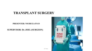
- 1. TRANSPLANT SURGERY PRESENTER: NSUBUGA IVAN SUPERVISOR: Dr. JOEL (SURGEON) De Pope
- 2. Introduction: Transplantation; Transfer of living cells, tissues and organs from one part of the body to another or from one individual to another. De Pope
- 3. INDICATIONS • When an organ or tissue becomes irreparably damaged (disease or injury) • When congenitally defective or absent. De Pope
- 4. TYPES OF TRANSPLANT; • Orthotopic transplants; Implantation in the same anatomic location in the recipient as it was in the donor. Require the removal of the diseased organ (heart, lungs, liver, or intestine). • Heterotopic transplants; implantation in another anatomic location. The diseased organ is kept in place (kidney, pancreas). • Autotransplant; From one body site to another in the same individual, no immunosuppression is required. Eg; skin • Allotransplant; Between genetically different individuals from the same species . • Isotransplant: Between isogenic individuals e.g identical twins • Xenotransplant; Between members of different species (still experimental). De Pope
- 5. GRAFT REJECTION • Allografts provoke a powerful immune response that results in rapid graft rejection unless immunosuppressive therapy is given. • Reason-allelic differences at polymorphic genes that give rise to the histocompatibility antigens (transplant antigens) of which ABO blood group antigens and HLA are the most important. De Pope
- 6. ABO blood group antigens • ABO blood group antigens are expressed not only by red blood cells but also by most other cell types. • For all types of organ allografts, its important to ensure that a recipient graft is ABO blood group compatible otherwise it will likely cause hyperacute graft rejection. • There is no need to take account of Rhesus antigen compatibility. De Pope
- 7. Human Leukocyte Antigen (HLA) System • Are the most common cause of graft rejection. • Their physiological function is to act as antigen recognition units. • They are highly polymorphic (amino acid sequence differs widely between individuals). Classes ; • Class I: located in nucleated cells, HLA-A-B and C. • Class II: located in dendritic cells, macrophages, B cells HLA-DR, DP and DQ. (antigen presenting cells) Located on the short arm of chromosome 6 • T cells recognize HLA molecules via their T-cell receptor • Each individual will inherit two half sets, one from each parent, called haplotypes, therefore individuals have a 25% chance of having an HLA identical sibling De Pope
- 8. Effector mechanisms of rejection • HLA antigens expressed by graft cells activate T cells and stimulate them to proliferate in response to IL-2 and other T-cell growth factor (TCGF). • The cellular effectors of graft rejection include: • Cytotoxic CD8 T cells; recognises donor HLA class I antigens and cause target cell death by releasing lytic molecules such as perforin and granzyme. • Graft infiltrating CD4 T cells; recognise donor HLA class II antigens, releases proinflammatory cytokines such as IFN-γ, which recruits and activates macrophages. • CD4 T cells also facilitate differentiation of B lymphocytes into plasma cells that produce alloantibodies that bind to graft antigen and induce target cell injury. De Pope
- 9. Types of allograft rejection: 1. Hyperacute rejection: • Occurs within minutes to hours after organ reperfusion. • Triggered by preformed antibodies against the donor’s HLA or ABO blood group antigens. • Causes: previous blood transfusion, a failed transplant and pregnancy. • After revascularization of the graft, these antibodies bind immediately to the vasculature and activate the complement system that result in diffuse intravascular thrombosis, causing ischemic necrosis of the graft within minutes and hours. • Kidney transplants are particularly vulnerable to hyperacute graft rejection, whereas heart and liver transplants are relatively resistant. Possibly because of dual blood supply • Can be avoided by ensuring ABO blood group compatibility and cross matching. De Pope
- 10. 2. Acute rejection: • Occurs during the first 6 months of transplantation but may occur later. • It is mediated predominantly by T lymphocytes, but alloantibodies may also play a role. • Characterised by mononuclear cell infiltration of the graft, consisting of cytotoxic T cells, B cells, NK cells and activated macrophages. • Antibody-mediated damage may also be present, evidenced by deposition of complement within the graft microvasculature. • All types of organ allograft are susceptible to acute rejection, most episodes can be reversed by additional immunosuppressive therapy. De Pope
- 11. 3. Chronic rejection • Occurs after the first six months, it is the major cause of allograft failure. • Alloantibodies are the major cause (transplant antibodies). • Characterised by myo intimal proliferation in graft arteries leading to ischaemia and fibrosis. • The risk factors for chronic rejection of a kidney transplant are: • previous episodes of acute rejection (most important) • poor HLA match • long cold ischaemia time • cytomegalovirus (CMV) infection • raised blood lipids • inadequate immunosuppression (including poor compliance). De Pope
- 12. De Pope
- 13. Graft-versus-host disease • The graft mounts an immune response against the antigens of the host • Essential components for GVHD The graft contains immunocompetent T cells. The recipient possesses transplantation antigens that are absent in the graft. The recipient must not reject the graft. • GVHD frequently involves the skin, causing a purpuric rash on the palms and soles. • Bone marrow transplant is the most affected. • May also occur after liver or small bowel transplant. • Acute GVHD • Characterized by epithelial cell death in the skin, GI tract, and liver • Chronic GVHD • Characterized by atrophy and fibrosis of one or more of these same target organs De Pope
- 14. Tissue Typing Laboratory • To determine the HLA type of blood for both donor and recipient by PCR • Lymphocyte cross matching to exclude circulating antibodies in recipient against HLA expressed by donor • HLA antibody screening and specificity in recipient before and after transplant to guide immunosuppressive therapy De Pope
- 15. Continua………. Positive cross matching, implies recipient antibodies attack donor’s and hence not suitable for transplant Negative cross matching; implies that recipient antibodies do not attack donor’s and hence suitable for transplant De Pope
- 16. Immunosupressive Therapy: • A range of agents that act at different sites during T-cell activation to prevent rejection. 1. Calcineurin inhibitors (ciclosporin and tacrolimus) • Block IL-2 gene transcription. 2. Corticosteroids: • Causes widespread anti-inflammatory effect 3. Antibody therapies ( anti CD25mAb, alemtuzumab and rituximab) • Directed against the IL-2 receptor on T lymphocytes 4. Antiproliferative agents (Azathioprine and mycophenolate) • Inhibit lymphocytes proliferation De Pope
- 17. Immunosuppressive Regimens Principles of Immunosuppression • The principles are the same for all types of organ transplant • Aim is to maximize graft protection and minimize side effects • Most regimens are based on calcineurin blockade and are steroids and an anti-proliferative agent • The need for immunosuppression is highest in the first 3 months but indefinite treatment is needed • It increases the risk of infection and malignancy De Pope
- 18. Regimens Immunosuppressive agents are given as 1. Induction therapy; started at the time of transplantation 2. Maintenance therapy; given for life 3. Rescue Agents; to reverse acute rejection De Pope
- 19. Induction regimen Most current CNI+ anti CD25 monoclonal antibody • Triple therapy: CNI plus antiproliferative agent like mycophenolate mofetil (MMF) plus steroid • Dual therapy: CNI+MMF alone or steroid alone De Pope
- 20. Maintenance therapy • mTOR-inhibitors these are not nephrotoxic as compared to CNI NB; In acute rejection, polyclonal or monoclonal antibody preparations are administered followed by triple therapy If acute rejection recurs, Rx with antilymphocyte globulin (ALG) De Pope
- 21. De Pope
- 22. De Pope
- 23. Complications of immunosuppression; • Infections; high risk of opportunistic infections. 1. Bacterial infections. These are common during first month after transplantation. Sources of infection may include wound infection, UTI which may be catheter related, community acquired infections • Pathogens involved include staph aureus, pseudomonas aeruginosa De Pope
- 24. Continua……… 2. Viral infections. Highest in first six months, include CMV, herpes simples virus, herpes zoster infection 3. Fungal infections. Pneumocystis jiroveci, candidiasis. Usually occurs in the first 3 months after transplantation. De Pope
- 25. Malignancy • Post transplant lymphoprolipherative disease (PTSD) • Squamous cell carcinoma. 50% of transplant patients would develop skin malignancy in 20 years • Basal cell Ca and malignant melanoma • Kaposi sarcoma (still uncommon) De Pope
- 26. Organ Donation • Most of the organs used for transplantation are obtained from brainstem dead and heart beating deceased donors. • However the number of organs required to satisfy the need of transplantation is high. De Pope
- 27. Preoperative donor evaluation • General evaluation; CVS, GIT, Renal Status, cancer screening • Immunologogic evaluation; Serology for Hepatitis, HIV, CMV Source of organs for transplantation • Living donors: • The donor remains alive and donates a renewable tissue or organ, eg; skin, blood, kidney, liver lobe, lung lobe. • Deceased donor: • The donor is declared brain dead • Organs are kept viable by ventilators or other mechanical mechanisms until they can be excised for transplantation. De Pope
- 28. Donation after brain death donors • Brain death is defined in terms of permanent functional death of the brain stem as neither consciousness nor spontaneous respiration is possible in the absence of a functional brain stem. • Occurs when severe brain injury causes irreversible loss of the capacity for breathing • Most deceased donor organs are obtained from patients in whom brain stem death has been diagnosed. De Pope
- 29. Clinical testing for brain stem death • Absence of spontaneous respiration After preventilation with 100% O2 for at least 5 minutes, the patient is disconnected from the ventilator for 10 minutes to confirm absence of respiratory effort • Absence of cranial reflexes (Pupillary reflex, Corneal reflex, Pharngeal (gag) and tracheal (cough) reflex, Oculovestibular (caloric) reflex) • Absence of motor response The absence of a motor response to painful stimuli applied to the head/face and the absence of a motor response within the cranial nerve distribution to adequate stimulation of any somatic area is an indicator of brainstem death De Pope
- 30. Criteria for brain death • Irreversible causes • Absence of cerebral blood flow • Isoelectric EEG • Sustained apnoea with elevated CO2 De Pope
- 31. Organ procurement (organ recovery from deceased donors) • When brainstem death has been confirmed, management of the donor is aimed at preserving the functional integrity of the organs. • Brainstem death produces profound metabolic and neuroendocrine disturbances leading to cardiovascular instability. • Careful monitoring and management of fluid balance is essential. De Pope
- 32. Organ procurement • Brain death is confirmed in the donor, after giving inotropic support drugs (triiodothyronine,T3 and argipressin) • Various organs are removed carefully, preserving their vessels • Organs are flushed with chilled preservative solution • Placed in sterile bags, containing saline and preservatives • Immersed in 0-40C box containing ice De Pope
- 33. Non Heart Beating Donors (NHBD) The Maastricht Classification • Category I : dead on arrival to hospital • Category II : Unsuccessful resuscitation • Category III: Awaiting cardiac arrest after support withdraw • Category IV: Cardiac arrest with brain death • Category V: Cardiac arrest and unsuccessful resuscitation in hospital De Pope
- 34. Non Heart Beating Donors (NHBD) • Maastricht category 1, 2 and 5 donors are sometimes referred to as uncontrolled NHBD. • The warm ischaemic time of organs from these three categories of donor is usually longer and less predictable than in the case of categories 3 and 4 (controlled) donors. • The majority of NHBD organs used for transplantation are from controlled (category 3) donors. De Pope
- 35. Non Heart Beating Donors (NHBD) • NHBD are not generally suitable for organs other than the kidney and liver. • To minimise additional ischaemic damage it is essential that organs from NHBD are transplanted with the minimum possible cold storage time. De Pope
- 36. Non Heart Beating Donors (NHBD) Maximum and optimal cold storage times in hours optimal time safe maximum storage time Kidney <24 48 Liver <12 24 Pancreas <10 24 Small I <4 8 Heart <3 6 Lung <3 8 De Pope
- 37. Renal Transplantation Evaluation of Living donor • Tissue typing, ABO, MHC typing • RFTs, BUN, serum creatinine • Selective renal angiogram • serology: HIV, Hepatitis N.B: usually the Lt Kidney is preferred because of the long Lt renal vein. It’s placed in the right iliac fossa and vasculature anastomosed. De Pope
- 38. Pretransplant evaluation • Identify any factors that would contraindicate a transplant or any risk factors that could be minimized pretransplant. • Divided into four parts: medical, surgical, immunologic, and psychosocial. • The purpose of the medical evaluation: identify risk factors for the surgical procedure. Mortality posttransplant usually is because of underlying CVS disease, so a detailed cardiac evaluation is necessary. De Pope
- 39. Continua…….. • Untreated malignancy and active infection are absolute contraindications to a transplant, bcoz of the requisite lifelong immunosuppression. After curative txt of malignancy, an interval of 2–5 years is recommended pretransplant. • The medical evaluation also should concentrate on GI problems such as peptic ulcer disease, symptomatic cholelithiasis, and hepatitis. Pts with serologic evidence of hepatitis C or B, but without evidence of active hepatic inflammation or cirrhosis, are acceptable transplant candidates. De Pope
- 40. Continua………. • Surgical evaluation: identify vascular or urologic abnormalities that may contraindicate or complicate a transplant. Evidence of vascular disease revealed by the history (claudication or rest pain) or the physical examination (diminished or absent pulse or bruit) should be evaluated further by Doppler studies or angiography. • Urologic evaluation shld exclude chronic infection in the native kidney, which may require nephrectomy pretransplant. Other indications for nephrectomy include huge polycystic kidneys, significant vesicoureteral reflux, or uncontrollable renovascular hypertension. De Pope
- 41. Continua……… • Immunologic evaluation: determining blood type, tissue type (HLA-A, -B, or -DR antigens), and presence of any cytotoxic antibodies against HLA antigens (because of prior transplants, blood transfusions, or pregnancies). If a living-donor transplant is planned, a cross-match should be performed early on during the initial evaluation. De Pope
- 42. Continua…….. • Psychosocial evaluation: ensure that transplant candidates understand the nature of the transplant procedure and its attendant risk. They must be capable of rigorously adhering to the medical regimen posttransplant. • Pts who have not been compliant with their medical regimen in the past must demonstrate a willingness and capability to do so before they undergo the transplant. De Pope
- 43. De Pope
- 44. Technique • Donor first receives IV mannitol, diuretics and Heparin • Azathioprine, cyclosporine and prednisolone should be started 3 days prior to surgery as diuretics and mannitol are continued • After removal, protamine sulphate is administered. • Removed kidney is then preserved ( Euro Collins solution / University of Wisconsin solution) N.B: Kidney will stand cold ischemia for 72hrs. De Pope
- 45. Continua…….. Bilateral nephrectomy in recipient is required only in; • Polycystic kidney disease • persistent symptomatic renal infection -Hematuria • Severe Hypertension resistant to medical therapy De Pope
- 46. De Pope
- 47. Early Postoperative Care The immediate postop care of all recipients involves: • Stabilizing the major organ systems (e.g., cardiovascular, pulmonary, and renal) • Evaluating graft function • Achieving adequate immunosuppression • Monitoring and treating complications directly and indirectly related to the transplant. De Pope
- 48. Continua…….. • Fluid and electrolyte management. • Recipients should be kept euvolemic or slightly hypervolemic. • If initial graft function is good, fluid replacement can be regulated by hourly replacement of urine. Half-normal saline is a good solution to use for urine replacement. • Aggressive replacement of electrolytes, including calcium, magnesium, and potassium, necessary, especially for recipients undergoing brisk diuresis. De Pope
- 49. Continua……… • Those with acute tubular necrosis (ATN) and fluid overload or hyperkalemia need fluid restriction and even hemodialysis. Magnesium levels: kept above 2 mEq/L to prevent seizures, and phosphate levels kept between 2 and 5 mEq/L for proper support of the respiratory and alimentary tracts. • NOTE: Repeated evaluation of graft function, which in fact begins intraoperatively, soon after the kidney is reperfused. Signs of good kidney function: appropriate color and texture, along with evidence of urine production. De Pope
- 50. Continua………….. • Postoperatively, urine output is the most readily available and easily measured indicator of graft function. Urine volume may range from none (anuria) to large quantities (polyuria). Some knowledge of base line urine output. • Labs: serum blood urea nitrogen (BUN) and creatinine levels. De Pope
- 51. Postoperative recipient classification Recipients can be divided into three groups (by initial graft function as indicated by their urine output and serum creatinine): (1) Immediate graft function (IGF), xtd by a brisk diuresis posttransplant and rapidly falling serum creatinine level; (2) Slow graft function (SGF), xtd by a moderate degree of kidney dysfunction posttransplant, with modest amounts of urine and a slowly falling creatinine level, but no need for dialysis at any time posttransplant (3) Delayed graft function (DGF), which represents the far end of the spectrum of posttransplant graft dysfunction and is defined by the need for dialysis posttransplant. De Pope
- 52. Continua……… • Decreased or minimal urine output is a frequent concern posttransplant. Most commonly, because of an alteration in volume status. • Other causes: blocked urinary catheter, vascular thrombosis, a urinary leak or obstruction, early acute rejection, drug toxicity, or DGF. • Early dx important, begins with an assessment of the recipient’s volume status. Urinary catheter is checked-exclude the presence of occlusion with clots or debris. Other diagnostic tests: Doppler ultrasound, nuclear medicine scan, or a biopsy. • N.B: overall survival is 90% in one year an 80% in 5 years De Pope
- 53. Late posttransplant care Goal of late posttransplant care: • Optimize immunosuppression • Carefully monitor graft function • Screen and monitor for complications that are directly or indirectly related to immunosuppressive medications • Detect compliance. • Manage hypercholestemia, hypertriglycerides, screen for malignancy(especially skin, colorectal, breast, cervical, and prostate), HTN. De Pope
- 54. complications Vascular complications • Bleeding • Renal Artery Thrombosis • Renal Vein Thrombosis • Renal Artery Stenosis • Arteriovenous Fistula • Lymphocele Urologic complications • Extravasation • Obstruction • Others: malignancy, GI complications De Pope
- 55. LIVER TRANSPLANTATION Indications • Primary biliary atresia, metabolic liver disease. • Cirrhosis. • Malignant disease of the liver. • Acute fulminant liver failure • Children respond better for liver transplantation. Tissue typing and cross matching are not that necessary and do not influence the results. De Pope
- 56. BONE MARROW TRANSPLANTATION Indications • Leukaemias. • Aplastic anaemias. • Immune deficiencies etc. • Recipients are initially treated with total body irradiation. As bone marrow is an active immune system, proper tissue typing is essential. Infant bone marrow is better marrow as a donor. Immunosuppression with cyclosporin-A is always needed. It will take few weeks to show the response De Pope
- 57. PANCREATIC TRANSPLANTATION • It is used in diabetic patients, taken from cadaver donor to replace insulin. • It can be combined with kidney transplantation to patients who have diabetes with end stage kidney disease. • Donor criteria are individuals without pancreatitis. • Entire pancreas with duodenum or part of the pancreas (body and tail of the pancreas) can be transplanted. • It is placed in the right iliac fossa with anastomosis done between portal vein and iliac vein, duodenum fixed to bladder. • Graft take up is 60-70%. De Pope
- 58. De Pope
- 59. Isolated Pancreatic Islet Transplantation • Islets of Langerhans are obtained by mechanical disruption of pancreas by injecting collagenase into the pancreatic duct. • Tissue disrupted is collected and purified by density gradient centrifugation. • These islet cells are injected into the liver through portal vein. • Islet cell rejection is prevented by covering them with semipermeable membrane which prevents antibodies reaching islet cells but allowing insulin to get secreted De Pope
- 60. SMALL BOWEL TRANSPLANTATION • Indication is short bowel syndrome following massive resection, atresia, necrotising enteritis, Crohn’s disease. • Bowel anastomosis with a stoma (ileostomy) is usual method. • As small bowel is rich in lymphoid tissue, graft versus host reaction is a major problem. So graft take up is poor. • It is an immunological challenge even though technically easier. De Pope
- 61. HEART AND LUNG TRANSPLANTATION Indications • Dilated cardiac failure • Intractable angina • Primary restrictive cardiomyopathy, • primary valvular disease and congenital heart disease • Pulmonary HTN Contraindications • Advanced age >60 • Obesity/Cachexia (>25% ideal body weight, <80% ideal body weight) • Active infections • Previous or Current Neoplastic Disease • Coexisting Systemic Disease De Pope
- 62. Continua……… indications • EisenMenger syndrome • Think of more others Contraindications • Dysfunction of Other Major Organ Systems • Unresolved Pulmonary Infarction • Psychosocial Instability/Non- compliance De Pope
- 63. postop care • Not so diff from others • Prohylaxis antibiotics (increased risk of infections than others) • Maintenance immunosuppressive therapy is started immediately posttransplant. • Cardiac output is sustained by establishing a heart rate of 90–110 beats per minute, using either temporary epicardial atrial pacing or low-dose isoproterenol. • For recipients who may suffer transient right-sided heart failure, adequate preload is important. De Pope
- 64. Continua……… • Use of an oximetric Swan-Ganz catheter can be helpful to monitor pulmonary artery pressure and measure cardiac output. • Urine output and arterial blood gases must be carefully monitored. Hypotension and a low cardiac output usually respond to an infusion of volume and to minor adjustments in inotropic support • Complications both surgical and medical De Pope
- 65. Privileged sites: • Sites where grafts are not likely to be rejected • Eg: • Brain; lacks lymphatic vessels, and its blood vessel walls are impermeable to lymphocytes such as T- cells • Cornea; lacks blood vessels • Eyes and testes; contain naturally high levels of immunosuppressive molecules • The fetus is considered an allograft but it is not rejected • Early embryos do not express MHC molecules • Placenta generates a hormone which is locally immunosuppressive • High concentration of AFP in fetal blood- immunosuppressive properties De Pope
- 66. SKIN GRAFTS • They are autografts (one body site to another) Types of skin grafts; • Partial thickness graft (Split Skin Graft/Thiersch graft) • Epidermis and varying portions of dermis from donor Indications well granulated ulcer(5Ps) Clean wound or defect which can not be apposed To cover the excision area after surgery e.g mastectomy De Pope
- 67. De Pope
- 68. Continua……… • It may be • Thin SSG. • Intermediate SSG, all depends on the amount of thickness of dermis taken. • Thick SSG De Pope
- 69. De Pope
- 70. Donor Site Selection and Graft Harvest • Selection of a graft donor site is based on three factors: 1) whether a full-thickness skin graft or a split-thickness skin graft is to be used 2) whether the intended donor site matches the recipient bed in color; 3) potential morbidity of graft harvest at that site. Appropriate color match is particularly important in head and neck reconstruction with skin grafts. Skin graft taken below the clavicles and applied above the clavicle will result in a lifelong color mismatch extremely difficult, if not impossible, to correct. De Pope
- 71. Continua……… • Common full-thickness graft donor sites: the groin, postauricular area, and clavicular region, preputium as a source of graft skin in children. • Splitskin grafts are usually harvested from the outer thigh because technical ease and convenience of intraoperative positioning and postoperative dressings. De Pope
- 72. Graft Sizing and Expansion • Preformed templates (latex and cardboards) • Wound is reepitheliased from edges-center hence the perimeter of the graft is the only contributor to epitheliasation process. • Pinch grafts: whole graft broken up into tiny pieces to increase edge area. Good in: small-medium size venous leg ulcer, radiodermatitis, pressure sores and small burns. • Relay transplantation: cutting a graft into strips 3–6 mm wide and 5– 10 mm apart. Epithelial growth becomes clinically obvious 5 to 7 days later, the original strips are removed and transplanted, leaving the epithelial explants in place. Process may be repeated up to 4 times De Pope
- 73. Continua……… • Meshing: is cutting slits into a sheet graft and stretching it open prior to transplantation. advantages over sheet grafts: (1) cover a larger area with less morbidity than non-meshed grafts; (2) the contour of the meshed graft can be adapted to fit in a regular recipient bed; (3) blood and exudate can drain freely through the interstices of a meshed graft; (4) in the event of localized bacterial contamination, only a small area of meshed graft will be jeopardized; (5) offers multiple areas of potential reepithelialization. • Disadvantages: considerable surface area of secondary healing and poor cosmetics De Pope
- 74. Graft Fixation • Adherence of the graft to its bed is essential for skin graft take. • A thin fibrin layer holds the graft to the bed and forms a barrier against potential infection. • Factors such as bleeding, infection, and shear force tend to work against graft take. • fixation methods: silicone rubber dressings and silicone gel sheets, rubber band stents, De Pope
- 75. Continua………… • Transparent gasbag tie-over dressings, Coban self-adherent wrap, thin hydrocolloid dressings, and assorted Silastic and foam dressings for grafts to the neck or hand. • Nonadherent gauze mesh + Reston self-adhering foam:skin grafts to the penis and scrotum. De Pope
- 76. Donor Site Management • Open-wound technique: associated with prolonged healing time, more pain, and a higher risk of complications than if the wound is covered. Most authors recommend dressing the donor site of a skin graft to protect it from trauma and infection. De Pope
- 77. GRAFT REINNERVATION • Nerves grow into skin grafts from wound margins and the graft bed. • The timing vary according to the graft thickness and recipient site. • Human skin grafts begin to show sensory recovery at 4–5 weeks postgrafting, can be delayed for up to 5 months. The return of normal sensation is usually complete by 12–24 months. De Pope
- 78. GRAFT FAILURE A meticulous surgical technique contributes greatly to the survival of a skin graft. Pay attention to ensure: • atraumatic graft handling • a well-vascularized, scar-free bed • careful hemostasis and removal of accumulated blood before dressing the wound • postoperative immobilization of the graft recipient site • use of a tourniquet during graft harvest and transfer • no proximal constricting bandages De Pope
- 79. Continua….. • Most common cause of autologous skin graft failure is hematoma. • Second most common cause is infection. (carefully preparing the wound bed, using quilting sutures, meshing or pie-crusting the graft surface to allow free egress of subjacent fluids, and applying wet saline dressings that are changed every 4 hours) • Fluid beneath the graft-graft necrosis. Areas rich in lymphatics: supraclavicular, inguinal, and axillary regions are particularly prone to develop seromas. Atraumatic tissue handling, cauterization of lymphatic vessels, limited use of electrocautery in the graft bed, and a light pressure dressing or VAC technique minimizes the risk of fluid accumulation under the graft. De Pope
- 80. Continua…….. • Excessive pressure on a fresh graft may also cause it to die. Never exceed 30 mmHg. Tie-over dressings immobilize the graft, reduce dead space, and prevent hematoma formation, but exert no significant pressure on the wound. • gravitational dependency, movement of the area, arterial insufficiency, venous congestion, lymphatic stasis, and surgeon error. De Pope
- 81. Prerequisite; Healthy granulation area B-hemolytic streptococci load< 105/g of tissue Contraindications; SSG cannot be done over bone, tendon, cartilage, joint. Technique; Donor area is commonly thigh, occasionally arm, leg, fore arm Humby’s knife and eschmann blade/Down’s blade Graft is taken and puctate bleeding observed at donor site De Pope
- 82. De Pope
- 83. continua…. Donor area is dressed and opened after 10 days Recipient area is scraped well and the graft is placed after making window cuts in the graft to prevent the development of seroma. Graft is fixed and tie-over dressing is placed. If graft is placed near the joint, then the part is immobilised to prevent friction which may separate the graft. On 5th day, dressing is opened and observed for graft take up De Pope
- 84. Continua……… Stages of graft intake; • Plasmatic imbibition: lasts 24-48hrs Thin, uniform, layer of plasma forms between recipient bed and graft. Allows grafts to survive the immediate postgraft ischaemic period until such time as graft vasculature is established. Thick FTSGs-3days, thin FTSGs-5days, STGs-4days. • Inosculation: from 48 hrs, Linking of host and graft which is temporary, capillary buds from the blood vessels in the receipt bed make contact with graft vessels, open channels are formed. Skin graft becomes pink. • Neovascularization: New capillaries proliferate into graft from the recipient bed which attains circulation later. De Pope
- 85. Advantages and disadvantages of SSG Advantages Disadvantages • Technically easier • Wide recipient area can be covered. Mesher can be used. • Graft take up is better • Donor area heals on its on • Contractures • Seroma and hematoma formation. • Infection • Loss of hair growth, blunted sensations • Dry, scaling skin (Rx; coconut oil) • Graft failure De Pope
- 86. Full Thickness Skin Graft • Includes both the epidermis and full dermis with variable amounts of subcutaneous tissue • Used over the face, eyelid, hands and over the joints • Removed using a scapel blade, and underlying fat is cleared • The deeper raw donor area is closed by suturing Common donor sites Post auricular area. Supraclavicular area. Groin crease area. De Pope
- 87. Advantages and disadvantages of FSG Advantages Disadvantages • Good color match • No contractures • Sensations, function of sebaceous glands all retained • Better function and cosmesis • Only for small areas • Wider donor area has to be covered with SSG • Cannot be used to cover ulcers De Pope
- 88. Other common grafts • Composite graft (skin+fat+other tissues) • Tendon graft • Bone graft • Nerve graft • Venous graft • Corneal graft • Combined graft (allograft + autograft) De Pope
- 89. FLAPS • Transfer of donor tissue with its blood supply to recipient area. Indications; Wider deep defects Over bone, tendon, cartilage If skin graft repeatedly fails De Pope
- 90. Types of flaps • 1. Random pattern flaps: Here vascular basis is subdermal plexus of blood vessels. No known blood vessel is supplying it. Rectangular flap with length to width ratios 1 : 1 or less than 1.5 : 1. • 2. Axial pattern flaps: Here superficial vascular pedicles pass along their long axes, e.g. forehead flap, deltopectoral flap, groin flap • 3. Muscle flap: Gluteus maximus muscle flap, gracilis flap, tensor fascia lata muscle flap De Pope
- 92. Continua………. 4. Myocutaneous flap: Pectoralis major myocutaneous flap, latissimus dorsi flap - composite flap. • 5. Osteomyocutaneous flaps: Radius with brachioradialis and skin, rib with intercostal muscles and skin - commosite flap. • 6. Local rotation flaps, transposition flaps: When the flap moves laterally it is called a transposition flap. When the flap rotates laterally towards defect it is called a rotation flap. De Pope
- 93. • 8. Free flaps: Vascular pedicle of the flap, both artery and vein are anastomosed to recipient vessels using operating binocular microscopes. • 9. Omental flaps. • 10. Island flap: Localised flap is swung around a stalk from the donor area to the recipient area often with the pedicle buried underneath the skin bridge in between De Pope
- 94. Advantages Disadvantages • Good blood supply, good take up • Gives bulk, texture, color to the area. • Allows required movements in the recipient area • Cosmetically better • Long term hospitalization • Infection • Kinking, rotation, flap necrosis (especially the tip) staged procedure is done to decrease necrosis( 10-14 days) De Pope
- 95. SSG De Pope
- 96. Flap De Pope
- 97. FTG De Pope
- 98. Mesher Can expand graft x6 original size De Pope
- 99. References • Bailey and Love's Short Practice of Surgery 27th Ed • Schwartz principles of surgery 10th edition • Transplantation surgey 5th edition • Operative techiniques in transplant surgery 1st edition KNOWLEDGE MUST BE FREE FOR ALL De Pope