36 focal or diffuse distortion of normal renal
•Download as PPTX, PDF•
2 likes•502 views

CLINICAL IMAGAGING AN ATLAS OF DIFFERENTIAL DAIGNOSIS EISENBERG

Recommended
More Related Content
What's hot
What's hot (20)
Viewers also liked
Viewers also liked (20)
Similar to 36 focal or diffuse distortion of normal renal
Similar to 36 focal or diffuse distortion of normal renal (20)
More from Dr. Muhammad Bin Zulfiqar
More from Dr. Muhammad Bin Zulfiqar (20)
Recently uploaded
Recently uploaded (20)
36 focal or diffuse distortion of normal renal
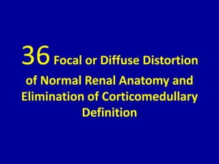
- 1. 36Focal or Diffuse Distortion of Normal Renal Anatomy and Elimination of Corticomedullary Definition
- 2. CLINICAL IMAGAGING AN ATLAS OF DIFFERENTIAL DAIGNOSIS EISENBERG DR. Muhammad Bin Zulfiqar PGR-FCPS III SIMS/SHL
- 3. • Fig GU 36-1 Focal acute bacterial nephritis. Prone parasagittal sonogram of the left kidney (LK) demonstrates acute focal bacterial nephritis (LN) as focal prominence of the renal parenchyma with poor definition of medullary pyramids in the upper pole. (H, head.)2
- 4. • Fig GU 36-2 Chronic atrophic pyelonephritis. Prone sonogram of the kidney (arrowheads) shows a focal loss of renal parenchyma and extension of the calyces peripherally from the renal sinus to the renal margin. Note the associated focal area of increased echogenicity due to fibrosis (arrow) in the upper pole.12
- 5. • Fig GU 36-3 Renal failure due to chronic glomerulonephritis. Parasagittal sonogram demonstrates a tiny right kidney (RK) with marked thinning of the renal parenchyma. The echogenicity of the renal tissue greatly exceeds that of the adjacent liver (L). The medullary pyramids are no longer distinguishable. Scans of the left kidney showed similar findings. (D, diaphragm; H, head; QL, quadratus lumborum muscle.)2
- 6. • Fig GU 36-4 Renal transplant rejection. Transverse sonogram shows that the renal transplant (RT) has become huge and has lost its corticomedullary definition. The renal vascular pedicle is compressed as it enters the renal hilum. An effusion (E), sometimes seen with acute transplant rejection, is noted medial to the kidney (R, right.).2