

Recommended
More Related Content
What's hot
What's hot (20)
Similar to Mucopolysaccharidoses
Similar to Mucopolysaccharidoses (20)
More from Dr M Sanjeevappa
More from Dr M Sanjeevappa (15)
Recently uploaded
Recently uploaded (20)
Mucopolysaccharidoses
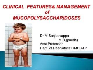
- 1. Dr M.Sanjeevappa M.D.(paeds) Asst.Professor Dept. of Paediatrics GMC,ATP.
- 2. SPHINGOLIPIDOSES GM2-gangliosidosis Tay–Sachs Sandhoff Gaucher disease Fabry disease Metachromatic leukodystrophy Krabbe disease Farber syndrome Mucolipidoses Disorders involving sialic acid
- 3. MPS-I: Hurler disease MPS-II: Hunter disease MPS-III: Sanfilippo disease MPS-IV: Morquio disease MPS-VI: Maroteaux-Lamy disease MPS-VII: Sly Syndrome MPS-IX : Hyaluronidase deficiency
- 4. Overall frequency is between 3.5 to 4.5 in 100,000 births. The most common subtype is MPS-III, followed by MPS-I and MPS-II. MPS III -80%. Inheritance –AR Except MPS II(Hunter’s syndrome )
- 5. Normal development initially. Symptomatic in infancy and early childhood. CNS : Hydrocephalus , Atlantoaxial dislocation MUSCULOSKELETAL : Short stature , Joint stiffness , peripheral nerve entrapment ,Tendon entrapment. CVS : Valvular dysfunction , Hypertension ,CHF , Anginal pains , sudden cardiac deaths .
- 6. RS : Obstructive airway disease Sleep apnea Cor pulmonale EYE : Corneal clouding ,Glaucoma ,Chronic papilledema , Retinal degeneration. EAR : Recurrent middle ear infections , Deformities of the ossicles. Deafness.
- 7. MPS IH(Hurler syndrome): mutations of the IDUA gene on chromosome 4p16.3 encoding α-L-iduronidase. Healthy at birth. Inguinal and Umbilical hernias. Corneal clouding Hepatosplenomegaly . Skeletal deformities . Course facial features Large tongue . Prominent forehead . Hirsutism . Mental retardation. Death usually occurs by 10 yrs of age
- 8. Intermediate form of mps I. Onset at 3 – 8 yrs of age. Normal intelligence. Survive into 3rd decade of life. Cardiac involvement and upper airway obstruction. Spondylolisthesis.
- 9. Onset is after the age of 5 yr. Mild disorder. Joint stiffness. Aortic valve disease. Mild dysostosis multiplex. Normal intelligence and stature. Ophthalmic features include corneal clouding, glaucoma, and retinal degeneration. Obstructive airway disease- sleep apnea
- 10. X-linked recessive disorder caused by the deficiency of iduronate 2-sulfatase (IDS). SEVERE CLINICAL PHENOTYPE: Major deletions or rearrangements of the IDS gene Manifests almost exclusively in males. it has been observed in females also. Manifest between 2 to 4 yr of age. Features similar of Hurler disease exceptions lack of corneal clouding, slow progression
- 11. Grouped skin papules. Extensive mongolian spots at birth. Chronic diarrhea. Communicating hydrocephalus and spastic paraplegia. MILD FORM: Point mutations of the IDS gene normal life span, minimal CNS involvement. slow progression of somatic deterioration. preservation of cognitive function in adult life. Survival to ages 65 and 87 yr.
- 12. Genetically heterogeneous. Characterized by slowly progressive, severe CNS involvement with mild somatic disease. Onset occurs between 2 to 6 yr. Developmental delay Hyperactivity with aggressive behavior. Coarse hair. Hirsutism. Sleep disorders Mild hepatosplenomegaly.
- 13. deficiency of N-acetylgalactosamine-6-sulfatase or β-galactosidase . defective degradation of keratan sulfate. short-trunk dwarfism. skeletal dysplasia. Intelligence preserved. genua valga, kyphosis. growth retardation with short trunk and neck. waddling gait with a tendency to fall. Atlantoaxial instability and dislocation. Extra skeletal manifestations: mild corneal clouding, small teeth with abnormally thin enamel. hepatomegaly Cardiac Valvular lesions.
- 14. mutations of the ARSB gene on chromosome 5q11-13 encoding N-acetylgalactosamine-4-sulfatase (arylsulfatase B). Intelligence preserved. somatic involvement resembles MPS I growth can be normal for the first few years of life virtually stop after age 6-8 yr. Spinal cord compression in upper cervical canal.
- 15. Caused by mutations of the GUSB gene located on chromosome 7q21.11. Deficiency of β-glucuronidase, intracellular storage of glycosaminoglycan fragments. Most severe form: Non-immune fetal hydrops. Some severely affected newborns survive for some months and develop thick skin, visceromegaly, and dysostosis multiplex.
- 16. Less-severe form: present during the first years of life with features of MPS-I but slower progression. Patients with manifestation after 4 yr of life have skeletal abnormalities of dysostosis Multiplex. normal intelligence and usually clear corneae. blood smear that shows coarse granulocytic inclusions.
- 17. The disorder is caused by a mutation in the HYAL1 gene on chromosome 3p21.2-21.2 encoding one of 3 hyaluronidases. Clinical findings: bilateral nodular soft-tissue periarticular masses. lysosomal storage of GAGs in histiocytes. mildly dysmorphic craniofacial features. short stature. normal intelligence. Small erosions in both acetabula.
- 18. skeletal survey. assay the urinary excretion of GAG. Quantitative analysis of single GAG by tandem mass spectrometry. Morquio disease-monoclonal antibodies to keratan sulfate. enzyme assay: Serum leukocytes, or cultured fibroblasts are used as the tissue source for measuring lysosomal enzymes.
- 19. Prenatal diagnosis is available for all MPSs and is carried out on cultured cells from amniotic fluid or chorionic villus biopsy. Prenatal molecular analysis in a male fetus of a proven female carrier of the IDS gene to prevent MPS II. MPSs I, II, and VI are candidates for neonatal blood spot screening by tandem mass spectrometry allowing early diagnosis and enzyme replacement therapy.
- 21. Hematopoietic stem cell transplantation and enzyme replacement therapy are performed in specialized institutions. Bone marrow transplantation and cord blood transplantation have resulted in significant clinical improvement of somatic disease in MPSs I, II, and VI. Transplantation does not significantly improve the neuropsychologic outcome. Stem cell transplantation does not correct skeletal and ocular anomalies
- 22. Enzyme replacement therapy : Recombinant α-L-iduronidase approved for patients with MPS-I. Recombinant iduronate-2-sulfatase ameliorates the non neurologic manifestations of Hunter disease. Recombinant N-acetylgalactosamine-4-sulfatase has been successfully tested in patients with MPS-VI.
- 23. NEUROLOGIC: Hydrocephalus- Ventriculoperitoneal shunt Disturbed sleep/wake circle MPS-III -Melatonin Seizures- anticonvulsants Odontoid hypoplasia MPS-IV -upper cervical fusion Spinal cord compression-Laminectomy, dural excision OPHTHALMOLOGIC: Corneal opacity-Corneal transplant Glaucoma-Medication, surgery
- 24. EARS, AIRWAYS: Recurrent otitis media- Ventilating tubes. Impaired hearing - Audiometry, hearing aids. Obstruction of RS - tonsillectomy, bronchodilator therapy, continuous positive airway pressure at night, laser excision of tracheal lesions, tracheotomy. CARDIAC: Cardiac valve disease - Endocarditis prevention, valve replacement Coronary insufficiency - Medical therapy Arrhythmias- Antiarrhythmic medication, pacemaker
- 25. ORAL, GASTROINTESTINAL: Hypertrophic gums, poor teeth -Dental care Chronic diarrhea MPS-II- Diet modification, loperamide MUSCULOSKELETAL: Joint stiffness All except MPS-IV- Physio therapy Weakness - Physio therapy, wheelchair Gross long bone malalignment -Corrective osteotomies Carpal tunnel syndrome- Electromyography, surgical decompression