1. Yr 12 AS Biology Revision Questions
1. The table below refers to three organic compounds found in cell organelles.
If the compound is found in the organelle, place a tick (√) in the appropriate box and if the
compound is not found in the organelle, place a cross (x) in the appropriate box.
O rg a n e lle P h o s p h o lip id D N A RN A
R ib o s o m e
C h lo ro p la s t
S m o o th e n d o p la s m ic
re tic u lu m
M ito c h o n d rio n
(Total 4 marks)
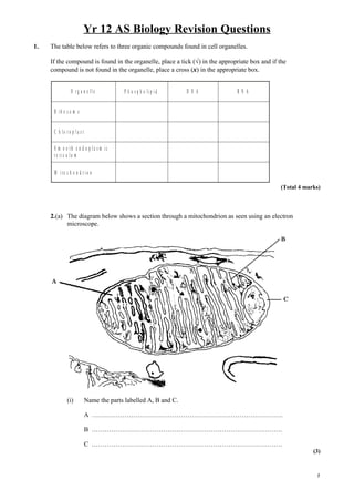
2.(a) The diagram below shows a section through a mitochondrion as seen using an electron
microscope.
(i) Name the parts labelled A, B and C.
A …………………………………………………………………………….
B …………………………………………………………………………….
C …………………………………………………………………………….
(3)
1
2. (ii) On the diagram, by means of an arrow, show the location of the electron
transport system.
(1)
(iii) The magnification of this diagram is × 70 000. Calculate the actual length of the
mitochondrion, giving your answer in suitable units. Show your working .
Answer ........................................................
(2)
(b) Active mitochondria can be isolated from liver cells. If these mitochondria are then
incubated in a buffer solution containing a substrate, such as succinate, dissolved oxygen
will be used by the mitochondria. The concentration of dissolved oxygen in the buffer
solution can be measured using an electrode.
An experiment was carried out in which a suspension of active mitochondria was
incubated in a buffer solution containing succinate, an intermediate of the Krebs cycle.
The concentration of dissolved oxygen was measured every minute for five minutes. A
solution containing sodium azide was then added to this preparation and the concentration
of dissolved oxygen was measured for a further five minutes. Sodium azide combines
with cytochromes and prevents electron transport.
The results are shown in the graph below.
7
6
5
C o n c e n tra tio n S o d iu m a z id e a d d e d
o f d is s o lv e d 4
oxygen
/ a r b itra ry u n its 3
2
1
0
0 5 10
T im e / m in s
(i) Explain why the concentration of oxygen decreased during the first five minutes.
..………………………………………………………………………………
..………………………………………………………………………………
..………………………………………………………………………………
(2)
3. Yr 12 AS Biology Revision Questions
(ii) Suggest what effect the addition of sodium azide will have on the production of
ATP and give an explanation for your answer.
..………………………………………………………………………………
..………………………………………………………………………………
..………………………………………………………………………………
..………………………………………………………………………………
..………………………………………………………………………………
(3)
(Total 11 marks)
3. The table below refers to features of prokaryotic and eukaryotic cells.
If the feature is present place a tick (√) in the appropriate box and if the feature is absent, place a
cross (X) in the appropriate box.
Feature Prokaryotic cell Eukaryotic
Endoplasmic reticulum
Mesosome
Ribosomes
Golgi apparatus
(Total 4 marks)
4. The photograph below shows a section through a mitochondrion as seen using an electron
microscope.
A
C B
Prof. R. Bellairs/Wellcome Photo Library
3
4. (a) Name the parts labelled A, B and C.
A ................................................................................................................................
B ................................................................................................................................
C ................................................................................................................................
(3)
(b) Describe the role of mitochondria.
....................................................................................................................................
....................................................................................................................................
....................................................................................................................................
....................................................................................................................................
(2)
(Total 5 marks)
5. An experiment was carried out to determine what happens to amino acids after they are absorbed
by animal cells. The cells were incubated for 5 minutes in a medium containing radioactively
labelled amino acids. The radioactive amino acids were then washed off and the cells were
incubated in a medium containing only non-radioactive amino acids.
Samples of the cells were taken at 5, 10 and 45 minutes after the start of the experiment and the
sites of radioactivity in the cells were determined.
The results are given in the table below. The figures show radioactivity in certain cell organelles
expressed as a percentage of the total radioactivity within the cells.
P e rc e n ta g e o f to ta l r a d io a c tiv ity
O rg a n e lle
A t 5 m in u te s A t 1 0 m in u te s A t 4 5 m in u te s
R o u g h e n d o p la s m ic
80 10 5
re tic u lu m
G o lg i a p p a ra tu s 10 80 30
S e c re to ry v e s ic le s 0 5 60
(a) Name ONE type of molecule synthesised from amino acids in cells.
……………………………………………………………………………….…..
(1)
5. Yr 12 AS Biology Revision Questions
(b) Explain why the radioactivity is associated mainly with the rough endoplasmic reticulum
after the first 5 minutes of the experiment.
……………………………………………………………………………….…..
……………………………………………………………………………….…..
……………………………………………………………………………….…..
……………………………………………………………………………….…..
(2)
(c) Explain the changes in the pattern of radioactivity in the cell during the remaining 40
minutes of the experiment.
……………………………………………………………………………….…..
……………………………………………………………………………….…..
……………………………………………………………………………….…..
……………………………………………………………………………….…..
……………………………………………………………………………….…..
(3)
(d) Suggest why the figures in the tables total less than 100%.
……………………………………………………………………………….…..
……………………………………………………………………………….…..
……………………………………………………………………………….…..
……………………………………………………………………………….…..
(2)
(e) If the experiment is continued for a further period of time, most of the radioactivity will
be found outside the cell.
Name and describe the process which brings about this result.
……………………………………………………………………………….…..
……………………………………………………………………………….…..
……………………………………………………………………………….…..
……………………………………………………………………………….…..
……………………………………………………………………………….…..
(3)
(Total 11 marks)
5
6. 6. The diagram below shows the structure of a bacterium, a typical prokaryotic cell.
B
C
A
(a) Name A, B and C as labelled on the diagram.
A ...............................................................................................................................
B ...............................................................................................................................
C ...............................................................................................................................
(3)
(b) Complete the table below to show three differences between a prokaryotic cell and a
eukaryotic cell.
Prokaryotic cell Eukaryotic cell
1
2
3
(3)
(Total 6 marks)
7. Yr 12 AS Biology Revision Questions
7. The photograph below shows human cells as seen using a light microscope. It has been
magnified 800 times.
A
B
(a) Calculate the actual diameter of the cell labelled A, expressing your answer in µm
(micrometres). Show your working.
Answer ……………………… µm
(3)
(b) In the space below, make an accurate drawing of the cells labelled A and B,
enlarge 2 ×. Do not label your drawing.
(4)
(Total 7 marks)
7
8. 8. The photograph below-shows part of an animal cell, as seen using an electron microscope. The
magnification is ×5000.
(a) Name the structures labelled A and B.
A............................................................................................................................................
B............................................................................................................................................
(2)
(b) Calculate the actual length of the structure labelled B in µm. Show your working.
Actual length of B .................. µm
(3)
9. Yr 12 AS Biology Revision Questions
(c) Describe how proteins synthesised on the rough endoplasmic reticulum are processed and
transported out of the cell.
...............................................................................................................................................
...............................................................................................................................................
...............................................................................................................................................
...............................................................................................................................................
...............................................................................................................................................
...............................................................................................................................................
...............................................................................................................................................
...............................................................................................................................................
...............................................................................................................................................
...............................................................................................................................................
(5)
(Total 10 marks)
9
10. 9. The following table refers to organelles found in eukaryotic cells. Complete the table by writing
the name of the organelle, two features of its structure or one function of the organelle in each of
the four empty boxes as appropriate.
Name of organelle Two features of structure One function
1. Stack of curved cisternae
Modification
of proteins
2. Surrounded by many vesicles
1.
Rough endoplasmic
reticulum
2.
1.
Site of
photosynthesis
Chloroplast
2.
(Total 6 marks)
11. Yr 12 AS Biology Revision Questions
10. The diagram below shows the structure of a bacterial cell as seen using an electron
microscope.
C e ll w a ll S to ra g e g ra n u le B
A
X Y
(a) (i) Name the parts labelled A and B.
A ............................................................................................................................
B ............................................................................................................................
(2)
(ii) Name the carbohydrate present in the storage granules.
...............................................................................................................................
(1)
(iii) Describe how the cell wall in this bacterial cell differs from that in a plant cell.
...............................................................................................................................
...............................................................................................................................
(1)
(b) The diagram has been magnified 6000 times. Calculate the actual length of the
bacterial cell between X and Y. Show your working, and give your answer in
micrometres.
Answer ............................. µm
(3)
(Total 7 marks)
11
12. 11. (a) Draw and label a diagram to show the structure of the Golgi apparatus as seen using
an electron microscope.
(3)
13. Yr 12 AS Biology Revision Questions
(b) The process of protein synthesis in cells and the secretion of proteins from the cells was
investigated using radioactively labelled amino acids.
The cells were incubated with radioactive amino acids for 30 minutes. The cells were then
removed and washed thoroughly to remove any radioactive amino acids on the cell
surfaces.
The washed cells were then incubated with non-radioactive amino acids for 120 minutes.
Every 20 minutes a sample of cells was removed and the level of radioactivity in the
rough endoplasmic reticulum and in the secretory vesicles was determined.
The graph below shows the levels of radioactivity in the rough endoplasmic reticulum and
the secretory vesicles.
80
70 S e c re to ry
v e s ic le s
60
L evel of 50
ra d io a c tiv ity
/ a rb itra ry 4 0
u n its
30
20 R ough
e n d o p la s m ic
re tic u lu m
10
0
0 20 40 60 80 100 120
In c u b a tio n tim e / m in
(i) Describe and explain the changes in the level of radioactivity in the rough
endoplasmic reticulum during the first 40 minutes of the incubation period.
...............................................................................................................................
...............................................................................................................................
...............................................................................................................................
...............................................................................................................................
...............................................................................................................................
...............................................................................................................................
(2)
13
14. (ii) Explain the shape of the curve for the secretory vesicles between 0 and 40 minutes.
...............................................................................................................................
...............................................................................................................................
...............................................................................................................................
...............................................................................................................................
...............................................................................................................................
...............................................................................................................................
...............................................................................................................................
...............................................................................................................................
(3)
(Total 8 marks)
12. Eukaryotic cells contain organelles, many of which are bound by a membrane. Some organelles
have a double membrane, often called an envelope.
(a) (i) Describe two structural differences between the double membrane
surrounding a mitochondrion and the double membrane surrounding a nucleus.
................................................................................................................................
................................................................................................................................
................................................................................................................................
................................................................................................................................
(2)
(ii) Name one other organelle that has a double membrane.
................................................................................................................................
(1)
15. Yr 12 AS Biology Revision Questions
(b) Centrioles are an example of organelles that are not membrane-bound. Describe the
structure and function of centrioles.
.......................................................................................................................................
.......................................................................................................................................
.......................................................................................................................................
.......................................................................................................................................
.......................................................................................................................................
.......................................................................................................................................
(3)
(Total 6 marks)
13. The diagram below shows some of the stages in protein synthesis and secretion in a mammalian
cell.
15
16. (a) (i) Name the processes taking place during stages A and B.
A ….................................................................................................................
B .....................................................................................................................
(2)
(ii) Name the process by which protein is secreted in stage F.
(1)
(b) Describe the part played by transfer RNA in the formation of the polypeptide chain during
stage B.
....................................................................................................................................
....................................................................................................................................
....................................................................................................................................
....................................................................................................................................
(3)
(Total 6 marks)