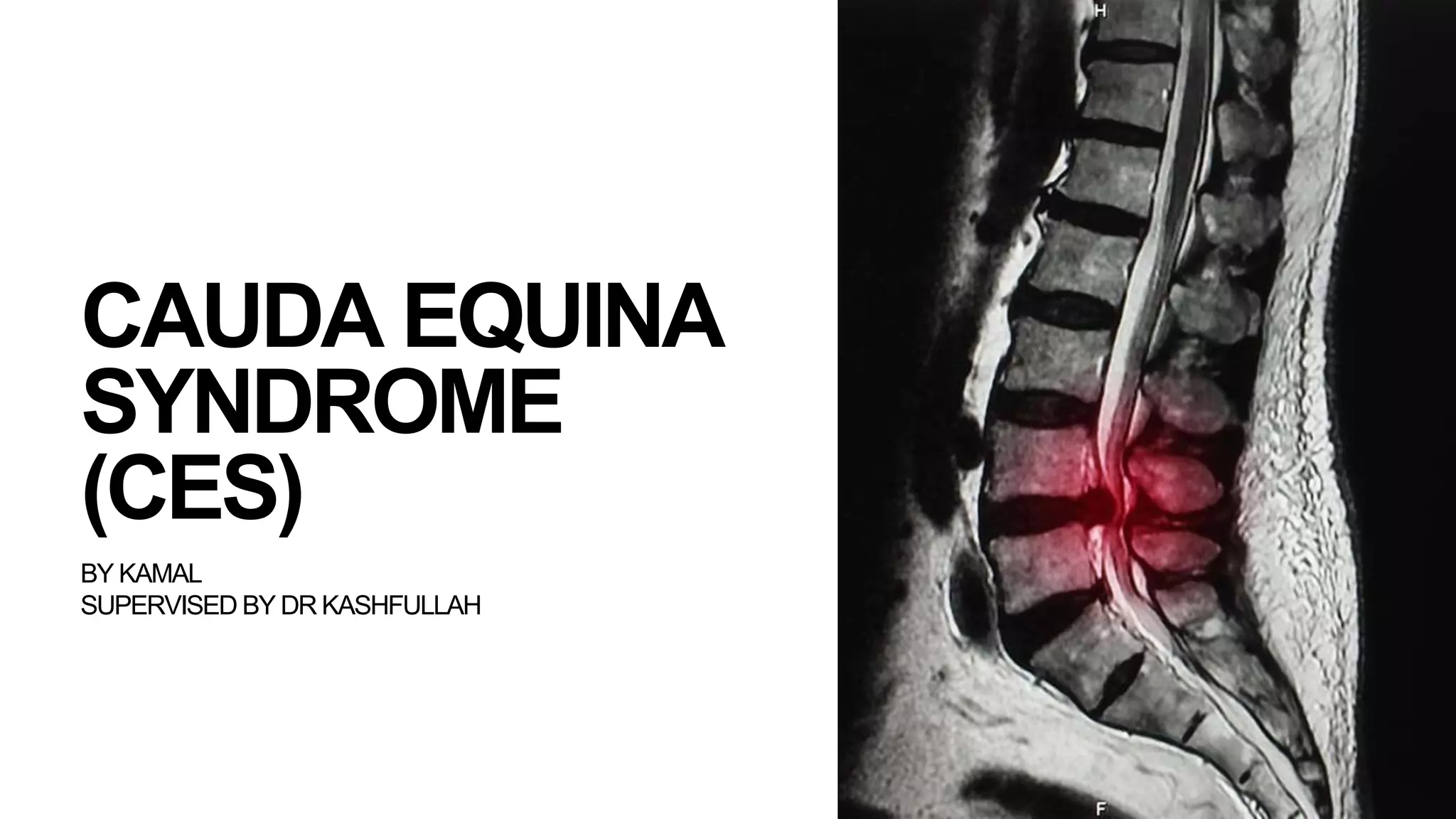
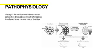
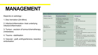
CAUDA EQUINA
SYNDROME
(CES)
BY KAMAL
SUPERVISED BY DR KASHFULLAH
- Cauda equina (CE) is a bundle of intradural nerve roots at the
end of the spinal cord, in the subarachnoid space distal to the
conus medullaris.
- Cauda Equina (latin) = horse tail
- Sensory innervation to the saddle area, motor innervation to the
sphincters, and parasympathetic innervation to the bladder and
lower bowel (ie, from the left splenic flexure to the rectum).
- Nerves including L2-S5
ANATOMY
- A retrospective review in Slovenia found an annual incidence of
cauda equina syndrome resulting from intervertebral disc
herniation of 1.8 per million population. Using US data on annual
incidence of symptomatic disc herniation (1500 per million popula-
tion), the author estimates that each year 0.12% of herniated discs
are likely to cause cauda equina syndrome.
EPIDEMIOLOGY
- Cauda equina syndrome (CES) is a relatively uncommon
condition typically caused by a compression within the canal of the
lumbosacral spine. It is one of the few spinal surgical emergencies
and its prompt diagnosis and surgical treatment are of paramount
importance as, otherwise, patients could be left with permanent
and disabling neurological deficits involving lower limb
sensorimotor function, bladder, bowel and sexual function.
- Classic clinical presentation : bilateral leg weakness, decreased
reflexes, saddle anaesthesia and bowel/bladder disturbance.
DEFINITION
- the cause of CES is basically anything that causes
compression to the CE :
1. Degenerative - disc herniation (most common), spinal
stenosis, spondylolisthesis
2. Inflammatory - ankylosing spondylitis
3.Vascular - aortic dissection, arteriovenous malformation
4. Infective - epidural abcess, arachnoiditis, Pott’s
disease
5. Tumours - primary. Metastasis
6. Trauma
AETIOLOGY
- injury to the lumbosacral nerve causes
conduction block (discontinuity of electrical
impulses) hence causes loss of function
PATHOPHYSIOLOGY
-Three classic patterns of presentation have been
described:
Type 1: Presents acutely as the first symptom of
lumbar disc prolapse
Type 2: Presents as the endpoint of a long history
of chronic lower back pain with or without sciatica
Type 3: Presents insidiously with slow progression
to numbness and urinary symptoms
SYMPTOMS AND SIGNS
Red Flag symptoms
- Severe lower back pain
- Motor weakness, sensory loss or radicular pain
(usually bilateral)
- Loss of lower limb reflexes
- Saddle anaesthesia
- Recent onset of bladder dysfunction (i.e. urinary
retention or incontinence)
- Recent onset of faecal incontinence
- Recent onset of sexual dysfunction
1. CESS (CES suspected) : Thecal sac compression with bilateral radiculopathy and/or
subjective sphincteric problems and/or subjective perineal sensory changes with no objective
evidence of CES
2. CESI (incomplete CES) : Thecal sac compression with subjective symptoms and objective
signs of CES
3. CESR (CES with retention) : Thecal sac compression with neurogenic urinary retention
CLASSIFICATION
- head to toe examination
- neurological examination :
Power, sensation, reflex, tone, clonus, anal tone,
bulbocavernosus reflex (BCR)
*LMN findings
- ASIA charting
PHYSICAL EXAMINATION
- Blood baseline investigation
- spine xray
- MRI/CT myelogram (if MRI contraindicated)
- further investigations depends on suspected
aetiology (blood C+S, tumour markers etc)
INVESTIGATION
Depends on aetiology :
1. Disc herniation (24-48hrs)
2. Infection/inflammation- treat underlying
infection/inflammation
3. Tumour - excision of tumour/chemotherapy
(metastasis)
4. Trauma - stabilisation
5. Vascular - graft, antihypertensive, resection,
embolisation
MANAGEMENT
Supportive :
- analgesia
- high dose steroids
- laxatives
- CBD insertion/pampers
- CRIB
- bone protection (malignancy)
- anticoagulants
- refer physio
- Early recognition and treatment of CES is associated
with improved outcome with respect to bladder
function.
- Outcomes in patients treated at the time of CESI are
generally favourable, whereas outcomes following
bladder paralysis (CESR) are less favourable. If male
patients present with erectile dysfunction, this usually
has a poor prognosis.
- For these reasons, failure to recognize and treat this
condition expediently may have outcome and
medicolegal implications.
PROGNOSIS
THANK YOU
1. Junseok W Hur et al (2019) Guidelines for Cauda Equina Syndrome Management
2. Pararajasingham J. And Deniz K (2011), Cauda Equina Syndrome
3. Gardner A et al (2011), Cauda equina syndrome: a review of the current clinical and medico-legal position
4. Lady C et al (2009), Cauda Equina Syndrome
5. Ebnezar J (2010), Textbook of Orthopaedics
REFERENCES
Q&A
1. Cauda Equina nerves include ?
2. What is CES ?
3. Common cause of CES ?
4. What are the classic clinical presentation of CES ?
5. Gold standard investigation for CES ?
6. Management for CESI ?

CES.pptx

  • 1.
  • 2.
    - Cauda equina(CE) is a bundle of intradural nerve roots at the end of the spinal cord, in the subarachnoid space distal to the conus medullaris. - Cauda Equina (latin) = horse tail - Sensory innervation to the saddle area, motor innervation to the sphincters, and parasympathetic innervation to the bladder and lower bowel (ie, from the left splenic flexure to the rectum). - Nerves including L2-S5 ANATOMY
  • 3.
    - A retrospectivereview in Slovenia found an annual incidence of cauda equina syndrome resulting from intervertebral disc herniation of 1.8 per million population. Using US data on annual incidence of symptomatic disc herniation (1500 per million popula- tion), the author estimates that each year 0.12% of herniated discs are likely to cause cauda equina syndrome. EPIDEMIOLOGY
  • 4.
    - Cauda equinasyndrome (CES) is a relatively uncommon condition typically caused by a compression within the canal of the lumbosacral spine. It is one of the few spinal surgical emergencies and its prompt diagnosis and surgical treatment are of paramount importance as, otherwise, patients could be left with permanent and disabling neurological deficits involving lower limb sensorimotor function, bladder, bowel and sexual function. - Classic clinical presentation : bilateral leg weakness, decreased reflexes, saddle anaesthesia and bowel/bladder disturbance. DEFINITION
  • 5.
    - the causeof CES is basically anything that causes compression to the CE : 1. Degenerative - disc herniation (most common), spinal stenosis, spondylolisthesis 2. Inflammatory - ankylosing spondylitis 3.Vascular - aortic dissection, arteriovenous malformation 4. Infective - epidural abcess, arachnoiditis, Pott’s disease 5. Tumours - primary. Metastasis 6. Trauma AETIOLOGY
  • 6.
    - injury tothe lumbosacral nerve causes conduction block (discontinuity of electrical impulses) hence causes loss of function PATHOPHYSIOLOGY
  • 7.
    -Three classic patternsof presentation have been described: Type 1: Presents acutely as the first symptom of lumbar disc prolapse Type 2: Presents as the endpoint of a long history of chronic lower back pain with or without sciatica Type 3: Presents insidiously with slow progression to numbness and urinary symptoms SYMPTOMS AND SIGNS
  • 8.
    Red Flag symptoms -Severe lower back pain - Motor weakness, sensory loss or radicular pain (usually bilateral) - Loss of lower limb reflexes - Saddle anaesthesia - Recent onset of bladder dysfunction (i.e. urinary retention or incontinence) - Recent onset of faecal incontinence - Recent onset of sexual dysfunction
  • 9.
    1. CESS (CESsuspected) : Thecal sac compression with bilateral radiculopathy and/or subjective sphincteric problems and/or subjective perineal sensory changes with no objective evidence of CES 2. CESI (incomplete CES) : Thecal sac compression with subjective symptoms and objective signs of CES 3. CESR (CES with retention) : Thecal sac compression with neurogenic urinary retention CLASSIFICATION
  • 10.
    - head totoe examination - neurological examination : Power, sensation, reflex, tone, clonus, anal tone, bulbocavernosus reflex (BCR) *LMN findings - ASIA charting PHYSICAL EXAMINATION
  • 11.
    - Blood baselineinvestigation - spine xray - MRI/CT myelogram (if MRI contraindicated) - further investigations depends on suspected aetiology (blood C+S, tumour markers etc) INVESTIGATION
  • 13.
    Depends on aetiology: 1. Disc herniation (24-48hrs) 2. Infection/inflammation- treat underlying infection/inflammation 3. Tumour - excision of tumour/chemotherapy (metastasis) 4. Trauma - stabilisation 5. Vascular - graft, antihypertensive, resection, embolisation MANAGEMENT
  • 14.
    Supportive : - analgesia -high dose steroids - laxatives - CBD insertion/pampers - CRIB - bone protection (malignancy) - anticoagulants - refer physio
  • 15.
    - Early recognitionand treatment of CES is associated with improved outcome with respect to bladder function. - Outcomes in patients treated at the time of CESI are generally favourable, whereas outcomes following bladder paralysis (CESR) are less favourable. If male patients present with erectile dysfunction, this usually has a poor prognosis. - For these reasons, failure to recognize and treat this condition expediently may have outcome and medicolegal implications. PROGNOSIS
  • 16.
  • 17.
    1. Junseok WHur et al (2019) Guidelines for Cauda Equina Syndrome Management 2. Pararajasingham J. And Deniz K (2011), Cauda Equina Syndrome 3. Gardner A et al (2011), Cauda equina syndrome: a review of the current clinical and medico-legal position 4. Lady C et al (2009), Cauda Equina Syndrome 5. Ebnezar J (2010), Textbook of Orthopaedics REFERENCES
  • 18.
    Q&A 1. Cauda Equinanerves include ? 2. What is CES ? 3. Common cause of CES ? 4. What are the classic clinical presentation of CES ? 5. Gold standard investigation for CES ? 6. Management for CESI ?