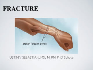
2. • A fracture is a break in the continuity of bone and is de
fi
ned
according to its type and extent. Fractures occur when the bone is
subjected to stress greater than it can absorb.
• When the bone is broken, adjacent structures are also affected,
resulting in soft tissue edema, hemorrhage into the muscles and
joints, joint dislocations, ruptured tendons, severed nerves, and
damaged blood vessels.
• Body organs may be injured by the force that caused the fracture or
by the fracture fragments.
3. Cause
s
Fractures are caused by
;
Direct blow
s
Crushing force
s
Sudden twisting motion
s
Extreme muscle contractions.
4. Types
Avulsion: a fracture in which a fragment of bone has been pulled away by a
ligament or tendon and its attachment
6. Compound: a fracture in which damage also involves the skin or mucous
membranes; also called an open fracture. Open fractures are graded according to the
following criteria:
Grade I is a clean wound less than 1 cm long.
Grade II is a larger wound without extensive soft tissue damage.
Grade III is highly contaminated, has extensive soft tissue damage, and is the
most severe.
11. Impacted: a fracture in which a bone fragment is driven into another bone fragment
12. Oblique: a fracture occurring at an angle across the bone (less stable than a
transverse fracture)
13. Pathologic: a fracture that occurs through an area of diseased bone (eg,
osteoporosis, bone cyst, Paget’s disease, bony metastasis, tumor); can occur without
trauma or a fall.
19. Clinical Manifestations
PAIN
The pain is continuous and increases in severity until the bone
fragments are immobilized. The muscle spasm that accompanies
fracture is a type of natural splinting designed to minimize further
movement of the fracture fragments.
LOSS OF FUNCTION
After a fracture, the extremity cannot function properly, because
normal function of the muscles depends on the integrity of the
bones to which they are attached. Pain contributes to the loss of
function. In addition, abnormal movement (false motion) may be
present.
DEFORMITY
Displacement, angulation, or rotation of the fragments in a fracture
of the arm or leg causes a deformity (either visible or palpable)
that is detectable when the limb is compared with the uninjured
extremity. Deformity also results from soft tissue swelling.
20. SHORTENING
In fractures of long bones, there is actual shortening of the
extremity because of the contraction of the muscles that are
attached above and below the site of the fracture. The fragments
often over- lap by as much as 2.5to5cm(1to2inches).
CREPITUS
When the extremity is examined with the hands, a grating
sensation, called crepitus, can be felt. It is caused by the rubbing
of the bone fragments against each other
.
NURSING ALERT Testing for crepitus can produce further
tissue damage and should be avoided.
SWELLING AND DISCOLORATION
Localized swelling and discoloration of the skin (ecchymosis)
occurs after a fracture as a result of trauma and bleeding into the
tissues. These signs may not develop for several hours after the
injury.
21. Emergency Management of Fractures
Immediately after injury, whenever a fracture is suspected, it is
important to immobilize the body part before the patient is
moved
.
Splints can be applied, the extremity is supported above and
below the fracture site to prevent rotation as well as angular
motion.
Adequate splinting, including joints adjacent to the fracture, is
essential. Movement of fracture fragments causes additional
pain, soft tissue damage, and bleeding.
Temporary, well-padded splints,
fi
rmly bandaged over clothing,
serve to immobilize the fracture.
Immobilization of the long bones of the lower extremities may
be accomplished by bandaging the legs together, with the
unaffected extremity serving as a splint for the injured one.
22. In an upper extremity injury, the arm may be bandaged to the
chest, or an injured forearm may be placed in a sling.
The neuromuscular status distal to the injury should be assessed to
determine adequacy of peripheral tissue perfusion and nerve
function.
With an open fracture, the wound is covered with a clean (sterile)
dressing to prevent contamination of deeper tissues. No attempt is
made to reduce the fracture, even if one of the bone fragments is
protruding through the wound. Splints are applied for
immobilization.
In the emergency department, the patient is evaluated completely.
The clothes are gently removed,
fi
rst from the uninjured side of
the body and then from the injured side. The patient’s clothing
may be cut away.
The fractured extremity is moved as little as possible to avoid
more damage.
23. Diagnosi
s
• Doctors can usually recognize most fractures by examining the injury and
taking X-rays. Sometimes an X-ray will not show a fracture. This is
especially common with some wrist fractures, hip fractures (especially in
older people), and stress fractures
.
• In some cases, such as a possible wrist fracture with an initially normal X-
ray, doctor may apply a splint to immobilize the area and order a second
X-ray 10 to 14 days later when healing can make the fracture visibe
.
• Occasionally, even after the fracture diagnosis has been made, may need
other tests (such as a CT scan, MRI, or angiogram, a special X-ray of
blood vessels) to determine whether other tissues around the bone have
been damaged
.
• A bone scan is a nuclear imaging procedure. In nuclear imaging, tiny
amounts of radioactive materials (tracers) are injected into a vein and
taken up in varying amounts at different sites in the body. Areas of the
body where cells and tissues are repairing themselves most actively take
up the largest amounts of tracer. Nuclear images highlight these areas,
suggesting the presence of abnormalities associated with disease or injury.
24. Medical Management of Fractures
The principles of fracture treatment include
reduction, immobilization, and regaining of normal
function and strength through rehabilitation.
REDUCTION
Reduction of a fracture (“setting” the bone) refers to
restoration of the fracture fragments to anatomic
alignment and rotation. Either closed reduction or
open reduction may be used to reduce a fracture.
Usually, the physician reduces a fracture as soon as
possible to prevent loss of elasticity from the tissues
through in
fi
ltration by edema or hemorrhage.
25. Closed Reduction.
• In most instances, closed reduction is accomplished by bringing
the bone fragments into apposition (ie, placing the ends in
contact) through manipulation and manual traction.
• The extremity is held in the desired position while the physician
applies a cast, splint, or other device. Reduction under anesthesia
with percutaneous pinning may be used.
• The immobilizing device maintains the reduction and stabilizes
the extremity for bone healing.
• X-rays are obtained to verify that the bone fragments are
correctly aligned.
• Traction (skin or skeletal) may be used to effect fracture
reduction and immobilization. Traction may be used until the
patient is physiologically stable and able to withstand surgical
fi
xation.
26.
27. Open Reduction.
• Some fractures require open reduction.
• Through a surgical approach, the fracture fragments are
reduced.
• Internal
fi
xation devices (metallic pins, wires, screws,
plates, nails, or rods) may be used to hold the bone
fragments in position until solid bone healing occurs.
• These devices may be attached to the sides of bone, or they
may be inserted through the bony fragments or directly into
the medullary cavity of the bone.
• Internal
fi
xation devices ensure
fi
rm approximation and
fi
xation of the bony fragments.
28.
29. IMMOBILIZATION
• After the fracture has been reduced, the bone fragments must be
immobilized, or held in correct position and alignment, until
union occurs.
• Immobilization may be accomplished by external or internal
fi
xation.
• Methods of external
fi
xation include bandages, casts, splints,
continuous traction, and external
fi
xators.
• Metal implants used for internal
fi
xation serve as internal splints
to immobilize the fracture.
30.
31. MAINTAINING AND RESTORING FUNCTION
• Reduction and immobilization are maintained as prescribed to promote bone and
soft tissue healing.
• Swelling is controlled by elevating the injured extremity as prescribed.
• Neuromuscular status (circulation, movement, sensation) is monitored, and the
orthopaedic surgeon is noti
fi
ed immediately if signs of neurovascular compromise
are identi
fi
ed.
• Restlessness, anxiety, and discomfort are controlled with a variety of approaches,
such as reassurance, position changes, and pain relief strategies, including use of
analgesics.
• Isometric and muscle-setting exercises are encouraged to minimize atrophy and to
promote circulation.
• Participation in activities of daily living (ADLs) is encouraged to promote
independent functioning and self-esteem.
• Gradual resumption of activities is promoted within the therapeutic prescription.
• With internal
fi
xation, the surgeon determines the amount of movement and
weight-bearing stress the extremity can withstand and prescribes the level of
activity.
32. Fracture Healin
g
Weeks to months are required for most fractures to heal.
The reduction of fracture fragments must be accurate and
maintained to ensure healing.
The affected bone must have an adequate blood supply. The
type of fracture also affects healing time.
In general, fractures of
fl
at bones (pelvis, scapula) heal
rapidly.
Fractures at the ends of long bones, where the bone is more
vascular and cancellous, heal more quickly than do
fractures in areas where the bone is dense and less vascular
(midshaft).
Weight bearing stimulates healing of stabilized fractures of
the long bones in the lower extremities.
33. Factors That Enhance Fracture Healing
Immobilization of fracture fragments
Maximum bone fragment contact
Suf cient blood supply
Proper nutrition
Exercise: weight bearing for long bones
34. Factors That Inhibit Fracture Healing
Extensive local trauma
Bone loss
Inadequate immobilization
Space or tissue between bone fragments
Infection
Local malignancy
Metabolic bone disease
Irradiated bone (radiation necrosis)
Avascular necrosis
Intra-articular fracture (synovial uid contains brolysins,
which lyse the initial clot and retard clot formation)
35. Complications (Early and Delayed)
Complications of fractures fall into two categories—early and
delayed.
Early complications include shock, fat embolism, com-
partment syndrome, deep vein thrombosis, thromboembolism
(pulmonary embolism), disseminated intravascular
coagulopathy, and infection.
Delayed complications include delayed union and nonunion,
avascular necrosis of bone, reaction to internal
fi
xation devices,
complex regional pain syndrome (formerly called re
fl
ex
sympathetic dystrophy), and heterotrophic ossi
fi
cation.
36. SHOCK (EARLY)
Hypovolemic or traumatic shock resulting from
hemorrhage (both visible and nonvisible blood loss)
may occur in fractured extremities, thorax, pelvis, or
spine.
Because the bone is very vascular, large quantities of
blood may be lost as a result of trauma, especially in
fractures of the femur and pelvis.
Treatmen
t
Treatment of shock consists of restoring blood
volume and circulation, relieving the patient’s pain,
providing adequate splinting, and protecting the
patient from further injury and other complications.
37. FAT EMBOLISM SYNDROME (EARLY)
• After fracture of long bones or pelvis, multiple fractures, or crush
injuries, fat emboli may develop.
• At the time of fracture, fat globules may move into the blood.
• The fat globules (emboli) occlude the small blood vessels that supply
the lungs, brain, kidneys, and other organs.
• The onset of symptoms is rapid, usually occurring within 24 to 72
hours, but may occur up to a week after injury.
Clinical Manifestations
• Presenting features include hypoxia, tachypnea and tachycardia. The
respiratory distress response includes tachypnea, dyspnea, crackles,
wheezes, precordial chest pain, cough, large amounts of thick white
sputum, and tachycardia.
• Occlusion of a large number of small vessels causes the pulmonary
pressure to rise. Edema and hemorrhages in the alveoli impair oxygen
transport, leading to hypoxia.
38. Prevention and Management
• Immediate immobilization of fractures (including early surgical
fi
xation), minimal fracture manipulation, adequate support for
fractured bones during turning and positioning are measures that
may reduce the incidence of fat emboli
.
• The objectives of management are to support the respiratory
system, to prevent respiratory and metabolic acidosis, and to
correct homeostatic disturbances.
• Respiratory failure is the most common cause of death.
Respiratory support is provided with oxygen given in high
concentrations
.
• Corticosteroids may be administered to treat the in
fl
ammatory
lung reaction and to control cerebral edema
.
• Because fat emboli are a major cause of death for patients with
fractures, the nurse must recognize early indications of fat
embolism syndrome and report them promptly to the physician.
39. COMPARTMENT SYNDROME (EARLY)
• Compartment syndrome is a complication that develops
when tissue perfusion in the muscles is less than that
required for tissue viability.
• The patient complains of deep, throbbing, unrelenting
pain, which is not controlled by opioids.
• The forearm and leg muscle are involved most
frequently.
• The pressure within a muscle compartment may increase
to such an extent as to decrease microcirculation,
causing nerve and muscle anoxia and necrosis.
• Permanent function can be lost if the anoxic situation
continues for longer than 6 hours.
40.
41. Assessment and Diagnostic Findings
• Frequent assessment of neurovascular function after fracture
is essential. Sensory de
fi
cits include paresthesia, unrelenting
pain, and hypoesthesia
.
• Peripheral circulation is evaluated by assessing color,
temperature, capillary re
fi
ll time, swelling, and pulses.
• As intracompartment pressure increases, the patient
complains of deep, throbbing, unrelenting pain, which is
greater than expected and not controlled by opioids.
42. Medical Management
• Prompt management of acute compartment syndrome
is essential.
• Delay may result in permanent nerve and muscle
damage or even necrosis.
• Compartment syndrome is managed by elevation of
the extremity, release of restrictive devices (dressings
or cast), or both.
• If conservative measures do not restore tissue
perfusion and relieve pain within 1 hour, a fasciotomy
(surgical decompression with excision of the
fi
brous
membrane that covers and separates muscles) may be
needed to relieve the constrictive muscle fascia.
43. OTHER EARLY COMPLICATIONS
• Deep vein thrombosis (DVT), thromboembolism, and
pulmonary embolus (PE) are associated with reduced skeletal
muscle contractions and bed rest.
• Disseminated intravascular coagulopathy (DIC) includes a
group of bleeding disorders with diverse causes, including
massive tissue trauma
.
• All open fractures are considered contaminated. Surgical
internal
fi
xation of fractures carries a risk for infection.
Antibiotic therapy must be appropriate and adequate for
prevention and treatment of infection.
44. DELAYED UNION AND NONUNION (DELAYED)
• Delayed union occurs when healing does not occur at a
normal rate for the location and type of fracture. Delayed
union may be associated with distraction (pulling apart) of
bone fragments, systemic or local infection, poor
nutrition, or comorbidity (eg, diabetes mellitus;
autoimmune disease). Eventually, the fracture heals.
• Nonunion results from failure of the ends of a fractured
bone to unite. Factors contributing to union problems
include infection at the fracture site, interposition of tissue
between the bone ends, inadequate immobilization,
excessive space between bone fragments (bone gap),
limited bone contact, and impaired blood supply resulting
in avascular necrosis.
45. Medical Management
The physician treats nonunion with internal
fi
xation, bone
grafting, electrical bone stimulation, or a combination of
these therapies.
Internal
fi
xation stabilises the bone fragments and
ensures bone contact.
• Bone grafts provide for osteogenesi
s
• Osteogenesis in nonunion may be stimulated by
electrical impulses; the effectiveness is similar to that of
bone grafting. Use of electrical impulses is not effective
with large bone gaps or synovial pseudarthrosis. The
electrical stimulation modi
fi
es the tissue environment,
which enhances mineral deposition and bone formation.
46. AVASCULAR NECROSIS OF BONE (DELAYED)
• Avascular necrosis occurs when the bone loses its
blood supply and dies.
• It may occur after a fracture with disruption of the
blood supply (especially of the femoral neck).
• The devitalized bone may collapse.
• The patient develops pain and experiences limited
movement.
• X-rays reveal calcium loss and structural collapse.
• Treatment generally consists of attempts to revitalize
the bone with bone grafts, prosthetic replacement, or
arthrodesis (joint fusion).
47. REACTION TO INTERNAL FIXATION DEVICES
(DELAYED)
• Internal
fi
xation devices may be removed after bony union
has taken place. In most patients, however, the device is
not removed unless it produces symptoms.
• Pain and decreased function are the prime indications that
a problem has developed. Problems may include
mechanical failure (inadequate insertion and
stabilization); material failure (faulty or damaged device);
corrosion of the device, causing local in
fl
ammation;
allergic response to the metallic alloy used.
• If the device is removed, the bone needs to be protected
from refracture related to altered bone structure, and
trauma.
48. COMPLEX REGIONAL PAIN SYNDROME (DELAYED)
Complex regional pain syndrome (CRPS) is a painful sympathetic
nervous system problem. It occurs infrequently. Clinical
manifestations of CRPS include severe burning pain, local edema,
hyperesthesia, stiffness, discoloration, vasomotor skin changes (ie,
fl
uctuating warm, red, dry and cold, sweaty, cyanotic), and trophic
changes. This syndrome is frequently chronic, with extension of
symptoms to adjacent areas of the body.
Management
• Prevention may include selection of an immobilization device (eg,
external
fi
xator) that allows for the greatest ROM and functional use
of the rest of the extremity.
• Early effective pain relief is the focus of management.
• With pain relief, the patient can participate in ROM exercises and
functional use of the affected area.
49. HETEROTROPHIC OSSIFICATION (DELAYED)
• Heterotrophic ossi
fi
cation (myositis ossi
fi
cans) is the abnormal
formation of bone, near bones or in muscle, in response to soft
tissue trauma after blunt trauma, fracture, or total joint replacement.
• The muscle is painful, and normal muscular contraction and
movement are limited.
• Early mobilization has been recommended.
• Indomethacin (Indocin) may be used prophylactically if deep
muscle contusion has occurred.
• Usually, the bone lesion resorbs over time, but the abnormal bone
eventually may need to be excised if symptoms persist.
50. NURSING DIAGNOSES
Based on the assessment data, the patient’s major nursing diagnoses may include
the following:
•Acute pain related to fracture, soft tissue damage, muscle spasm, and surgery
Goal: Relief of pain
Nursing Interventions
1.Assess type and location of patient’s pain.
2.Acknowledge existence of pain; inform patient of available analgesics; record
patient’s baseline discomfort.
3.Handle the affected extremity gently, supporting it with hands or pillow.
4.Use pain-modifying strategies.
5.Position for comfort and function.
6.Assist with frequent changes in position.
51. •Impaired physical mobility related to fracture
Goal: Achieves pain-free, functional, stable movements
Nursing Interventions
1.Maintain neutral positioning of affected area.
2.Instruct and assist in position changes and transfers.
3.Instruct in and supervise isometric exercises.
4.In consultation with physical therapist, instruct in and supervise
progressive safe ambulation within limitations of weight- bearing
prescription.
5.Offer encouragement and support exercise regimen.
6.Instruct and supervise safe use of ambulatory aids.
52. •Impaired skin integrity related to surgical incision
Goal: Achieves wound healing
Nursing Interventions
1.Monitor vital signs.
2.Perform aseptic dressing changes.
3.Assess wound appearance and character of drainage.
4.Assess report of pain.
5.Administer prophylactic antibiotic if prescribed, and
observe for side effects.
53. •Risk for impaired urinary elimination related to
immobility
Goal: Maintains normal urinary elimination patterns
Nursing Interventions
1.Monitor intake and output.
2.Avoid/minimise use of indwelling catheter.
3.Perform intermittent catheterisation for urinary
retention
54. •Risk for ineffective coping related to injury, surgery, and
dependence
Goal: Uses effective coping mechanisms to modify stress
Nursing Interventions
1.Encourage patient to express concerns and to discuss the possible
impact of fracture.
2.Support use of coping mechanisms. Involve significant others and
support services as needed.
3.Contact social services, if needed.
4.Explain anticipated treatment regimen and routines to facilitate
positive attitude in relation to rehabilitation.
5.Encourage patient to participate in planning.