
Recommended
More Related Content
Similar to Action Potential Generation and Propagation in Excitable Cells
Similar to Action Potential Generation and Propagation in Excitable Cells (20)
More from WallerianDegenration
More from WallerianDegenration (20)
Recently uploaded
Recently uploaded (20)
Action Potential Generation and Propagation in Excitable Cells
- 2. All cells for example muscle cells , neuron cells ( nerve cells ) and cardiac cells posses electrical excitability, the ability to response to a stimulus and convert it into an action potential Stimulus :- Any change in the environment that is strong enough to initiate an action potential , for e.g. sound & pressure wave etc.
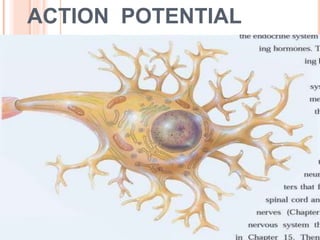
- 3. Action potential :- An electrical signal that propagates along the surface of the membrane of a neuron (nerve cell ) due to the movement of ions ( sodium & potassium) between interstitial fluid and the inside of a neuron through specific ion channels in its plasma membrane
- 4. ELECTRIC SIGNAL Two types Graded potential :- short distance communication (e.g. mainly in the dendrites and cell body of a neuron) Action potential :- communication over long distance within the body Graded potential :- on the basis of site of stimulus Post synaptic potential :- dendrites or cell body of a neuron in response to a neurotransmitter . Receptors & generator potential :- sensory receptors and sensory neurons .
- 6. The production of these types of potential depends on few basic features of the plasma membrane of excitable cells a) Existence of membrane potential b) Presence Of specific types of ion channels
- 7. MEMBRANE POTENTIAL An electrical potential difference across the membrane It is like voltage stored in battery In living cells the flow of ions rather than electrons constitutes the electrical current/ signal Because the lipid bilayer of the plasma membrane is a good electrical insulator . The main path for current to flow across the membrane are through ion channels.
- 8. ION CHANNELS Ion channels open and close due to presence of gates ( gate is a part of channel protein that can seal the channel, pore shut or move aside to open the pore) When ion channels are opened, they allow specific ions to move across the plasma membrane, down their electrochemical gradient. As ions move , they create a flow of electrical current that can charge the membrane potential
- 10. The electrical signal produced by nerve and muscle fiber rely on four types of ion channels a) Leakage channel :- randomly open and close b) Ligand - gated channel :- open in response to the binding of a ligand (chemical) stimulus c) Mechanical- gated channel :- mechanical stimulus ( touch, press ,vibration, tissue stretching ) d) Voltage- gated channel:- voltage stimulus
- 11. RESTING MEMBRANE POTENTIAL (RMP) RMP exists because of a small build of negative ions in the cytosol along the inside of the membrane and an equal build up of positive ions is the extra-cellular fluid (ECF) Such separation of positive and negative electrical charges is a form of potential energy which is measured in volts or millivolts.
- 12. In neurons the RMP ranges from -40 to -90 mv ( typically -70mv). here negative sign indicates that the inside the cell is negative relative to the outside. A cell that exhibits a membrane potential is said to be polarized. Resting membrane potential arises from three major factors. 1. Unequal distribution of ions in the ECF and cytosol. ECF is rich in sodium and chloride ions Cytosol - potassium , phosphate and amino acids
- 13. Because the plasma membrane typically has more potassium leakage channels than sodium leakage channel , the numbers of potassium ions that diffuse down their concentration gradient out of the cell into the ECF in greater than the number of sodium ions that diffuse down their concentration gradient from the ECF into the cells.
- 14. As more and more positive potassium ions exit , the inside of the membrane becomes increasingly negative and the outside of the membrane becomes increasingly positive. 2. Inability of most anions to leave the cell some anions cannot follow potassium out of the cell because they are attached to non – diffusible molecules such as ATP and large proteins.
- 15. 3. ELECTRO GENIC NATURE OF THE SODIUM/POTASSIUM ATPASE SODIUM – POTASSIUM PUMP Small inward sodium leak and outward potassium leak are offset by the sodium/potassium ATPase ( sodium – potassium pump ) Uneven distribution of sodium and potassium channel if (sodium channel increase then more positivity inside the cell ) Pumps out sodium ions as fast as it leaks in and at the same time , the sodium –potassium ATPase bring in K+ . Na+/K+ ATPase expel 3 Na+ for each 2 K+ imported. These pumps remove more positive charges from the cell than they bring into the cell , they are electro genic , which means they contribute to the negativity of the resting membrane potential .
- 16. Na+- K+ pump
- 17. GENERATION OF ACTION POTENTIAL Action potential / impulse is a sequence of rapidly occurring events that decreases and reverse the membrane potential and then eventually restore it to the resting state . Two main phase 1. Depolarizing phase 2. Repolarizing phase
- 18. Action potential
- 19. ALL OR NONE PRINCIPLE . Supra – threshold And sub - threshold stimulus doesn't cause an action potential because it does not bring the membrane potential to the threshold . Threshold stimulus is just enough to depolarize the stimulus is strong enough to depolarize the membrane above threshold . Action potential either occurs completely or it doesn't occur at all.
- 21. 1. DEPOLARIZING PHASE Negative membrane potential becomes less negative , reaches to zero and then positive . Stimulus – membrane of the axon to depolarize to threshold ( -55mV ) , voltage gated Na+ channel open rapidly . Inward movement of Na+ changes the membrane potential from -55mV to +30mV.
- 23. Voltage gated Na+ channel 1. Resting state :- inactivation gate is open , but the activation gate is closed. 2. Threshold state :- both activation and inactivation gate are open . As more channels open , Na+ inflow increases , the membrane depolarizes further and more Na+ channel open – positive feedback mechanism .
- 25. 2. REPOLARIZING PHASE K+ channels are opening , accelerating K+ outflow , slowing of Na+ inflow and acceleration of K+ outflow causes the membrane potential to change from +30mV to -70mV .
- 28. AFTER – HYPERPOLARIZING PHASE Voltage gated K+ channels open Membrane potential becomes even more negative ( - 90mV ) As the voltage gated k+ channel close , the membrane potential returns to the resting level of -70mV .
- 29. REFRACTORY PERIOD The period of time after an action potential begins during which an excitable cell cannot generate another action potential in response to a normal threshold stimulus . During the absolute refractory period , even a very strong potential cannot initiate second action potential . During the relative refractory period second action potential can be initiated , but only by a larger than normal stimulus .
- 30. Action potential
- 31. Because an action potential travels from point to point along the membrane without getting smaller, it is useful for long- distance communication. Nerve impulse propagation in which the impulse “leaps” from one node of Ranvier to the next along a myelinated axon is saltatory conduction. Saltatory conduction is faster than continuous conduction. Axons with larger diameters conduct impulses at higher speeds than do axons with smaller diameters. The intensity of a stimulus is encoded in the frequency of action potentials and in the number of sensory neurons that are recruited.
- 32. CARDIAC ACTION POTENTIAL The action potential initiated by the SA node travels along the conduction system and spreads out to excite the “working” atrial and ventricular muscle fibers, called contractile fibers. An action potential occurs in a contractile fiber by :- 1. Depolarizing phase 2. Plateau phase 3. Repolarizing phase
- 33. 1. DEPOLARIZING PHASE When a contractile fiber is brought to threshold by an action potential from neighboring fibers, its voltage gated fast Na channels open. Opening of these channels allows Na inflow because the cytosol of contractile fibers is electrically more negative than interstitial fluid and Na concentration is higher in interstitial fluid. Inflow of Na down the electrochemical gradient produces a rapid depolarization. Within a few milliseconds, the fast Na channels automatically inactivate and Na inflow decreases.
- 34. 2. PLATEAU PHASE a period of maintained depolarization. It is due in part to opening of voltage-gated slow Ca2 channels in the sarcolemma. When these channels open, calcium ions move from the interstitial fluid (which has a higher Ca2 concentration) into the cytosol. This inflow of Ca2 causes even more Ca2 to pour out of the sarcoplasmic reticulum into the cytosol through additional Ca2 channels in the sarcoplasmic reticulum membrane. The increased Ca2 concentration in the cytosol ultimately triggers contraction.
- 35. voltage-gated K channels are also found in the sarcolemma of a contractile fiber. Just before the plateau phase begins, some of these K channels open, allowing potassium ions to leave the contractile fiber. Therefore, depolarization is sustained during the plateau phase because Ca2 inflow just balances K outflow.
- 36. 3. REPOLARIZING PHASE After a delay (which is particularly prolonged in cardiac muscle), additional voltage-gated K channels open. Outflow of K restores the negative resting membrane potential (90 mV). At the same time, the calcium channels in the sarcolemma and the sarcoplasmic reticulum are closing, which also contributes to repolarization.
- 37. The mechanism of contraction is similar in cardiac and skeletal muscle , Propagation of a muscle action potential along the sarcolemma & into the T-tubule system initiates the events of muscle contraction. The electrical activity (action potential) leads to the mechanical response (contraction) after a short delay.
- 38. As Ca2 concentration rises inside a contractile fiber, Ca2 binds to the regulatory protein troponin, which allows the actin and myosin filaments to begin sliding past one another, and tension starts to develop. Substances that alter the movement of Ca2 through slow Ca2 channels influence the strength of heart contractions. Epinephrine, for example, increases contraction force by enhancing Ca2 flow into the cytosol.
- 39. The refractory period of a cardiac muscle fiber lasts longer than the contraction itself . As a result, another contraction cannot begin until relaxation is well underway. For this reason, tetanus (maintained contraction) cannot occur in cardiac muscle as it can in skeletal muscle. ventricles pumping function depends on alternating contraction (when they eject blood) and relaxation (when they refill). If heart muscle could undergo tetanus, blood flow would cease.
- 41. Comparison of excitable cells Cell RMP Duration of impulse Neuron / nerve fibres -70mV 0.5-2 mSec Skeletal muscle fibres -90mV 1-5mSec Cardiac & smooth muscle fibres -90mV 10-300mSec