
Recommended
More Related Content
Similar to Urinalysis Hands on Lab AssignmentIntroductionThe million nephr.docx
Similar to Urinalysis Hands on Lab AssignmentIntroductionThe million nephr.docx (20)
More from jessiehampson
More from jessiehampson (20)
Recently uploaded
Recently uploaded (20)
Urinalysis Hands on Lab AssignmentIntroductionThe million nephr.docx
- 1. Urinalysis Hands on Lab Assignment Introduction: The million nephrons in each of your kidneys form urine and which allows the body to get rid of metabolic wastes from blood and maintain homeostasis by regulating blood pH, water volume and ion concentrations in blood. Urinalysis is a standard test that can determine if the urine formation is normal or abnormal. In doing this lab we can determine the volume of urine, chemical and physical properties, and microscopic composition along with levels of some metabolic waste materials in blood. Pre-activity There are three processes for making urine, glomerular filtration, tubular reabsorption, and tubular secretion. Below, for each of the processes below, define them and describe where they occur in the nephron. 1. Glomerular Filtration 2. Tubular Reabsorption 3. Tubular Secretion Activity 1: The Nephron Make a hand drawn picture of the nephron, make sure to include all of the structures and information listed below. A. Renal corpuscle B. Renal tubule C. Location of glomerular filtration D. Location and directional arrow of tubular reabsorption E. Location and directional arrow of tubular secretion F. Proximal convoluted tubule G. Distal convoluted tubule H. Glomerular capsule (Bowman’s capsule) I. Collecting duct J. Ascending loop of Henle K. Descending loop of Henle
- 2. Post your labeled picture here. Activity 2: Renal Reabsorption and Secretion Using pages 970-988 in your lecture book and these videos Urinary System 1 (opens window) and Urinary System 2 (opens window) use the table below to predict the difference in osmolarity between the filtrate in the glomerular capsule and the other regions of the nephron. Will it be higher, lower, the same as, or dependent on hormones? For example, looking at the first structure in the table, does the filtrate found at the beginning of the proximal convoluted tubule have a higher/lower/same as the filtrate found in the glomerular capsule or does it depend on the hormones present? Nephron structure Glomerular Capsule Beginning of proximal convoluted tubule End of proximal convoluted tubule End of the descending limb of the Loop of Henle End of the ascending limb of the Loop of Henle Distal convoluted tubule Collecting duct Table 1: Filtrate Osmolarity Comparison Activity One and Two Conclusion Questions 1. What is filtrate? What does it contain? 2. There are several substances in blood that are not filtered in the glomerulus. What are these substances? What prevents them from being filtered out? (Hint: think about the type of capillary makes up the glomerulus.)
- 3. 3. What factors play a part in glomerular filtration rate? What is the main factor that determines the rate of glomerular filtration? How is the glomerular filtration rate regulated? 4. Explain the countercurrent mechanism for urine concentration. Make sure you address 1. What is being added to or removed from the filtrate in the Loop of Henle and how that happens. 2. Compare the osmolarity of the filtrate at the beginning and end of the Loop of Henle, explain why you go that answer. Make sure this is in your own words. 5. What does ADH determine in regards to the filtrate osmolarity? Where do we see this effect most in the nephron? Explain how ADH causes this effect to occur. Activity 3: Urinalysis Normal urine is composed of water, urea, creatinine, uric acid, electrolytes and occasionally small amounts of hormones, pigments, carbohydrates, fatty acid, mucous and some enzymes. In this activity you will examine the physical characteristics of your own urine sample. You will observe color, cloudiness and odor. You will then compare your sample to a chart showing normal characteristics of urine. Materials Needed: · Urinalysis Test Strip · Test Strip Analysis Key · Paper cup for urine sample · Paper Towel Procedure: 1. Get a small cup to in which to collect your urine sample. 2. Before collecting, void a small amount of urine from your bladder to clear the urethra of any contaminants such as bacteria or menstrual blood. 3. Void into the small cup and wash your hands. a. Observe the physical characteristics of your urine sample and record the information into the data table (below). i. The color of your urine
- 4. ii. Turbidity (is it cloudy) 4. To test the chemical properties of your urine. a. Submerge the dip stick into your urine for approximately 10 seconds. b. Record the following characteristics in the data table (below). i. pH ii. specific gravity iii. glucose iv. blood v. protein vi. bilirubin vii. leukocytes 5. After you have finished the analysis of the test strip Images Make sure you provide at least two pictures of you doing the lab and your results. Data Table For the data table, you must research what the normal values or properties for each characteristics. Input your results in the analyzed values column. Finally, you must explain the significance of each characteristic and why these values are used during to analyze urine. Characteristics Normal Values Analyzed Values Significance Urine color Turbidity
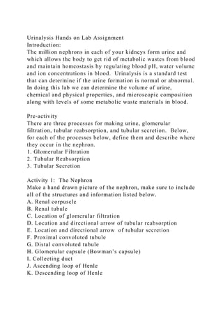
- 5. pH Specific gravity Glucose Blood Protein Bilirubin Leukocytes Table 2: Urine AnalysisResources to help you complete your analysis: If you are using the urinalysis strip we provided, below is the key to read the results.
- 6. Figure 1: 10 Parameter Urinalysis Strip Key Urinalysis Conclusion Questions 1. What makes urine have a higher specific gravity than distilled water? 2. If student’s urine shows positive for glucose, is this normal? Explain what could be the cause of the excess glucose? 3. Is normal daytime urine output diluted or concentrated?