Feasibility of CT scan studies with triple split bolus intravenous contrast ...
Somatom session 27
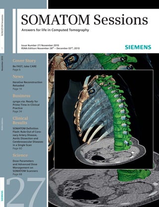
1. SOMATOM Sessions
Answers for life in Computed Tomography
Issue Number 27/ November 2010
RSNA-Edition /November 28th – December 03rd, 2010
Cover Story
Be FAST, take CARE
Page 6
News
Iterative Reconstruction
Reloaded
Page 14
Business
syngo.via: Ready for
Prime Time in Clinical
Practice
Page 34
Clinical
Results
SOMATOM Defi nition
Flash: Rule-Out of Coro-nary
Artery Disease,
Aortic Dissection and
Cerebrovascular Diseases
in a Single Scan
Page 60
Science
Dose Parameters
and Advanced Dose
Management on
SOMATOM Scanners
Page 68
27
RSNA-Edition November 2010 27 SOMATOM Sessions
2. Editorial
2 “With FAST CARE we address
todays’ challenges of our
customers, accelerate CT
workfl ows and reduce patient
exposure even further.”
Sami Atiya, PhD, Chief Executive Office,
Business Unit Computed Tomography, Siemens Healthcare, Forchheim, Germany
Cover Page: Courtesy of University of Erlangen- Nuremberg, Erlangen, Germany
SOMATOM Sessions · November 2010 · www.siemens.com/healthcare-magazine
3. Editorial
Dear Reader,
Recent improvements in healthcare have
created a serious backlog of patients at
many medical facilities, creating a con-tradictory
situation: the medical care is
better but it has become more difficult
to be treated as medical facilities stagger
under an ever-increasing workload.
Adding to the contradictory matrix is a
medically well-informed public con-cerned
with radiation exposure. An effi-cient,
faster throughput of patients while
maintaining quality care has be come
the critical issue in modern health care.
The creative and innovative products
developed by Siemens to deal with this
situation are truly amazing. The revolu-tionary,
single-source SOMATOM
Definition AS (and AS+) scanner that
reduces many scans to a one click op-eration
at extremely low dose. The
second noteworthy is the unique
SOMATOM Definition Flash scanner that
scans an entire thorax in less than one
second with sub-mSv dose and can
“freeze” even the fastest beating heart,
producing diagnostic quality cardiology
images in minutes.
We then introduced the syngo.via*,
multi-modality imaging software. With
syngo.via*, the reading physician can
observe and analyze CT, MR, PET,
Radio graphy, Fluroscopy and Angio-graphy
simultaneously on a single
monitor – eliminating many trips from
the regular reading workplace to various
workstations. Another great advantage
of syngo.via* is the pre-processing
André Hartung,
Vice President
Marketing and Sales
Business Unit CT,
Siemens Healthcare
system. When a case is opened, many
pre-processing tasks such as table re-moval,
bone removal, curved planar re-for
mat ting, naming of vessels, ejection
frac tion calculations and orthogonal cuts
are already done. The reading physician
can start the interpretation and diagnosis
immediately.
The challenge now became combining
these (and many other) systems to re-lieve
pressure on hospitals and clinics by
increasing throughput while maintaining
quality medical care. This goal resulted in
the introduction of our new FAST CARE
platform at the recent RSNA convention
in Chicago. When it comes to the FAST
CARE plat form, incorporating “Fully
Assisting Scanner Technology” (FAST) and
“Com bined Applications to Reduce Ex-posure”
(CARE), the name says it all.
This new platform for the SOMATOM
Definition family, guides the user through
a CT scan in just a few intuitive steps,
starting with planning, through the ac-tual
scanning process, to recon struction
and evaluation of clinical images. In this
way, FAST prio ritizes considerations of
efficiency and focuses on patient-centric
productiv ity.
The CARE standard combines a variety of
Siemens’ innovations, like CARE kV, CARE
Child or the next generation of Iterative
Reconstruction, SAFIRE** that we have
intro duced at this years’ RSNA.
patients – including trauma or young
children – from head to toe without
having to repeat the scan. In addition you
now have the possibility to reduce dose
even further.
Additionally, in keeping with our tradi-tional
cooperation with out-of-house
experts, – radiologists and others who
are confronted daily with challenges
in their daily scanning practice – we have
launched the Siemens Radiation Reduc-tion
Alliance (SIERRA). This panel of
highly respected experts in the medical
imaging field will track and provide
valuable feed back and make recommen-dations
on dose-related subjects to
Siemens, infor mation that will mean
even healthier examinations for your
patients. Our ultimate goal with this
prestigious group is to reduce dose
exposure in CT to a level below 2.4 mSv,
the annual natural level of radi ation
always present in our environement.
More complete information and valuable
links on all these new and exciting deve-lop
ments can be found in the pages of
this SOMATOM Sessions issue. And
invisibly em bed ded in every page is a
factor that is not new here at Siemens…
better health care for all patients.
We wish you enjoyable and profitable
reading.
Sincerely,
Using these powerful tools enables you
to quickly examine your most challenging André Hartung
** syngo.via can be used as a standalone device or together with a variety of syngo.via based software options, which are medical devices in their own rights.
** The information about this product is being provided for planning purposes. The product is pending 510 (k) review, and is not yet commercially available in the U.S.
4. Content
Cover Story
6 Be FAST, Take CARE
News
12 CEO Corner: Excellence in Clinical
Practice
12 Working with syngo.via – an
In- Practice Report
14 Iterative Reconstruction Reloaded
16 Flash Spiral Dual Source CT for
Precise and Patient-Friendly
Transcatheter
Aortic Valve Implantation (TAVI)
Procedure Planning.
18 Siemens Launches SIERRA, the
Siemens Radiation Reduction
Alliance
19 Siemens CT Stroke Management:
Helping to Save Brain and Quality
of Life
20 A Pediatric Breakthrough: Auto-mated
Adaptation of CT Dose Levels
22 Expanding Radiodiagnostics:
University Hospital Hradec Králové,
Czech Republic
24 Full Cardiac Assessment with
syngo.via – Maximal Significance,
Minimal Dose
Contents
Cover Story
6 Technology should serve
the physician, not vice
versa. The true task of the
doctor is caring for the
patient, not handling
apparatus. Therefore,
FAST CARE is set to raise
the standard for patient-centric
productivity and
intro duces innovations for
patient dose reduction.
The result: safe, reprodu-cible
examinations that
involve less exposure and
are therefore more
effective and efficient.
4 SOMATOM Sessions · November 2010 · www.siemens.com/healthcare-magazine
20
A Pediatric Breakthrough
6
Be FAST, Take CARE
26 Advanced Imaging for Four-Legged
Patients
27 SOMATOM Definition AS Open –
Dedicated High-end CT for Radiation
Therapy Planning
27 Among Europe’s Best
28 SOMATOM Scanners: Ahead of the
Innovative Curve
Business
30 1,000th SOMATOM Definition AS
Installed – A Success Story
32 Time is Brain – A Comprehensive
Stroke Program at the University
of Utah Considerably Improves
Patients’ Outcome
34 syngo.via: Ready for Prime Time in
Clinical Practice
36 SOMATOM Spirit: A Choice That
Paid Off
All articles mentioned on the cover are
designated in orange.
5. Content
54 Volume Perfusion CT Neuro as a Reli-able
Tool for Analysis of Ischemic
Stroke Within Posterior Circulation
Acute Care
56 Dual Source, Dual Energy CT:
Improvement of Lung Perfusion
Within 5 Hours in a Patient With
Acute Pulmonary Embolism
58 Differentiation of Pulmonary Emboli
and Their Effect on Lung Perfusion
Determined With a Low-Dose Dual
Energy Scan
60 SOMATOM Definition Flash: Rule-Out
of Coronary Artery Disease, Aortic
Dissection and Cerebrovascular
Diseases in a Single Scan
62 SOMATOM Definition Flash: RIPIT to
the Rescue – Fast CT Examination
for Trauma Patients
Pulmonology
64 Xenon Ventilation CT Scan Demon-strates
an Increase in Regional
Ventilation After Bullectomy in a
COPD Patient
Orthopedics
66 SOMATOM Definition: Dual Energy
Locates Progressive Wrist Arthritis
SOMATOM Sessions · November 2010 · www.siemens.com/healthcare-magazine 5
Clinical Results
Cardio-Vascular
38 SOMATOM Definition Flash Ruling
out Coronary Artery Disease with
0.69 mSv
40 SOMATOM Definition Flash:
Low-Dose Abdomen Pediatric Scan:
Follow-Up Study of Fibromuscular
Dysplasia
42 CT Dynamic Myocardial Stress
Perfusion Imaging – Correlation
with SPECT
Oncology
44 SOMATOM Definition Flash: Motion-free
Thoracic Infant Scan: Follow-Up
Study After Chemotherapy
46 SOMATOM Definition Flash:
Dual Energy Carotid Angiography
for Rapid Visualization of
Paraganglioma
48 Total Occlusion of the Left Superior
Pulmonary Vein by a Metastasis
Detected with Dual Energy CT
50 SOMATOM Spirit: Follow-Up Exami-nation
of Cerebral Meningioma
Neurology
52 SOMATOM Definition Flash: Improv-ing
Image Quality of Brain Scans
With IRIS, X-CARE and Neuro
BestContrast
Science
68 Dose Parameters and Advanced
Dose Management on SOMATOM
Scanners
72 IRIS and Flash: Cardio CT with
Minimum Radiation Exposure
Delivers Precise Images
Life
74 Clinical Fellowship: Learning From
the Experts in the Field
76 STAR: Specialized Training in
Advances in Radiology
76 Evolve Update Facilitates Dose
Savings
77 Frequently Asked Questions
77 Siemens Healthcare is Proud to
Present a New Series of Live Clinical
Webinars
78 News at Educate Homepage:
Recommended CT Literature
78 Clinical Workshops 2011
79 Upcoming Events & Congresses
80 Corporate Magazines
81 Imprint
32
Time is Brain
60
SOMATOM Definition Flash:
Rule-Out of Coronary Artery Disease
6. Coverstory
Be FAST, Take CARE
FAST CARE reduces the complexity of
CT scans to just a few clicks and facilitates
even more reduction of dosage.
Technology should serve the physician, not vice versa. The true task of the
doctor is caring for the patient, not handling apparatus. Therefore, FAST
CARE is set to raise the standard for patient-centric productivity and introduces
several innovations for patient dose reduction. The result: safe, reproducible
examinations that involve less exposure and are therefore more effective and
effi cient. Dr. Michael Lell shared his observations and expectations with us.
By Hildegard Kaulen, PhD
The new generation of the FAST CARE software will be availabe for all SOMATOM Definition scanners spring 2011.
7. The medical profession is changing.
As patient numbers increase, budgets
are ever-decreasing. At the same time,
patients seek the assurance and the
advice of the physician. In the University
Clinic at Erlangen, Germany, too, the
numbers of examinations have been
skyrocketing, while the residence time
at the clinic has been going down. Less
and less resources for diagnostics are
available. Associate Professor Dr. med.
Michael Lell, Senior Physician at the Insti-tute
of Radiology, feels the pinch, espe-cially
when it comes to staff. This is why
he is particularly appreciative of soft-ware
solutions that not only leave him
more time for his obligations as a doctor
and researcher, but also optimizes the
utilization of staff. When it comes to
Siemens’ new FAST CARE technology,
incorporating “Fully Assisting Scanner
Technologies” (FAST) and “Combined
Applications to Reduce Exposure” (CARE),
the name says it all. The new platform
for the SOMATOM Definition product
family guides the user through a CT scan
in just a few intuitive steps, starting with
planning, through the actual scanning
process, to reconstruction and evalua-tion
of clinical images. In this way, FAST
prioritizes considerations of efficiency
and focuses on patient-centered produc-tivity.
Standardization ensures that all
examinations follow the same pattern,
avoiding errors and uncertainty. So,
scans that erroneously fail to depict
parts of the target organ can be avoided
in the future. At the same time, FAST
CARE also offers the user new solutions
for reducing the applied radiation dose
and supports the consistent use of
al ready available solutions. The entire CT
scan thus not only becomes more intui-tive
and reproducible, but also safer for
the patients.
Reducing users’ workloads
FAST Planning, one of the new function-alities
of FAST CARE, provides sugges-tions
for the scan and reconstructions
that are appropriate for the selected
mode based on the characteristics of the
organ, including the length of the exam-ination
volume. Thus, for example, in
the case of a cranial CT, the isocenter is
automatically adapted to the position of
the skull. CT scans are complex proce-dures
and operating the equipment is
demanding, even with standardized pro-tocols.
Lell agrees: there will always be
situations where the standard protocol
must be adapted to the stature of the
patient or the problem being investi-gated.
Also, the technical staff operates
not just one, but many modalities. The
constant back and forth between indi-vidual
applications makes high demands
of staff members’ expertise and concen-tration.
A program that guides users
intuitively through the entire CT scan
makes the task simpler, safer, more repro-ducible
and more efficient. “In view of
the fact that well-trained staff is increas-ingly
difficult to find,” Lell continues,
“this is an important aspect.” He has
high expectations for the automatic cou-pling
of the contrast agent injection
with the scanning protocol, which will
be offered as a special add-on feature
for the standard package under the des-ignation
CARE Contrast III. “Currently,
two staff members work on examina-tions
involving contrast agents,” says
Lell. “One of them injects the contrast
agent, while the other prepares the scan
. If the injection and the scan are linked,
Coverstory
“A program that
guides users intu-itively
through
the entire CT scan
makes the task
simpler, safer,
more reproduc-ible
and more
effi cient.”
Michael Lell, MD, PD, Departement
of Radiology, University of
Erlangen-Nuremberg, Erlangen,
Germany
we can do the same work with one less
staff member. Since we have less and
less staff available due to cost reasons,
that would be a major economization.”
If the selected scan parameters create
conflicts, FAST CARE resolves them
through a single click on the FAST Adjust
button. On occasion, Dr. Lell explains,
a selected scan protocol could combine
different parameters in such a manner,
that scanner will prevent the scan
in order to avoid a faulty result. Currently,
University of Erlangen-Nuremberg,
Erlangen, Germany.
8. Coverstory
such situations have to be resolved man-ually,
which costs time. With FAST CARE,
the FAST Adjust function suggests the
ideal solution. But the focus is also on
faster diagnostics. This is where the
strengths of syngo.via,* Siemens new,
leading-edge imaging software, come
into effect. The software automatically
loads the images into the appropriate
application and segments them in such
a way that they can be adjudged with-out
further ado. The physician can arrive
at a final diagnosis with just a few clicks
of the mouse as the images have already
been pre-processed for him. The applica-tion
is determined by the disease-specific
criteria of the case at hand and no longer
needs to be independently selected.
Since syngo.via handles all preparatory
steps, the physician can focus com-pletely
on his actual task, namely diag-nostics.
This, too, saves time and
enhances diagnostic reliability.
Improved image reconstruction
FAST CARE also introduces SAFIRE,**
Siemens’ first raw-data-based iterative
reconstruction. This technique removes
noise and artifacts in iterative steps in
the image and raw data domain, with-out
compromising image sharpness. The
procedure can be used in two different
ways. Either the image quality of the
standard reconstruction is maintained,
and the dose can be reduced, or the dose
level is maintained and clinical images
of noticeably higher quality are gener-ated.
Until now, however, calculation of
the projection data required significantly
more time than the standard reconstruc-tion.
For FAST CARE, the image space
algorithm was enhanced and a new
reconstruction computer was specially
developed for this purpose. This now
also allows use of raw data in the recon-struction
process to further enhance
image quality and reduce dose. In this
way, users can take advantage of the
potential for dose reduction in a notice-ably
greater number of examinations
during routine clinical application, signifi-cantly
reducing the average dose. (For
further information, see the article “Iter-ative
Reconstruction Reloaded” on page
14 in this issue.) Using the potential of
SAFIRE, 72% of all Siemens standard pro-tocols
apply dose of below the average
annual natural background radiation of
2.4 mSv.***
Michael Lell has performed clinical
studies with the previous version of the
software. He describes the results: “For
research purposes, we always perform
both the standard reconstruction and the
iterative reconstruction. With the previ-ous
algorithm, iterative reconstruction
takes about four to five times longer than
standard reconstruction. Here, I expect
a clear improvement with the new algo-rithm.
With the previous algorithm an
abdominal CT can be performed using
half the dosage without compromising
image quality. Our work on thoracic CT
has not yet been concluded, but the
potential for dosage reduction is ex pec-ted
to be of a similar order of magni-tude.
These are considerable reductions
of dose that should be used. If the new
algorithm is faster and offers better
image quality, it is ready for routine
application.”
Optimal scan parameters
for everyone
When it comes to the sensitive issue
of radiation exposure, Siemens follows
the ALARA principle: “As Low As Reason-ably
Achievable.” FAST CARE comes
with CARE kV, an expansion of CARE
Dose4D, which modulates the tube cur-rent
according to the patient’s anatomy.
In addition, CARE kV now automatically
identifies the optimal tube voltage and
adapts the tube current accordingly.
This change is useful, for instance,
when contrast agents are used. Because
2
“If the new algorithm
is faster and offers
better image quality,
it is ready for routine
application.”
Michael Lell, MD, PD, Departement of Radiology,
University of Erlangen- Nuremberg, Erlangen, Germany
*** syngo.via can be used as a standalone device or together with a variety of syngo.via based software options, which are medical devices in their own rights.
*** SAFIRE: The information about this product is being provided for planning purposes. The product is pending 510 (k) review, and is not yet commercially available in the U.S.
*** Data on fi le.
9. 1 Manually setting the scan range too
short in the topogram can cut off relevant
parts of the examined organ.
2
3 FAST Planning uses the defined anatomical landmarks to set the correct ranges. When
applied manually without FAST CARE, only based on the coronal view the lower part of the
lung could be easily be missed (indicated by the reference line).
4 Direct setting of the scan range in with FAST Planning assures covering the entire
organ without overscanning
SOMATOM Sessions · November 2010 · www.siemens.com/healthcare-magazine 9
the higher iodine contrast more than
makes up for the higher absorption of
iodine, a lower tube voltage can be
applied. In this case, however, the mAs
value should be adapted. This requires
quite a bit of familiarity with the tech-nology.
Many users are not confident
enough to make that adaptation and
therefore do not exploit the potential to
be gained from changing the tube volt-age.
CARE kV takes this insecurity out by
preparing the appropriate kV and mAs
value, thus taking the burden off the
user. Also, CARE Dashboard can be used
to display which dose-reducing mea-sures
are available for the scan regions
selected in the scanning protocol and
whether these have been activated. Lell
explains: “We have a legal and moral
obligation to protect patients from
unnecessary radiation. The Medical Ser-vice,
tasked with providing the radiation
protection of supervisors and physicians
involved with suggestions for improving
radiation protection, reducing radiation
exposure and enhancing image quality,
routinely checks whether we adhere to
this obligation. CARE kV and CARE Dash-board
give us further support in this
area. Many users, however, do not use
the available solutions consistently
enough. Automation is useful, but we
also need better training. The various
options for dose reduction must be cho-sen
suitably.” For instance, Lell has
found that caution is required when
using specific solutions on children.
Therefore, new parameter sets were
developed for CARE Dose4D that take
into account the specific anatomy of the
child. Also, the STRATON tube
was developed further so that in case
of pediatric scans, the voltage can be
reduced to 70 kV.
The issue of dose cannot be discussed
independently of the diagnostic evalua-tion
when it comes to CT. A clear deci-sion
is always required as to when the
clinical necessity of a CT examination is
greater than the potential risks of radia-tion
exposure. Lell believes dose can
also be reduced by ensuring that the
selected examination area is defined as
narrowly as possible, which FAST CARE
does automatically. Furthermore, the
1
2 Manually setting the scan range too
long in the topogram could potentially
over-radiate the patient
3
4
10. Coverstory
5
6
10 SOMATOM Sessions · November 2010 · www.siemens.com/healthcare-magazine
requirements for image detail should be
limited to what is necessary for resolving
the problem at hand. In planning a lung
biopsy, less detail is required than when
searching for metastases. “Therefore,”
emphasizes Dr. Lell, “all radiologists
should ask themselves what degree of
quality is in the best interests of the
patient.” This, too, is an important con-tribution
to reducing radiation exposure.
Improving visualization and
management of dose
FAST CARE also offers a number of
functionalities that serve to visualize the
radiation given to the patient during
the scan. Before the start of the exami-nation,
CARE Profile displays the course
of the dose to be applied according
to the patient’s anatomy. The user can
also determine reference values and
upper limits for the individual protocols
and request notification when the scan
approaches these limits, as required
under a new IEC standard. Furthermore,
the software includes applications for
quality control. Currently, the CTDIvol
and DLP data specified in the patient
protocol must still be entered manually
into a quality control monitoring pro-gram.
This is arduous and time-consum-ing
work. FAST CARE stores the data into
the DICOM Dose SR with CARE
Analytics that then can be evaluated.
Lell explains: “Automatic data export
offers unforeseen opportunities for qual-ity
control. It would be possible to review
the average dosage distribution values
for every day and to check which scans
exceed or fall below a certain value.
Currently, such a degree of quality con-trol
is still unattainable.”
5 FAST Cardio
Wizard: It is an
intuitive guid-ance
software,
integrated
in the Cardio
workflow.
6 Anatomically
correct spine
reconstructions
are typically
very time con-suming
proce-dures,
as every
spinal cord and
disc needs to
have an own
recon layer
depending on
its individual
position. With
FAST Spine,
these manual
steps can be
simplified to
ideally just a
single click.
Assistant Professor Dr. med. Michael Lell studied at the University of Regensburg and Technische
Universität München. He is specialized in diagnostic radiology. Currently, he is Senior Physician at
the Institute of Radiology, Erlangen University Clinic, Erlangen, Germany, where he has been working
since 1997. He was a visiting researcher at the David Geffen School of Medicine at the University
of California, Los Angeles, and is a member of various national and international professional bodies.
He is also a peer reviewer of several medical journals.
11. Coverstory
Dr. Hildegard Kaulen is a molecular biologist.
After sojourns at the Rockefeller University in
New York and Harvard Medical School in Boston,
USA, she has been working as a freelance sci-ence
journalist for prestigious daily newspapers
and science journals since the mid-1990s.
SOMATOM Sessions · November 2010 · www.siemens.com/healthcare-magazine 11
Dr. Sodickson, in the past three years,
concerns have been raised about
cumulative exposure by repetitive CT
imaging. How serious is the problem?
SODICKSON: There is persistent contro-versy
over the risk models that exist for
radiation exposure of the magnitude
used in CT. We attempted to quantify the
levels of risk using the most common
Linear-No-Threshold risk model used in
the 7th Biological Effects of Ionizing
Radiation (BEIR-VII) report. We studied
32,000 patients undergoing CT at our
institution, using the BEIR-VII model to
estimate cumulative cancer risks from
CT exposures. We found that 7% of our
cohort had undergone enough previous
CT radiation exposure to increase their
cancer risk by at least 1% or more above
baseline. As a result, we believe that
patients undergoing recurrent imaging
over time warrant heightened radiation
protection efforts.
Many CT users don’t take full advan-tage
of the available dose reduction
tools and work with protocols that are
not fully optimized. Is active assis-tance,
such as that provided by FAST
CARE, the key to a more universal
adoption?
SODICKSON: Active assistance is one of
many excellent solutions. Any automa-tion
that makes scanning easier and
helps to create reproducible results
across the wide range of patient sizes
and technologist skill levels is extremely
valuable. But we also need better default
protocols that are dose-optimized and
“We Need Better
Default Protocols.”
Dr. Aaron Sodickson, MD, PhD, Assistant Director of
Emergency Radiology, Brigham and Women’s Hospital,
Harvard Medical School, Boston, spoke to journalist
Dr. Hildegard Kaulen for SOMATOM Sessions:
robust in order to ensure adequate diag-nostic
image quality for every patient.
We need close collaboration between CT
manufacturers, radiologists, technolo-gists,
and medical physicists. By com-bining
our different areas of expertise,
we can best reach consensus about
what works and what doesn’t, and what
represents adequate image quality for
the particular diagnostic task at hand.
What are the essentials for a radiation
risk assessment program?
SODICKSON: We should routinely review
the imaging history of our patients. We
are working to implement a decision
support system that alerts ordering phy-sicians
in real time of the magnitude of
a patient’s radiation risk. Our goal is to
bring appropriate perspective to the risk/
benefit decision by providing the best
risk estimates possible. We hope this will
enhance an active and critical review of
the imaging order and an assessment of
how the scan fits into the longitudinal
medical history of the patient.
Will risk assessment interfere with
the workflow and lengthen the deci-sion
making and scanning process?
SODICKSON: That depends on how it is
implemented. We need solutions that
create an efficient workflow without
frustrating delays. Otherwise they might
not be accepted in clinical routine.
An exciting feature for dose reduction
is lowering kV. You had the chance to
test CARE kV, which is a part of FAST
CARE. Did the tool meet your expecta-tions?
SODICKSON: We assessed an early proto-type,
which worked quite nicely. Based
on the patient’s size, the system automat-ically
suggests kV and effective mAs set-tings
that minimize the applied dose
without compromising image quality.
This tool takes a great deal of guesswork
out of low kV scanning, making it feasi-ble
for all technologists.
As Assistant Director of Emergency
Radiology, where do you see addi-tional
potential for increasing patient
care further, besides the ever-present
topic of continuous dose reduction?
SODICKSON: We need dose-optimized
default protocols that work in fast-paced,
sometimes chaotic settings such
as the ED, and can be used reliably by
technologists of all skill levels. We need
streamlined workflow to scan even our
sickest patients with reliably low dose
and high quality results every time. We
need improved education to ensure that
every user is aware of the excellent
dose-reduction tools that are available,
and knows how to use them correctly.
And finally, we need improved methods
to capture patient- and exam-specific
dose information from every scan, both
for real-time quality control and for
longitudinal dose-monitoring efforts.
12. News
CEO Corner: Excellence in Clinical Practice
Excellence in Clinical Practice through
innovation & responsibility remains the
cornerstone of Siemens’ leadership in
the CT medical imaging field. A constant
source of strength as aging markets in
industrial countries, and dynamic mar-kets
in rapidly developing countries,
demand better health care at lower cost.
We help you meet these challenges in
four key areas:
■ You can depend on us, as undisputed
trendsetter in CT technology, for the
industry’s fastest and healthiest single
and Dual Source scanners – today and
into the future.
■ To improve your clinical efficiency, we
support you with workflow excellence,
ease of use and high reliability.
■ As your caring partner, we maintain
highest industrial standards in cus-tomer
relationship & care.
■ To make state-of-the-art CT affordable
– and financeable – for you, we have
introduced the new Excel Editions of
our highly efficient 16- and 64- slice
scanners.
Reducing our vision to its essence:
As a caring partner of our customers, we
create CT-innovations that lift clinical
practice to a higher level of excellence
and enable wide access to better patient
care. Our ambitious global team contin-uously
Working with syngo.via – an In-Practice Report
Physicians and technologists at the department of radiology at the University
of Pennsylvania Hospital (HUP) have been evaluating the syngo.via*
software for two years now. Harold I. Litt, MD, PhD, assistant professor of
radiology and chief of the cardiovascular imaging section, reports on his
experiences with syngo.via in his daily routine.
By Michaela Spaeth-Dierl, medical editor, Spirit Link Medical, Erlangen
The Hospital of the University of Penn-sylvania
has a reputation as a world
leader in medical research and clinical
care. Since 1765, it has been dedicated
to the care of patients, the education of
physicians and development and imple-
mentation of new medical knowledge.
HUP therefore seemed to be the right
place to evaluate one of the first
research systems of the new syngo.via
software from Siemens, and the radiol-ogy
department there has now been
12 SOMATOM Sessions · November 2010 · www.siemens.com/healthcare-magazine
sets the trend in an always
changing environment… providing
answers for life.
We are looking forward, that in the years
ahead, you will continue to work with us
in our efforts to uphold excellence in CT’s
clinical practice.
evaluating it for two years. All cardiovas-cular
CT and MRI exams, neurovascular
CT, and body CT studies requiring addi-tional
processing (e.g. CT urography and
colonography) are automatically routed
to the syngo.via server, and six radiolo-
Dr. Sami Atiya, CEO Business Unit CT,
Siemens Healthcare, Forchheim, Germany
13. “Looking at curved MPR’s used
to take a lot of clicks and usually
wasn’t worth it. Since you now get it
automatically, I’m looking at them
in almost every case.”
Harold I. Litt, MD, PhD, Assistant Professor of Radiology and Medicine,
Chief, Cardio vascular Imaging Section, Depart ment of Radiology,
University of Pennsylvania School of Medicine, Philadelphia, PA, USA
SOMATOM Sessions · November 2010 · www.siemens.com/healthcare-magazine 13
gists and four 3D technologists regularly
work with the system. In his section,
Harold Litt mainly interprets cardio-vascular
studies with syngo.via. “With
syngo.via, the daily routine has changed.
Compared to a stand-alone workstation,
a thin-client system like syngo.via* has
benefits for both workflow and time,”
he summarizes his experiences.
A great advantage of syngo.via is the
automated pre-processing. When a case
is opened, many pre-processing tasks
such as table removal, bone removal,
curved planar reformatting, naming of
vessels, ejection fraction calculations
and orthogonal cuts are already done.
So, the radiologists can start their inter-pretation
immediately.
“My experience with syngo.via* in car-diac
CT is that the pre-processing of data
is very accurate and requires few edits.
This means fewer corrections and faster
reading,” says Dr. Litt. Compared to
other thin client technology, there are
also differences. Previously the workflow
involved the following: the data from
the scanners was sent to dedicated
workstations, where the cases were
post-processed by dedicated 3D technol-ogists.
The techs captured screenshots
of their results, saving them on the PACS
and manually transcribing any numeric
results into a web-based system. Radiol-ogists
would review the captured images
on PACS, another workstation, or a
thin-client system, then copy and paste
results from the web-based system to
their reports in the RIS. If the radiologist
wanted to review the technologist’s
work directly, it would mean a walk to
the 3D lab and reloading the case on a
workstation.
Now, and in the future with syngo.via,
all users access the same database.
Technologists prepare the cases and
forward their results to the radiologists
through “shared reading.” Radiologists
can start reviewing each case where
they are sitting and do not need to walk
to the workstations anymore, and tech-nologists
no longer need to type their
measurements into a separate system.
Furthermore, syngo.via allows its users
to load cases from different modalities
such as echocardiography or CT angio-graphy.
The series navigator shows all
images related to the opened patient, so
radiologists don’t have to search for the
right series from the right patient in the
entire patient list.
“Concerning several of the dedicated
features available, the right ventricular
analysis (RVA) within the syngo.CT
Cardiac Function – Right Ventricle** is
very much appreciated.” says Harold I.
Litt. “We study many patients with
congenital heart disease as well as those
undergoing electrophysiology ablation
procedures. Being able to calculate RV
ejection fraction without manual con-touring
saves half an hour per case.
Now you get the LV and RV wall motion
analysis and EF automatically as soon as
you open a case – without any waiting
or interaction.”
Experience that testers of syngo.via have
gained in the department of radiology at
HUP shows that the use of this software
provides a simplification of clinical work-flows
and time savings.
News
** syngo.via can be used as a standalone device or
together with a variety of syngo.via based software
options, which are medical devices in their own
rights.
** syngo.CT Cardiac Function- Right Ventricle is not
commercially available in the US.
Dr. Litt has received grant funding from Siemens
for research related to this product.
14. News
Iterative Reconstruction
Reloaded
For the fi rst time, SAFIRE* introduces the usage of raw-data information
within iterative reconstruction for everyday use in clinical practice.
By Jan Freund, Business Unit CT, Siemens Forchheim, Germany
For quite some time, iterative recon-struction
has been heavily discussed in
the CT community as a highly promising
method to achieve significant dose
reduction without compromising image
quality. Essentially, iterative recon-struction
introduces a correction loop in
the image generation process that
cleans up artifacts and noise in low-dose
images. The proposed approach is, that
after the initial reconstruction using the
weighted filtered back projection
(WFBP), the measured data of the
acquired image (in the so-called image
space) is compared to the data (raw-it
1A 1B 1C 1D
14 SOMATOM Sessions · November 2010 · www.siemens.com/healthcare-magazine
had to conquer the challenge of per-formance.
In order not to do so at the
expense of image quality – a “plastic-like”
image impression was one of the
major drawbacks of other solutions –
Siemens found a smart alternative:
The innovative first step was the recon-struction
of a super-high resolution
image that had virtually no image loss.
This was achieved by not applying the
filtering that typically reduced image
noise, taking into account that the
resulting image was then accordingly
very noisy, but contained all inform-ation.
The iteration loops to reduce the
data). But until now, the implemen-tation
of this method for clinical practice
was limited as the necessary re-trans-formation
of data from the image to the
raw-data space was very time-consuming
and the computational power required
to make it feasible for everyday use was
not available. Therefore, vendors found
several different approaches to handle
this limitation in their first individual
solutions.
The fi rst step – IRIS
At RSNA 2009, Siemens introduced its
solution – IRIS. Like all other vendors,
1A Plain FBP 1B Standard Siemens’ WFBP 1C IRIS 1D SAFIRE
15. SOMATOM Sessions · November 2010 · www.siemens.com/healthcare-magazine 15
noise in the image were then per formed
completely in image space, which was
the key to achieve the recon struction
performance and keep a well-known
image impression. This unique approach
then even found its way into the product
name: IRIS – Iterative Reconstruction in
Image Space. Several publications
proved IRIS to be highly effective when
it comes to reducing dose while main-taining
diagnostic image quality. The
University of Erlangen for example,
achieved average dose reductions of
50%** for abdomen examinations by
taking Dual Source datasets done with
the SOMATOM Definition Flash and
reconstructing the images based only
on data from one source. The resulting
images – now naturally utilizing only
half the dose – showed the same image
quality after being reconstructed with
IRIS compared to those reconstructed
without IRIS and utilizing the data from
both sources.
The next generation – SAFIRE
But now, Siemens actually shifted into a
higher gear and introduced the successor
at this year’s RSNA: SAFIRE – (Sinogram
Affirmed Iterative Recon struction)*. For
the first time, the use of raw data (which
is visualized in the so-called sinogram) is
actually being utilized in the image
im provement pro cess. Here, the current
set of CT images is transformed back into
raw data which models all relevant geo-metrical
pro perties of the CT scanner.
This step produces a CT raw-data set that
again resembles a virtual CT system. By
com paring the synthetic raw data with
the acquired data, differences are identi-fied.
This procedure can be regarded as
validating (or affirming) the current
images compared with the measured raw
data. The detected deviations are then
again reconstructed using WFBP, yielding
an updated image.
With this step, the images can be analy-zed,
subtracting image noise from the
previous images without loss of sharp-ness.
The same applies for potential arti-facts
that every vendor is confronted with
when using the WFBP and which often
remain in conventional CT images. Using
multiple iterations of these steps, geo-metrical
imperfections of the WFBP are
corrected in addition to incremen tally
reducing image noise. With this, SAFIRE –
Sinogram Affirmed Iterative Recon struc-tion
– can achieve a radiation dose re-duction
of up to 60%** at improved
image quality (contrast, sharpness and
noise), even surpassing the already
impressive image quality realized with
IRIS. This amazing achievement resulted
mainly from two measures: First, the
algorithms used in the iterations were
redesigned to make them more efficient.
And second, new image reconstruction
systems (IRS) – were developed and
intro duced parallel now finally providing
the compu tational means for the complex
calculations required. SAFIRE of course
also works with the former IRS but
naturally at a reduced performance.
With the new high performance IRS –
the FAST IRS – the performance is en -
han ced even further. The result: With
SAFIRE, the potential to reduce radiation
dose is up to 60%,** but at an signifi-cantly
improved image quality. The big
dif ference is now, that this potential is
accessible to a much larger number of
examinations, meaning that the average
dose saving over all examinations will be
significantly higher. Using the potential
of SAFIRE* 72% of all Siemens standard
protocols, apply dose of below the
average annual natural background
radiation of 2.4 mSv.** SAFIRE will be
com mercially available for all SOMATOM
Definition AS in March 2011 and for
SOMATOM Definition Flash in May 2011.
2 Improved noise reduction and workflow with SAFIRE*
2
** The information about this product is being pro-vided
for planning purposes. The product is pend-ing
510 (k) review, and is not yet commercially
available in the U.S.
** Results may vary. Data on file.
News
16. Topic
Flash Spiral for Precise and Patient Friendly
Transcatheter Aortic Valve Implantation (TAVI)
Planning.
By Peter Aulbach
Business Unit CT, Siemens Healthcare, Forchheim, Germany
Transcatheter heart valve implantation is
considered a technology with enormous
clinical potential. The percutaneous
implantation of a pulmonary valve was
reported for the first time in 2000. Since
then, these procedures have recorded
constant double-digit annual growth,1
since it presents a new option to candi-dates
for whom conventional surgery
was not suitable.
Clinical needs and challenges
The recent PARTNER trial, published in
the New England Journal of Medicine,2
demonstrates that transcatheter aortic
valve implantation (TAVI), in comparison
with standard therapy, resulted in signif-icantly
lower rates of death among
those patients. Patients who undergo
TAVI show a 45% reduction in the rate of
death in comparison with those receiv-ing
standard therapy.
Exact knowledge of the aortic root anat-omy,
including the proximal coronary
arteries, and the entire aorta up to the
femoral artery bifurcation, is necessary to
allow accurate pre-procedural planning.
After scanning with conventional proto-cols,
CT imaging requires relatively large
amounts of contrast which can be a prob-lem
in older patients, especially those
with concomitant renal disease. Prospec-tively
triggered high-pitch Flash Spiral
Dual Source CT (Flash Spiral), with up to
458 mm/s table feed, is able to obtain all
important anatomic information in one
single scan. Because of the extremely
rapid data acquisition, completed in less
than 2 seconds (Fig. 1B), the amount of
contrast agent can be reduced signifi-cantly.
In conventional aortic valve surgery, the
access route to the aortic valve is stan-dardized.
Normally the sizing of the
utilized valve prosthesis is done directly
under visual control at the surgical site.
In contrast, in TAVI procedures all these
points need to be meticulously addres-sed
during pre-operative planning, since
annulus size, access route or distance of
the coronary ostia to the aortic root will
influence the procedural strategy and
the appropriate selection of the artificial
heart valve.
Moreover, large amounts of contrast
agent have to be used in addition to the
16 SOMATOM Sessions · November 2010 · www.siemens.com/healthcare-magazine
contrast exposure during the aortic
valve implantation itself. In the TAVI
population, more than 50% of patients
show impairment of renal function
(elevated serum creatinine levels).
It is known that up to one third of all
patients undergoing catheter-based
aortic valve implantation develop acute
renal failure in the shortly following
post-operative course.3 Therefore the
application of contrast dye needs to be
reduced to a minimum.
Benefi ts of Flash Spiral CT
The latest Dual Source CT system, the
SOMATOM Definition Flash, allows the
use of prospectively triggered high-pitch
spiral data acquisition, called Flash
Spiral. This mode allows a significant
reduction of radiation dose compared to
other CT technologies. Effective radia-tion
doses of only 3-5 mSv are now only
needed to visualize all relevant thoraco-abdominal
structures (Fig. 1). Even
more importantly, within this patient
population, this new scan mode allows
an extremely rapid data acquisition in
less than 2 seconds (other CT technolo-
1A 1B
17. Topic
1 80-year old patient with severe aortic valve stenosis prior to trans-catheter
aortic valve implantation (TAVI). Pre-procedural Flash Spiral
angiography was performed using high-pitch spiral data acquisition pro-spectively
triggered at 60% of the R-R interval (128 x 0.6 mm slices,
100 kV, 320 mAs, SOMATOM Definition Flash). For thoraco-abdominal
angiography including the coronary arteries (Arrowhead) only 40 ml of
contrast agent was used (flow rate 4 ml /s). Estimated effective radiation
dose was 4.3 mSv. at a scan time of 1.7 seconds.
Images show assessment of aortic annulus diameters in syngo.via (Fig.
1A dotted line) as well as distances between the aortic annulus and the
coronary ostia. In addition, peripheral arteries have been evaluated for
significant stenosis (Fig. 1B). The red arrow indicates an occluded iliac
artery, making transfemoral access impossible here. The same data also
shows pronounced calcification along the whole thoracic aorta (Fig. 1C).
1 Cardiovascular News, Transcatheter heart valve
replacement: A European perspective,
www.cxvascular.com, Jan 2010
2 Valve Implantation for Aortic Stenosis in Patients
Who Cannot Undergo Surgery, N Engl J Med
2010
3 Aregger F, Wenaweser P, Hellige GJ, et al. Risk of
acute kidney injury in patients with severe aortic
valve stenosis undergoing transcatheter valve
replacement. Nephrol Dial Transplant 2009; 24:
2175–2179.
4 Vahanian A, Alfieri OR, Al-Attar N, et al. Transcath-eter
valve implantation for patients with aortic
stenosis: a position statement from the European
Association of Cardio-Thoracic Surgery (EACTS)
and the European Society of Cardiology (ESC), in
collaboration with the European Association of
Percutaneous Cardiovascular Interventions
(EAPCI). EuroIntervention 2008; 4: 193-199.
2 Up to 60%
less contrast
media by use of
high-pitch spiral
DSCT angio-graphy
of the
complete aorta
– compared to
other CT tech-nologies.
Courtesy of
University
of Erlangen-
Nuremberg,
Erlangen,
Germany
140 ml*
100 ml#
SOMATOM Sessions · November 2010 · www.siemens.com/healthcare-magazine 17
gies need about 6–9 seconds). This per-mits
a tremendous reduction of contrast
agent by 50–60%, which is crucial for
patients with renal insufficiency under-going
a subsequent TAVI procedure.
Compared to approximately 100–140 ml
of contrast agent needed in the past for
a CT angiography of the entire aorta, it
is now possible to use only 40 ml (flow
rate 4 ml/s) for the same examination,
which poses a significantly reduced risk
of Contrast Induced Nephropathy (CIN)
in this patient population (Fig. 2).
Accurate and fast planning
with syngo.via
The decision whether a patient is suit-able
for a catheter-based procedure and
the pre-operative planning with the
selection of the access route are based
upon results of the CT angiography. The
size of the aortic annulus for selection of
the valve prosthesis and the angulation
of the invasive fluoroscopy which allows
for simulating the optimal projection of
the aortic valve during the TAVI proce-dure
can be predicted from the same
DSCT angiography data with the support
of syngo.via.* This leads to further con-trast
media savings during the invasive
procedure since the syngo.via* software
automatically provides the correspond-ing
C-arm position.
On the basis of this protocol and ana-tomical
measurements by Flash Spiral
CT, physicians are able to quickly per-form
more patient friendly and precise
catheter-based procedures.
The time consuming planning of the
procedure is very well supported by the
many automated pre-processing steps
in the new syngo.via* software which
in early tests could show to reduce plan-ning
time by more than 33% (10 min.
versus 15 min.).
In a nutshell: Flash Spiral
and syngo.via
In conclusion the Definition Flash,
combined with the highly automated
syngo.via* workflow modules, provide
the most possible patient friendly and
accurate pre-operation planning solution
available. The high potential for cost
reduction coming from fewer patients
suffering acute CIN and therefore
requesting less of the expensive aftercare
is not yet taken into account herein.
SOMATOM Definition Flash:
www.siemens.com/SOMATOM-Definition-
Flash
CT Cardiovascular Engine:
www.siemens.com/CT-cardiology
Single-Source CT
for Abdominal
Aorta
160
140
120
100
80
60
40
20
0
*Loewe C, Eur Radiol 2010; #Wu W, AJR 2009; §Flash Thorax Protocol
40 ml§
Amount of Contrast Agent [mL]
Single-Source CT
for Triple Rule Out
Dual Source CT
SOMATOM
Definition Flash
2
News
* syngo.via can be used as a standalone device or
together with a variety of syngo.via based soft-ware
options, which are medical devices in their
own rights.
1C
18. “I am happy and proud to embark on this initiative together
with Siemens and my colleagues from around the globe in
order to ensure that Siemens’ powerful tools for dose reduc-tion
are used to their fullest extent.”
U. Joseph Schoepf, MD, Medical University of South Carolina, U.S.
Siemens Launches SIERRA, the Siemens
Radiation Reduction Alliance
SIERRA’s expert panel proposes its fi rst recommendations
on patient care and radiation reduction
By Stefan Ulzheimer, PhD, Business Unit CT, Siemens Healthcare, Forchheim, Germany
In a continual commitment to patient
care and radiation reduction in Com-puted
Tomography (CT), Siemens Health-care
has launched SIERRA, the Siemens
Radiation Reduction Alliance and has
established an expert panel to advance
the cause of dose reduction in CT. The
new Low Dose Expert Panel includes 16
specialists in radiology, cardiology and
physics, who are internationally recog-nized
for their publications on the sub-ject
of CT dose. The panel’s objective is
to generate proposals on how Siemens
can continue to develop their technology
and to help users better adapt their pro-cedures
in order to bring about further
dose reduction in CT. One of the most
important suggestions from the first
meeting of the Low Dose Expert Panel in
May 2010 concerns methods to recog-nize
and increase utilization in clinical
practice of the many CT dose reduction
technologies that are already available.
Siemens will pursue the following,
concrete, first recommendations
together with its partners:
Q To establish a baseline of dose levels
for the 10 most commonly performed
CT exams, the group agreed to estab-lish
and contribute to an international,
multi-institutional dose registry.
Q The participating, renowned institu-tions
will share their CT scan protocols
for the 10 most commonly performed
examinations on a central web site as
a first step to promote best practice
sharing in the field.
Q Siemens will develop a dedicated low
dose educational program in close
collaboration with the involved insti-tutions.
18 SOMATOM Sessions · November 2010 · www.siemens.com/healthcare-magazine
The Panel will meet twice a year to dis-cuss
new ideas and investigate whether
measures already agreed upon are hav-ing
a positive impact. The next meeting
takes place at RSNA 2010.
www.siemens.com/low-dose-CT
Current Members of SIERRA’s expert panel:
Hatem Alkadhi, MD, University Hospital Zürich, Switzerland
Christoph Becker, MD, Ludwig Maximilians University, Germany
Elliot Fishman, MD, Johns Hopkins University, U.S.
Donald Frush, MD, Duke University, U.S.
Jörg Hausleiter, MD, German Heart Center, Munich, Germany
Brian Herts, MD, Cleveland Clinic Foundation, U.S.
Willi Kalender, PhD, Erlangen University, Germany
Harold Litt, MD, PhD, Pennsylvania University, U.S.
Cynthia McCollough, PhD, Mayo Clinic, U.S.
Alec Megibow, MD, NYU-Langone Medical Center, U.S.
Michael Recht, MD, NYU-Langone Medical Center, U.S.
Dushyant Sahani, MD, Harvard Medical School, MGH, U.S.
U. Joseph Schoepf, MD, South Carolina Medical University, U.S.
Marilyn Siegel, MD, Mallinckrodt Institute of Radiology, U.S.
Aaron Sodickson, MD, PhD, Brigham and Women’s Hospital, U.S.
Kheng-Thye Ho, MD, Tan Tock Seng Hospital, Singapore
19. Siemens CT Stroke Management
Siemens Healthcare recently has started a new CT Stroke Management
Online Resource for healthcare professionals highlighting new diagnostic
opportunities by synergizing with latest Siemens CT scanners and post-processing
solutions – Helping to Save Brain and Quality of Life.
By Stefan Wünsch, PhD, Business Unit CT, Siemens Healthcare Forchheim, Germany
SOMATOM Sessions · November 2010 · www.siemens.com/healthcare-magazine 19
When diagnosing and treating stroke
patients, time is critical. Stroke is one of
the diseases where diagnosis, prognosis
and treatment drastically changes within
a short period of time. Every minute in
which a large vessel ischemic stroke is
untreated, the average patient loses 1.9
million neurons, 14 billion synapses, and
12 km (7 miles) of axonal fibers. Each
hour in which treatment fails to occur,
the brain loses as many neuron as it does
in almost 3.6 years of normal aging*.
Therefore, the need for faster diagnosis
and faster treatment is central to acute
stroke management care. Providing the
right information in every step of the
treatment is crucial in order to save
brain and thus save quality of life for
stroke patients. Siemens CT Stroke
Management moves beyond just ruling
out the bleed by helping to establish a
personalized treatment plan. Using the
possibilities of extended brain coverage,
Siemens has radically improved the
stroke workflow uniquely adding value
to stroke management. In order to share
these approaches, Siemens has pub-lished
a new information platform www.
siemens.com/CT-stroke-management to
share clinical outcomes. Dr. Schramm
from the University of Göttingen, Ger-many,
for example, shares his workflow
www.siemens.com/CT-stroke-management
of a certified stroke unit from the arrival
of a stroke patient in the emergency
department until the decision for further
treatment is made together with the
neurologist. In his institute, the door-to-needle
time is less than 20 min. Further-more,
leading stroke specialists share
their experience and protocols in webi-nars
and presentations. Trial versions are
offered to Siemens’ customers to test
the latest software solutions in stroke
imaging in actual clinical practice.
This campaign is meant to improve the
knowledge of stroke diagnosis with
extended brain coverage and Siemens
CT solutions and is also designed to inte-grate
experiences of other customers
worldwide.
If you are interested in sharing your
results with other colleagues on this
homepage, please contact
stefan.wuensch@siemens.com
* Time is brain-quantified. Saver JL. Stroke. 2006
Jan;37(1):263-6.
News
20. News
A Pediatric Breakthrough: Automated
Adaptation of CT Dose Levels
If only Siemens could re-engineer people like it does CT scanners. For
more than a decade, Siemens has been at the forefront of dose reduction
in computed tomography. New technology is coming on the market at
breakneck speeds, with each generation making scans safer and faster.
By Ron French
Dose levels of CT scans have fallen
dramatically in recent years and will
continue to drop with Siemens’ latest
scanners. Yet even as CT scans become
safer for patients, the variation of dose
from facility to facility can still be
unac ceptably high, says Dr. Marilyn
Siegel, Professor of Radiology and
Pediatrics at Washington University
School of Medi cine in St. Louis, Missouri
(USA) and Pediatric Radiologist at the
affiliated St. Louis Children’s Hospital.
Siegel is delighted at the advancements
in CT technology, allowing individual
organs to be shielded and automatically
adjusting the dose level in real time as
the patient moves through the scanner.
That technology must now be coupled
with education, to assure that radiolo-gists
and technologists across the globe
are aware of – and using – proper pro-tocols
for each patient.
A decade ago, the average CT dose was
15 to 20 mSv. As the use of CTs explod -
ed (more than 70 million scans are per-formed
annually in the U.S. alone),
does it, or you move and you do it
yourself,” Siegel explains. “Siemens”,
she adds, “has been at the forefront
of dose reduction”.
SOMATOM Defi nition AS:
The Adaptive Scanner
At St. Louis Children’s Hospital, the
volume of CT scans is declining, but it
is still the tool of choice for many neuro-lo
gical exams, chest and abdominal
scans including lung transplants,
tumors, trauma and abscess infection.
To limit radiation exposure, the hospital
invests in the latest CT technology.
The newest scanner at St. Louis Children’s
Hospital is a SOMATOM Definition AS.
The AS is the first scanner to intelligently
adapt to the patient, changing dose
levels automatically as it scans thicker
and thinner parts of the body. Instead of
setting a dose level that will offer clear
images in a thick part of the body such
as the shoulders and maintaining that
level throughout the scan, dose levels
rise and fall throughout the scan.
radiation exposure to the population,
especially in industrialized countries,
increased. The National Council on
Radiation Protection and Measurements
reported in March 2009 that radiation
exposure per capita more than doubled
in the United States in the past two
decades, largely due to increased use
of CT, nuclear medicine imaging and
interventional radiology.
Because the potential risk of repeated
radiation exposure accumulates over
time, and because the tissues of children
are particularly sensi tive to radiation,
dose levels are an even bigger concern
for pediatric radiologists like Siegel.
“Effective dose in children is 3–5 times
greater than in adults at comparable
exposure levels, and you have very
sensitive tissues, especially the breasts
and gonads, in children who are
growing,” Siegel clarifies. “The younger
the patient, the more is the potential
risk from radiation. There are two things
you can do when there is a challenge:
You can hide and hope somebody else
“Siemens has been in the forefront of dose-reduction.” Marilyn J. Siegel, MD, Pediatric Radiologist, Washington University School of Medicine and
St. Louis Children’s Hospital, Missouri, USA
21. SOMATOM Sessions · November 2010 · www.siemens.com/healthcare-magazine 21
The Definition AS also reduces dose level
in spiral scanning by eliminating radia-tion
in pre- and post-spiral areas that
won’t be reconstructed.
Siegel watches on a computer monitor
as a CT scan is performed on a young
cancer patient. “Before, we’d set one
dose level for the entire body,” Siegel
says, “a dose level high enough for good
image quality in the thickest part of the
body. Now, the automated adaption of
dose level cuts radiation.”
The scanner also incorporates an Adap-tive
Dose Shield to limit radiation to
cli nically relevant parts of the body. The
result is an average dose of 2 mSv to
3 mSv in young pediatric patients, a 10-
fold decrease in dose from a decade ago.
Though the St. Louis Children’s Hospital
installed the SOMATOM Definition AS in
January 2010, the hospital already has
on order the next generation of Siemens
CT scanner – the SOMATOM Definition
Flash. The Flash will offer scans at less
than 1 mSv – possibly as low as 0.5 mSv.
“That’s incredible,” Siegel explains.
“With the Flash, we can lower the dose
without the need of sedation for patients
under five (because of the speed of the
scan). “It’s a win-win situation. The older
scanners – yes, they were fast, and yes,
you could reduce the dose, but not like
you can now,” Siegel says. “It’s really
about patient care and affecting patient
outcomes, reducing the risk, and
in creasing the benefit for these kids.”
Siegel also published groundbreaking
work on how dose can be reduced,
especially in children and small patients,
by not only adapting the tube current
but also the tube voltage. Siemens has
been providing dedicated pediatric pro-tocols
using low tube voltages of 80 kV
since 2002 but now they take this
method to the next level. The latest
scanners will come with CARE kV, a fea-ture
that automatically recommends the
ideal tube voltage for the individual.
Additionally, Siemens will be the first CT
vendor to offer a tube voltage setting of
70 kV which allows for additional dose
savings in the youngest patients.
Education and certifi cation
is key
Siemens’ willingness to listen to the needs
of physicians and continue to improve
their scanners is why Siegel’s pediatric
radiology department uses Siemens
equipment.
Siegel was instrumental in the develop-ment
of CT protocols for Siemens, and
serves on an expert panel organized by
the company to brainstorm ways to reduce
dose levels in CT. “One of the things that
we discussed and that Siemens already
implemented is a warning system that
alerts the user if certain pre-set dose
limits are exceeded,” Siegel emphazies.
“If you choose a protocol and it’s really
way off, you get a warning to reconsider
your choices.”
Siegel does CT accreditation for the
American College of Radiology. “I am
sometimes surprised at what I see out
there,” she says. There is a lot of varia-tion
in radiation dose among sites. One
published study found a dose variation
of 13-fold. “There is a lot of education to
do, not only for radiologists but also
technologists,” Siegel says.
“We know we’re not there yet, but we’re
making progress.”
Newer dose reduction scanner technology
is one part of the solution for dose reduc-tion,
Siegel says, but another important
factor is education. Siegel is sold on
Siemens scanners, but also on the com-pany’s
commitment to education.
Siemens personnel are always available
to answer questions and have helped
train the hospital’s technologists.
While the number of CT scans continues
to rise for adult patients, scan levels
have stabilized among children and are
actually going down at academic centers
such as St. Louis Children’s Hospital.
Siemens has been a pioneer in reducing
CT dose level for more than a decade,
with each new generation of scanners
breaking barriers. At St. Louis Children’s
Hospital, Siemens helps train technolo-gists
to operate the scanners in ways
to get the best possible images and keep
radiation dose as low as reasonably
achievable (the ALARA principle), which
is what is all about when scanning
children.
What’s the future for pediatric radiology
at St. Louis Children’s Hospital? Faster
scans. Safer scans. Lower radiation
doses. More arm-in-arm innovation with
Siemens. “I feel like I’m lucky to work
with them,” says Siegel.
Ron French is a healthcare writer based
in Detroit, Michigan (USA).
1 6 weeks old pediatic case after congenital heart surgery (utilizing 3 mSv)
1
News
22. Topic
Expanding Radiodiagnostics: University
Hospital Hradec Králové, Czech Republic
The University Hospital in the Czech district capital Hradec Králové has been
able to increase its radiodiagnostic activities considerably, thanks to the installa-tion
of a Siemens CT scanner from the SOMATOM Emotion 6 range. Dr. Pavel
Ryska, principally highlights the device’s performance: reliability, application
range and image quality.
By Rudolf Hermann
With 23 clinical departments, 1,500
beds and an annual volume of around
40,000 patients, the University Hospital
(Fakultni nemocnice) in Hradec Králové,
the capital of Eastern Bohemia, is one of
the most important healthcare facilities
in the Czech Republic. Although, as a
university hospital, research forms a
prime focus of activity, the establish-ment
also fulfills the function of a
general hospital as Hradec Králové
has no separate city clinic. This results
22 SOMATOM Sessions · November 2010 · www.siemens.com/healthcare-magazine
in slightly different requirements and
prerequisites in the day-to-day running
of the hospital, setting it apart from
traditional university hospitals which
are not obliged to fulfill this additional
function.
Dr. Pavel Ryska performs up to 40 patients a day on the SOMATOM Emotion 6.
23. market for self-paying private patients
is virtually non-existent and it is thus
impossible to receive extra remuneration
for additional services. The SOMATOM
Emotion 6 CT scanner’s increased
efficiency over its HiQ predecessor is
used primarily for better, more complex
diagnostic assessments as opposed to
more examinations. “We could certainly
utilize another CT device to capacity on
the basis of potential patient figures
alone. At present, we treat patients from
our catchment area only. The SOMATOM
Emotion 6 is so efficient that we are able
to reduce waiting periods for examina-tions
during day-to-day operations”, says
Dr. Ryska.
Indispensable workhorse
Ryska believes that, as a university
hospital, his establishment should be at
the forefront of technical progress.
However, he knows only too well that,
the Czech healthcare system has limited
resources. With its excellent speed- and
examination quality ratio, the highly
efficient SOMATOM Emotion 6 blends
into this medical landscape with con-summate
ease. In fact, it could be
termed the indispensable workhorse,
while the Definition AS+ is called on to
perform more challenging tasks.
A particular benefit of the CT devices at
the hospital in Hradec Králové high-light
ed by Ryska is the variable and
therefore reduced patient radiation
exposure, achieved by state-of-the-art
technology (ultra-fast ceramic detectors
and CARE Dose4D technology). Exposure
is reduced by between 30 and 40 per-cent
on average in comparison with
earlier models. Physicians are
particularly pleased by this development
since patients do not tend to address the
issue as frequently. However, parents of
children undergoing examinations are
displaying increasing interest in the
ques tion of radiation exposure.
Improvements made via the use
of the SOMATOM Emotion 6
Clinical:
Q broader, more complex diagnostics for
routine examinations
Q a clear reduction in radiation dose by
an average of 30–40%
Workflow:
Q its outstanding capability to combine
high througput with high quality for a
large range of applications makes the
SOMATOM Emotion 6 a “workhorse”
for the majority of mainstream exami-nations
Q a user-friendly interface permits
synergies with other radiological
facilities at the hospital
Q high system reliability without signif-icant
downtime or maintenance
periods
Patient contact:
Q the highly efficient SOMATOM
Emotion 6 allows patient needs at a
public hospital funded by health
insurance firms to be met to the
required quality standards without
significant waiting periods.
Rudolf Hermann is a journalist based in Prague
with extensive experience of political and
economic developments in Central and Eastern
Europe.
SOMATOM Sessions · November 2010 · www.siemens.com/healthcare-magazine 23
The radiodiagnostics department at
Hradec Králové has been using a
Siemens SOMATOM Emotion 6 CT scan-ner
for around six years. The scan ner
replaced a previous model, also by
Siemens, from the HiQ range. The
hospital also recently installed another
CT scanner the SOMATOM Definition AS+,
which is used in the emer gency depart-ment.
Highly cost-effective
According to Dr. Pavel Ryska, responsible
for the SOMATOM Emotion 6, the deci-sion
to purchase Siemens scanners was
based both upon positive experiences
with the previous range and on the high
service level offered. Ryska values the
Emotion 6 range as it facilitates a high
examination density in line with manda-tory
medical standards for a large num-ber
of applications, making procedures
extremely cost-effective. Moreover, the
device is easy to install and has no
specific spatial demands. In Ryska´s
view, a further benefit is the system’s
reliability, which results in high eco-nomic
efficiency.
The head of department particularly
appreciates the syngo user interface,
which not only facilitates fast orienta-tion,
but also functions in a manner
similar to other radiological devices
from the same manufacturer (such as
magnetic resonance), with the result
that staff from other departments
quickly become familiar with its
operation (so-called multi-modality
workplaces).
In the light of the fact that Czech
hospitals conclude fixed fee contracts
with health insurance providers, the
“The scanner is an indispensable workhorse.
We examine up to 40 patients a day with
30–40% lower dose on average than before.”
Dr. Pavel Ryska
News
24. News
Full Cardiac Assessment with syngo.via –
Maximal Signifi cance, Minimal Dose
Siemens has once again succeeded in taking another step forward in the fi eld
of CT diagnostics. By combining SOMATOM Scanners with the new syngo.via**
imaging software, cardiac function assessments can now be carried out
using very low radiation doses.
By Michaela Spaeth-Dierl, medical editor, Spirit Link Medical, Erlangen
Assessment of cardiac function with CT
is still a challenging procedure for radio-logists.
Siemens has now managed to
solve some critical issues. A full cardiac
function evaluation requires multi-phase
CT data which previously led to high
patient doses.
Engineers at Siemens took up the chal-lenge.
Aiming at turning a difficult pro-cedure
into a routine task, they devel-oped
MinDose and syngo.CT Cardiac
Function*.
MinDose – about 50% reduc-tion
of radiation exposure
Conventional ECG multi-phase datasets
are usually acquired with a radiation
dose of 8–10 mSv. MinDose mode has
now reduced this dose by half. This
means that a full cardiac function
assessment is available with approx.
4 mSv.
The dose-saving effect of MinDose
mode is achieved by ECG-controlled
tube current modulation. Sharp images
are most likely to be obtained during
the diastolic phase, when there is mini-mal
movement in the heart. Therefore,
the tube output is raised to the maxi-mum
level during these intervals.
During the remaining, predominant
phase of the cardiac cycle, the tube
current can be reduced to 4%. This is
a unique plus for Siemens tubes since
other tubes only allow a current de -
crease down to 20%.
24 SOMATOM Sessions · November 2010 · www.siemens.com/healthcare-magazine
This benefit, however, can only be
achieved by combining SOMATOM CT
scanner MinDose data with syngo.CT
Cardiac Function,* an application run-ning
on the basis of the syngo.via**
imaging software.
syngo.CT Cardiac Function
optimally handles MinDose
data
During a multi-slice CT examination of
the heart, large amounts of data are
obtained, but only very few of them are
used for image reconstruction. With
the new syngo.CT Cardiac Function, it
is now possible to use MinDose data for
a full functional assessment.
The syngo.CT Cardiac Function software
Evaluation of cardiac function based on high quality images.*
25. The assessment of cardiac function also works with noisy MinDose images.
(30% dose savings in comparison with normal ECG Pulsing with 20% plateau)*
“Having the possibility to quantify
and evaluate a stenosis with one
click while moving through axial
slices tremendously improves my
workfl ow.”
Prof. Stephan Achenbach, MD, Erlangen University Hospital, Erlangen, Germany
17 manual steps with a single click and
to complete a full cardiac assessment
within four minutes.
SOMATOM CT scanners with
syngo.via – more than the sum
of its parts
The combination of Siemens SOMATOM
CT scanners and syngo.via** adds a
new dimension to cardiac assessment.
For the first time ever, radiologists can
perform full, highly precise “zero click”
News
SOMATOM Sessions · November 2010 · www.siemens.com/healthcare-magazine 25
defines “landmarks” in images taken
during a diastole and adapts these ana-tomic
regions for images taken during
other phases of the cardiac cycle. These
intelligent algorithms can perform
highly reliable cardiac anatomy seg-mentation
even with noisy low-dose
data. So in effect, not a single image is
wasted.
CT Cardio-Vascular Engine
offers automated workfl ows
Siemens looked at the concerns of
SOMATOM CT users and has also
addressed clinical challenges such as
time management, cost pressure and
work sharing. Based on syngo.via,**
Siemens has released a completely ren-ewed
CT Cardio-Vascular Engine that
almost entirely automates clinical work-flows.
Radiologists can immediately
start diagnosing – thanks to automated
performing pre-processing, the clear
arrangement of physiological parame-ters.
In cardiac function evaluation,
these pre-settings and supportive
evaluation tools enable the user to skip
full cardiac assessments with MinDose
CT data. This unique combination allows
them to reduce the dose by up to 50%
and to save a great amount of time and
effort. Thus, workflow optimization has
been taken a step further – benefitting
both the radiologist and the patient.
** syngo.CT Cardiac Function – Right Ventricle is not
commercially available in the US.
** syngo.via can be used as a standalone device or
together with a variety of syngo.via based software
options, which are medical devices in their own
rights.
26. News
26 SOMATOM Sessions · November 2010 · www.siemens.com/healthcare-magazine
Advanced
Imaging for
Four-Legged
Patients
Installing the SOMATOM
Spirit has brought a new
level of patient care to Croft
Veterinary Hospital in Cram-lington,
Northumberland,
UK, while also increasing
referrals.
By Sameh Fahmy
In the same way that tertiary care hospi-tals
provide the most advanced medical
care for humans, Croft Veterinary Hospital
in Cramlington, Northumberland, UK,
provides companion animals with
specialized care using state-of-the-art
equipment. Co-founder Malcolm Ness,
BVetMed, says that he and his col-leagues
wanted to build a referral center
where patient care would not be com-promised
by technological limitations.
This is why they chose to install Siemens
SOMATOM Spirit multi-slice CT scanner
when they moved to a new and larger
facility in 2008. “We just wanted to do
things better and to continue to improve,
largely for the good of the patients,
but also for our own academic and intel-lectual
satisfaction,” Mr. Ness says.
While the use of CT in veterinary prac-tices
is still relatively rare, Mr. Ness
explains that the Spirit technology has
allowed him and his colleagues to work
more efficiently while improving patient
outcomes. Metastases from mammary
cancers in dogs that were once visual-ized
with conventional radiography
taken from three different views are
now rapidly imaged using CT. Mr. Ness
points out that, in addition to saving
time, CT is much more sensitive and
routinely detects tumors less than
1 millimeter in diameter. “Cases that
were really quite complex and challeng-ing
from a diagnostic imaging point of
view are now very straightforward,
quick and affordable,” he says. Planning
spinal surgeries using radiographic
myelography used to require multiple
views and routinely took up to an hour,
whereas a single CT myelography scan
can give surgeons all of the information
they need in minutes. CT also improves
surgical planning for severely commi-nuted
fractures and allows for the visual-ization
of stress fractures in complex
anatomy, such as the hock (the equiva-lent
of the human ankle) in greyhounds.
One feature of the Spirit that is parti-cularly
useful, Mr. Ness reports, is the
ability to create three-dimensional
reconstructions almost instantaneously.
In addition to helping plan surgeries
such as pelvis reconstruction following
a vehicle collision, three-dimensional
images allow him and his colleagues to
better communicate treatment needs
and goals to their clients, the pets’ own-ers.
He says the Spirit offers the ideal
combination of image quality, reliability
and ease of use.
Leasing through Siemens Financial
Services allowed Mr. Ness to reduce his
upfront financial investment and made it
easier to plan his cash flow, and his
investment has already resulted in
increased referrals. “We get a number of
cases specifically because we have the
CT,” Mr. Ness says, “and when we’re out
talking to referring veterinarians, they
never cease to be amazed by the images
and are intensely jealous of the fact that
we have something that can give us such
brilliant pictures at the drop of a hat.”
Sameh Fahmy is an award-winning freelance
medical and technology journalist based in
Athens, Georgia, USA
In 2008 Croft Vets has opened the doors to its state-of-the-art flagship
veterinary hospital.
27. * The information about this product is being provided for planning purposes. The product is pending 510 (k) review, and is not yet commercially available in the U.S.
Among Europe’s Best
By Doris Pischitz, Corporate Communications, Siemens Healthcare,
Erlangen Germany
www.siemens.com/healthcare-magazine
www.siemens.com/healthcare-eNews
SOMATOM Defi nition AS Open* –
Dedicated High-end CT for
Radiation Therapy Planning
By Jan Freund, Business Unit CT, Siemens Healthcare, Forchheim, Germany
News
SOMATOM Sessions · November 2010 · www.siemens.com/healthcare-magazine 27
At this year’s annual meeting of the
American Society for Therapeutic Radio-logy
and Oncology (ASTRO), Siemens
introduced the new SOMATOM Defini-tion
AS Open* – the first and only dedi-cated,
high-end CT system to efficiently
cover both diagnostic radiology and
Radiation Therapy (RT) needs. Because of
its base in diagnostic CT, it delivers cut-ting
edge radiation image quality. In RT,
a precise diagnosis and location of the
tumor is key to an accurate planning,
positioning of the patient and finally to
a successful therapy. For example, the
capability to freeze motion is of highest
importance in order to easily and accu-rately
contour the tu mor. The SOMATOM
Definition AS Open is now a fully dedi-
Siemens Healthcare Publications received
the Silver Award in the category “Best
Crossmedia Solution” at the BCP Best of
Corporate Publishing Congress in Ham-burg,
Germany. Under the topic “Health-care
Publications,” Siemens Healthcare
submitted its crossmedia publications
portfolio, which consists of the business-to-
business magazine Medical Solutions,
the expert magazines SOMATOM Sessions
(computed tomography), AXIOM Innova-tions
(angiography, radiography, and flu-oroscopy),
MAGNETOM Flash (magnetic
resonance imaging), Perspectives (labora-tory
diagnostics), and the Healthcare
Newsletter.
The new SOMATOM Definition AS Open*
with its extra large bore.
Siemens Healthcare offers a variety of publica-tions
tailored to the customers’ needs.
cated RTP system due to its new, specific
RT options and modifications: its bore
diameter was increased to 80 cm. Next
to the regular Field of View (FOV) of
50 cm and the extended FOV of 80 cm,
it now also features an innovative High-
Definition (HD) FOV of 65 cm delivering
the required accuracy to reliably plan
radiation treatments. The dedicated,
multi-purpose table offers a patient load
capacity of 227 kg with a deflection of
less than 2 mm and the new Reference-
Fix function takes care of aligning the
relation bet ween the different coordi-nate
systems of the CT system and the
Linac. And even more so, the SOMATOM
Definition AS Open is available as a slid-ing
gantry solution,* so that the patient
can be kept on the table at all times.
In addition, Tspace View allows proper
motion management for safe, fast and
easy contouring for non-gated conven-tional
treatments and an open interface
for respiratory gating is also available.
The SOMATOM Definition AS Open will
be available starting March 2011.
The jury of the largest corporate publish-ing
contest in Europe honored the best
publications out of over 600 entries. We
hope you are just as satisfied with our
media as the jury. Don’t hesitate to tell us
your opinion at editor.medicalsolutions.
healthcare@siemens.com.
If you would like to subscribe to any of
our periodicals, please visit our websites.
28. SOMATOM Scanners Ahead of
the Innovative Curve
New Siemens technologies in Computed Tomography lead
to a wider spectrum of indications, providing additional infor-mation
for generating a more precise diagnosis. Advantages
of these new developments have been scientifi cally validated:
“Investigative Radiology” published two special issues
dedicated to “Advances in CT Technology”.
By Heidrun Endt and Stefan Ulzheimer, PhD , Business Unit CT, Siemens Healthcare,
Forchheim, Germany
“Investigative Radiology,” a world-renowned
journal, published two special
issues in June and July 2010 titled,
“Advances in CT Technology”. In these
two special issues, 16 out of the 21
studies were done on SOMATOM Scan-ners
which once more exemplifies
Siemens continuous commitment to
improve patient care and highlights
Siemens innovation leadership.
Perfusion Imaging and CT –
Angiography
The Adaptive 4D Spiral allows for whole
organ perfusion studies and long-range,
phase-resolved CT-Angiography (CTA). In
a phantom study, the tissue flow values
measured with the use of the Adaptive
4D Spiral correlated very well with those
measured with the standard dynamic
scan modes.1 Morhard et al. from Gross-hadern,
Munich report on the advantages
of the Adaptive 4D Spiral for brain perfu-sion
CT with the SOMATOM Definition
AS+ in 72 patients. The coverage was
extended to 9.6 cm. Using this new tech-nique,
“resulted in a different final diag-nosis
in 34.7% of all exams”2 and “led to
an augmentation of clinically important
information in the imaging of acute
stroke.”2 Helck et al. assessed morphology
and function in kidney grafts with the
SOMATOM Definition AS+ simultane-ously.
3 Qualitative and quantitative per-
28 SOMATOM Sessions · November 2010 · www.siemens.com/healthcare-magazine
1 Dual Energy CT
provides all the infor-mation
needed for the
characterization of
renal masses in a single-phase
scan. Diagnosis of
angiomyolipoma in the
left kidney:
1A: information of both
tubes;
1B: virtual non-contrast
image;
1C: iodine image;
1D: overlay of B and C
fusion information was acquired
in 21 patients with liver metastases
by researchers from Zurich with the
SOMATOM Definition AS and the
SOMATOM Definition Flash.4 A future
indication could be the evaluation
of perfusion patterns after anti-angio-genetic
treatment.
Dual Energy CT
Dual Energy CT (DECT) allows for the
acquisition of a virtual non-enhanced
image and an iodine image with a single
scan, whereas the conventional method
would need a dual-phase scan: a true
non-enhanced scan and one with the
application of contrast media. Research-ers
from Grosshadern, Munich evaluated
CT examinations of 202 patients with
renal masses comparing these two exam-ination
modes. “DECT allows for fast and
accurate characterization of renal masses
in a single-phase acquisition.”5 A total
radiation dose of 4.95 mSv was applied
for the DECT enabling a “48.9% ± 7.0%
dose reduction over the dual-phase pro-tocol.”
5 The Selective Photon Shield for
the SOMATOM Definition Flash makes an
News
1B
1D
1A
1C
29. 1 Haberland U. et al. Performance assessment of
dynamic spiral scan modes with variable pitch
for quantitative perfusion computed tomogra-phy.
Invest Radiol. 2010 Jul;45(7):378-86.
2 Morhard D. et al. Advantages of extended brain
perfusion computed tomography: 9.6 cm coverage
with time resolved computed tomography-angiog-raphy
in comparison to standard stroke-computed
tomography. Invest Radiol. 2010 Jul;45(7):363-9.
Dual Energy CT with the SOMATOM
Defi nition on the Cover of “Radiology”
By Heidrun Endt and Bernhard Krauss,
Business Unit CT, Siemens Healthcare, Forchheim, Germany
A new approach to bone imaging with
Dual Energy CT on the SOMATOM
Definition is shown on the cover of
“Radiology”, August 2010.
SOMATOM Sessions · November 2010 · www.siemens.com/healthcare-magazine 29
A new approach to bone imaging with
Dual Energy CT (DECT) is reported in an
article published in the August 2010
issue of “Radiology”. The internationally
recognized journal chose the cover
image for this issue from the study
done by Pache et al. on the SOMATOM
Definition.1 Researchers from Freiburg
revealed specific lesions of the bone
marrow, also known as bone bruise, with
a DECT virtual non-calcium technique.
Until now, the diagnosis, “bone bruises,”
was acquired only from magnetic reso-nance
(MR) imaging. Bone bruise is dis-cussed,
“to predict associated soft-tissue
injuries”1 and to, “be a precursor of early
degeneration changes.”1
Twenty-one patients with acute knee
traumas, were scanned with an MR as
well as a DECT scan. The applied post-processing
algorithms enabled the
scientists to subtract calcium from the
DECT images so that the marrow space
of the bones could be assessed.
The authors concluded that DECT
”might constitute an option for those
patients who have contraindications
to MR imaging or for whom MR imaging
will not be available”.1 Potentially, “other
pathologic processes (...), such as meta-static
spread, could also be detec ted by
using DECT with higher accuracy or in
earlier stages than with single-energy
CT alone.”1
This study shows once again that Dual
Energy CT on SOMATOM Scanners pro-vides
a lot of new possibilities waiting
to be discovered.
1 Pache G. et al. Dual-energy CT virtual noncalcium
technique: detecting posttraumatic bone marrow
lesions-feasibility study. Radiology. 2010 Aug;
256(2):617-24.
improved separation of the energy spectra
possible and allows for DECT scanning with-out
additional dose. With this technique
Thomas et al. from Tuebingen differentiated
urinary calculi reliably, while Dual Energy con-trast
was increased.6 The authors suggest:
“Also other applications as bone and plaque
removal from DECT-angiographic datasets can
be expected to benefit (…) because a higher
DE contrast will be advantageous for the sep-aration
of iodine and calcium.”6
Myocardial Perfusion
Myocardial perfusion imaging is one indica-tion
to which the spectrum of Computed
Tomography is extended due to the innova-tive
technology of the SOMATOM Definition
Flash. Mahnken et al. from Aachen report on
initial experience in “quantitative whole heart
stress perfusion CT imaging”7 in an animal
model. They assume that “this technique is
able to show the hemodynamic effect of high
grade coronary stenosis”7 and that “it exceeds
the present key limitation of cardiac com-puted
tomography.”7 First clinical experience
is shown in a study by Bastarrika et al.:
http://journals.lww.com/
investigativeradiology
http://radiology.rsna.org/
content/256/2.toc
Scanning with the SOMATOM Definition
Flash allows for “the evaluation of quali-tative
and semi quantitative parameters
of myocardial perfusion in a comparable
fashion as with MRI.”8
Outlook
Further publications are expected to
come, showing how these new tech-niques
are applied in clinical practice.
The editors of these two special issues
are convinced and conclude: , “For sure,
innovative research on imaging technol-ogy
(…) will contribute to advances in
clinical medicine and patient care.”9
Siemens Computed Tomography will
proceed and will stay committed to its
innovation leadership.
3 Helck A. et al. Determination of glomerular filtra-tion
rate using dynamic CT-angiography: simulta-neous
acquisition of morphological and functional
information. Invest Radiol. 2010 Jul;45(7):387-92.
4 Goetti R. et al. Quantitative computed tomogra-phy
liver perfusion imaging using dynamic spiral
scanning with variable pitch: feasibility and ini-tial
results in patients with cancer metastases.
Invest Radiol. 2010 Jul;45(7):419-26.
5 Graser A. et al. Single-phase dual-energy CT allows
for characterization of renal masses as benign or
malignant. Invest Radiol. 2010 Jul;45(7):399-405.
6 Thomas C. et al. Differentiation of urinary calculi
with dual energy CT: effect of spectral shaping
by high energy tin filtration. Invest Radiol. 2010
Jul;45(7):393-8.)
7 Mahnken AH. et al. Quantitative whole heart
stress perfusion CT imaging as noninvasive
assessment of hemodynamics in coronary artery
stenosis: preliminary animal experience. Invest
Radiol. 2010 Jun;45(6):298-305.
8 Bastarrika G. et al. Adenosine-stress dynamic
myocardial CT perfusion imaging: initial clinical
experience. Invest Radiol. 2010 Jun;45(6):306-13.
9 Fink C. et al. Advances in CT technology. Invest
Radiol. 2010 Jun;45(6):289.
News
30. Business
1,000th SOMATOM Defi nition AS
Installed – A Success Story
Following its introduction at the RSNA 2007, the fi rst SOMATOM Defi nition AS
was installed in May 2008. Since then, this unique, single-source CT system –
the world’s fi rst Adaptive Scanner – has written an unparalleled success story.
In September 2010, it was crowned with the 1,000th installation. And there
are many more to come.
By Jan Freund, Business Unit CT, Siemens Healthcare, Forchheim, Germany
The updated appearence of the new SOMATOM Definition AS, now with a clear resemblence that it inherited together with multiple features from
the SOMATOM Definition Flash.
31. Business
Right after its introduction, the manufacturing lines of the SOMATOM Definition AS
were filled and have remained filled since then.
SOMATOM Sessions · November 2010 · www.siemens.com/healthcare-magazine 31
With the introduction of the SOMATOM
Definition AS – the world’s first Adaptive
Scanner – in 2007, Siemens opened
a new chapter in single-source CT tech-nology.
The revolutionary idea was to
combine high-end CT imaging for any
clinical task at lowest possible dose with
a scanner design that didn’t exclude
patients because of the system’s geome-try.
And all this with a footprint small
enough to fit it into literally minimum
space. The result: for the first time, a
system actively adapts itself to virtually
every clinical situation. Offering a 128-
slice CT system with a pitch-independent
isotropic resolution of 0.33 mm, a rota-tion
time of 0.3 seconds and 100 kW
generator power, it delivers enough
reserves to meet virtually all clinical
tasks. With a 78 cm bore diameter, a scan
range of 200 cm that can be acquired in
approximate 10 seconds at highest reso-lution
and a table load capacity of up to
300 kg, whole body examinations in
acute care or bariatric imaging were
turned into clinical routine. Groundbreak-ing
innovations introduced new dimen-sions
in CT: the Adaptive 4D Spiral over-came
the limitations of a static detector
design and allowed covering whole
organs in 4D – and the still unique 3D
interventional suite provided 3D guided
intervention support. This was all realized
within a system that could be fit nearly
everywhere with only an 18 m² footprint,
freely selectable air or water cooling and
full on-site upgradeability.
After the first installations, users were
immediately excited. Among the first was
Prof. Joe Schoepf from the Medical Uni-versity
of South Carolina. In an interview,
he commented that the “Definition AS
will effectively overcome a number of
limitations we face today. […] All the
guess work is taken out” and it “has all
the power […] to capture clear images
unmarred by excess noise, even in obese
patients.” Following this excitement,
many publications proved that the
SOMATOM Definition AS kept the prom-ises
given. In 2009, a new software ver-sion
was rolled out to all customers,
underlining Siemens’ dedication to cus-tomer
care. With innovative features like
Neuro BestContrast, it boosted the
already outstanding image quality even
further and made IRIS – the Iterative
Reconstruction in Image Space – avail-able
for the SOMATOM Definition AS.
Naturally, this convinced the market and
the result was the fastest ramp-up in
Siemens CT’s history. After the first
installation in May 2008, the SOMATOM
Definition AS surpassed 500 installations,
in September 2009, and then achieved
the 1,000th installation in September
2010 in Washington DC, USA.
Now, Siemens has taken the SOMATOM
Definition AS to the next level with the
introduction of FAST CARE at this year’s
RSNA. For decades, Siemens has spear-headed
dose reduction and has intro-duced
many innovations following the
“As Low as Reasonably Achievable”
(ALARA) principle. For this, Siemens’ initi-ated
its CARE (Combined Applications to
Reduce Exposure) philosophy more than
15 years ago. Additionally, the SOMATOM
Definition AS brought many innovations
like the Adaptive Dose Shield that, for the
first time, virtually eliminated unneces-sary
over-radiation in every spiral scan.
The new FAST (Fully Assisting Scanner
Technologies) philosophy now aims to
give customers the possibility to maxi-mize
clinical outcome – meaning to
achieve best clinical results, but with
significantly less resources bound to the
CT system. The ultimate goal: provide
medical professionals more time for
patients – or patient-centric productivity.
The new FAST features, like FAST Plan-ning
or FAST Spine, simplify typically time
consuming and complex procedures. The
scanning process gets more structured
and results become more reproducible.
Integrating the capabilities of syngo.via,*
Siemens’ revolutionary, new imaging
software, the complete examination –
from scan preparation to data evaluation
– is streamlined. This gives medical pro-fessionals
significantly more time for
what is of utmost importance: the diag-nosis
and interaction with their patients,
leading ultimately to improved clinical
results with less patient burden. This
combination of highest image quality at
lowest dose and highest patient-centric
productivity is the lever to maximizing
clinical outcomes. The new SOMATOM
Definition AS with FAST CARE will be
available from March 2011.
* syngo.via can be used as a standalone device or
together with a variety of syngo.via based soft-ware
options, which are medical devices in their
own rights.
32. Time is Brain – A Comprehensive Stroke
Program at the University of Utah Helps
Improve Patients’ Outcome
In the event of a stroke, every minute counts. Therefore, recognizing a stroke
and treating it quickly and properly takes top priority. With its comprehensive
stroke program, the University of Utah is leading the way.
By Michaela Spaeth-Dierl, Medical Editor, Spirit Link Medical, Erlangen, Germany
and Jakub Mochon, Business Unit CT, Siemens Healthcare, Malvern, PA, USA
Stroke is the second leading cause of
death worldwide and the most common
cause for serious, long-term disability
and care dependency. On average,
795,000 persons suffer a new or a
recurrent stroke every year and every
three minutes someone dies of a stroke.
Saving lives and time through
close collaboration
“The more time that elapses between
the event of a stroke and the beginning
of therapy, the more brain tissue is
destroyed – with corresponding conse-quences
for the affected person,”
ex plains neuro-interventionalist Edwin
A. “Steve” Stevens, professor and chair-man
of the department of radiology at
the University of Utah Health Sciences
Center. Thus, an initially small team
consisting of a neuro-interventionalist –
Steve Stevens – a neuro-surgeon and a
stroke neurologist committed to saving
precious time, developed a stroke pro-gram
that provides fast and appropriate
treatment of the stroke patient. Part of
this program is the foundation of a
stroke center with a “Brain Attack Team”
available 24/7. This multi-disciplinary
team now consists of emergency physi-cians,
neurologists, neurosurgeons,
radiologists, and specially trained nurses
and medical staff. This team is notified
as soon as a stroke is suspected, often
even before the patient reaches the
hospital.
Staying ahead of the stroke
A crucial factor for activating the Brain
Attack Team is recognizing a stroke for
32 SOMATOM Sessions · November 2010 · www.siemens.com/healthcare-magazine
what it is. Thus, the stroke program
aims at educating people who are
involved with stroke in order to raise
awareness for its symptoms. This
includes training programs for physi-cians,
rescue workers and nurses, as
well as information events for lay
people since the latter are often the
first to arrive at the scene.
Advanced capabilities for
an accurate diagnosis and
effective therapy
A great advantage of the stroke center
is that it provides the latest in stroke
technology, including CT angiography
as well as diffusion and perfusion MR
imaging for an accurate diagnosis.
Therapies include interventional radi-ology
and advanced neurosurgical
“CT perfusion plays a tremendous
role in assessing what tissue is at risk,
which is why performing the study
quickly is so important.”
Edwin A. “Steve” Stevens, MD, Professor and Chairman of Radiology
Business