
Recommended
More Related Content
What's hot
What's hot (20)
Similar to Dr.CSS_Medical X ray Imaging Equipments in Healthcare
Similar to Dr.CSS_Medical X ray Imaging Equipments in Healthcare (20)
Recently uploaded
Recently uploaded (20)
Dr.CSS_Medical X ray Imaging Equipments in Healthcare
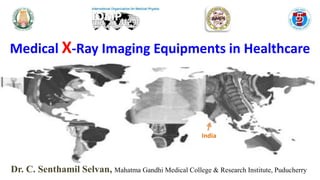
- 1. Dr. C. Senthamil Selvan, Mahatma Gandhi Medical College & Research Institute, Puducherry Medical X-Ray Imaging Equipments in Healthcare India
- 2. Outline What is Healthcare? Radiology - Basic Physics & Principles Commonly using Imaging Equipments Radiation Regulatory Bodies
- 3. The set of services provided by a country or an organization for the treatment of the physically and the mentally ill What are Healthcare Services? Mental health care Dental care Laboratory and diagnostic care Substance abuse treatment Preventative care Physical and occupational therapy Nutritional support Pharmaceutical care Transportation Prenatal care
- 4. https://www.uclahealth.org/medical-services Anesthesiology & Perioperative Medicine Dental Services Emergency Medicine Family Medicine Head and Neck Surgery Medicine Neurology Neurosurgery Obstetrics and Gynecology Ophthalmology Cardiology Orthopaedic Surgery Pathology & Laboratory Medicine Pediatrics Psychiatry & Biobehavioral Sciences Radiation Oncology Radiology Rehabilitation Services Surgery Urology
- 5. On November 8, 1895, physicist Wilhelm Conrad Roentgen (1845-1923)
- 6. A beam of x-rays may be: Transmitted: pass through unaffected or with a lower energy Absorbed: transfer all energy to matter and not pass through the patient to the film Scattered: diverted with or without energy loss Photons Interaction Mechanisms Photoelectric Effect Compton Scattering Pair Production
- 7. Radioactive Waste Radon X-Rays Consumer Products Nuclear Power Nuclear Medicine Solar Radiation Cosmic Rays Terrestrial Radiation Food & Drink Each Other
- 8. Radiography Nuclear Medicine Diagnostic Teletherapy Brachytherapy Radiotherapy Nuclear Medicine Therapy Radiation in Medicine
- 11. Radiographic images are recorded on a sheet of X- ray film. Composition of Film: Film consists of polyester base which is coated on both side by emulsion. Emulsion is suspension in gelatine of silver iodobromide crystals. Bromide (90%) Iodide (10%)
- 12. It is a process of capturing radiographic data from a conventional X-ray machine and processing the data digitally to produce high quality radiographic images. CR also commonly known as Photostimulable phosphor (PSP) imaging, employs reusable imaging plates and associated hardware and software to acquire and to display digital projection radiographs. It involves the PSP plate detector handling between two sequential stages of exposure and data acquisition. Imaging plate in a cassette must be processed in CR reader after X-ray exposure for conversion to digital images in raster pattern.
- 13. Direct radiography (DR) the image is produced directly from the image detector and is displayed on the screen. The image is divided into a matrix of individual cells and pixels. Each pixel has an assigned value that is related to the intensity of the signal in the corresponding part of the image. Digital radiographic images visible and storable on a personal computer or a hospital-wide system
- 14. Mobile radiography is bedside radiography examination performed on patients who are unable to be transported to the radiology These machines are self propelled and battery dependent Chest X-ray is the most requested mobile radiography examination Mobile imaging equipments offer less processing speed and eliminate long wait-times.
- 15. Bone fractures due to Road Traffic Accident (RTA) Infections (such as pneumonia) Calcifications (like kidney stones or vascular calcifications) Some tumors Bone loss (such as osteoporosis) Dental issues Heart problems (such as congestive heart failure) Blood vessel blockages Digestive problems Foreign objects (such as items swallowed by children)
- 16. 1890’s - Thomas Edison An imaging technique used to obtain real time moving images of the internal structure of a patient Components of Fluoroscope x-ray generator x-ray tube collimator filters patient table grid image intensifier optical coupling television system image recording
- 18. DIGITAL FLUROSCOPY - Dynamic flat panel Indirect detector - cesium iodide phosphor Direct detector - amorphous selenium TFT Barium X-rays and enemas (to view the gastrointestinal tract) Catheter insertion and manipulation (to direct the movement of a catheter through blood vessels, bile ducts or the urinary system) Placement of devices within the body, such as stents (to open narrowed or blocked blood vessels) Angiograms (to visualize blood vessels and organs) Orthopedic surgery (to guide joint replacements and treatment of fractures)
- 19. X-ray was first used to examine the breast by German surgeon Albert Salomon in 1913 Modern mammography came into existence since late 1960. Mammography is a radiographic examination that is designed for detecting breast pathology (particularly breast cancer). Breast cancer screening with mammography assists in detecting cancer at an earlier, and is an important clinical procedure because approximately one in eight women will develop breast cancer over their lifetime.
- 20. Mammography machine mainly constructed to examine the breast tissues Heel effect place an major role into it • Generator • Support system • X-Ray tube • Collimator / cone • Breast compressor • Anti- scatter grids • Cassette holder • Automatic exposure control • Photo timers • Detector system
- 21. Screening mammography Non-symptomatic patients with above 40 years Diagnostic mammography Symptomatic patients undergoes examination Digital detector technology affects cancer detection rate, dose and image quality. Digital detectors have facilitated new technologies such as tomosynthesis. 3-D techniques reduce superimposition and increase cancer detection in dense breasts. Contrast-enhanced mammography demonstrates improved sensitivity and specificity. Digital Mammography J.L. Diffey A comparison of digital mammography detectors and emerging technology, Published:July 17, 2015
- 22. CT is a well accepted imaging modality for evaluation of the entire body. CT has undergone several evolutions and nowadays dual CT scanners have been evolved which have better application in clinical field. Godfrey Hounsfield and Cormack got the 1979 Nobel Prize for their contribution to CT
- 27. 3-D imagines of CT and recently advanced retro processing techniques for accurate diagnosis. Unlike other imaging technique such as conventional x-ray imaging and CT enable direct imaging and differentiation of soft tissue structure such as liver, lung tissue, fat Therefore CT is valuable tools, for instance in searching for large space occupying lesion, tumors, and metastasis CT scan can not only reveal the presence but also the size spatial location and extent of tumor
- 28. Gantry can be used without the table to scan the head It is a multisection CT scanner intended for imaging in ear, nose, and throat settings and can be used to assess bone and soft tissue of the head.
- 29. Interventional Radiology I. C- Arm II. Catheterization Laboratory III. Digital Subtraction Angiography
- 30. C-arm is an imaging scanner intensifier C-shaped arm used to connect the X-ray source & X-ray detector to one another C-arms have radiographic capabilities, though they are used primarily for fluoroscopic intraoperative imaging during surgical, orthopedic and emergency care procedures. It provide high-resolution X-ray images in real time, thus allowing the physician to monitor progress and immediately make any corrections.
- 32. Angiography studies (peripheral, central and cerebral) Therapeutic studies (Line placements, transjugular biopsies, TIPS stent, embolizations) Cardiac studies Orthopedic procedures If there is a need to place needles or stents during a complicated surgery, C-Arm will be a handy choice. During surgeries, the real-time view of the gallbladder, liver, bone and several structures can be obtained. Multiple views of the same part are possible, thus enabling the systems to reconstruct a 3D model of the inner parts later. Surgical navigation is one of the primary applications and they aid in verifying the placement of all types of implant devices in the patient. C-Arm systems can also guide a needle placement procedure mainly while injecting anesthetic medicines. They can identify the joints and medication can be inserted perfectly onto the required shoulders and knees without damaging the other structures.
- 33. Not all heart problems require open-heart surgery Involve tiny and flexible tubes called catheters, which can be use instead of surgery, to access the heart and blood vessels Procedures performed in Cath Lab Balloon angioplasty Coronary and left ventricular digital angiography Coronary intravascular ultrasound Right and left heart catheterization Stent implantation Left Main Coronary Artery Stenosis – Angiogram, courtesy: Ivan Stankovic MD
- 34. Dental Radiography Orthopantamography (OPG) Intraoral X-ray
- 35. Intraoral X-rays are the most common type of dental X-ray These X-rays provide a lot of detail and allow your dentist to find cavities, check the health of the tooth root and bone surrounding the tooth, check the status of developing teeth, and monitor the general health of your teeth and jawbone.
- 36. Components of Intraoral X-ray Machine X-Ray tube Beam restriction devices (Cone and cylinders) Control panel Extension arm Miniature x-ray film
- 37. Ortho – straight Panoramic - complete view of the object in every direction Tomography – X-ray technique for making radiographs of layers of tissue in depth, without the interference of tissue above and below that level Film and x-ray tube head move around the patient in opposite directions in panoramic radiography Panoramic X-ray procedure requires less time causes no discomfort is radiation hygienic is painless and non-traumatic OPG was first described in Japan and Finland
- 38. X-Ray tube head Patient head rester Chin rest Bite rod Forehead rest Lateral head support Exposure controls
- 40. Lithotripsy is a medical procedure that uses shock waves or a laser to break down stones in the kidney, gallbladder, or ureter. The remaining particles of small stone will exit the body when a person urinates. Sound waves break down the stones into small pieces. Waves only affect stones & will not harm muscle, bone, or skin. 1st locate the stone with Fluoroscopy
- 41. A bone densitometry scan is an enhanced form of X-ray technology used to measure the bone mineral (calcium) content or bone loss and density of specific skeletal sites or whole body. Examination is also called a DEXA Scan DEXA is mostly performed on the lower spine and hips
- 42. What is Healthcare? Radiology - Basic Physics & Principles Commonly using Imaging Equipments Radiation Regulatory Bodies Radiation Safety
- 43. INTERNATIONAL International Commission on Radiation Units and Measurements (ICRU) International Commission on Radiological Protection (ICRP) National Council on Radiation Protection and Measurements (NCRP) International Atomic Energy Agency (IAEA) Atomic Energy Regulatory Board (AERB)
- 44. AERB was constituted on November 15, 1983 by the President of India by exercising the powers conferred by Section 27 of the Atomic Energy Act, 1962. Mission of the AERB is to ensure the use of ionizing radiation and nuclear energy in India does not cause undue risk to the health of people and the environment. Chairman : Shri. G. Nageswara Rao Headquarters : Anushaktinagar, Mumbai
- 45. Develop safety policies in nuclear, radiation and industrial safety areas. Develop Safety Codes, Guides and Standards Grant consents for siting, construction, commissioning, operation and decommissioning etc. Prescribe the acceptance limits of radiation exposure to occupational workers and members of the public and acceptable limits of environmental releases of radioactive substances. Review the training program, qualifications and licensing policies for personnel of nuclear and radiation facilities and prescribe the syllabi for training of personnel in safety aspects at all levels. Promote research and development efforts in the areas of safety.
- 46. AERB is the regulatory control authority on, Approval of new models of x-ray equipment and layout of any new proposed x- ray installation. Registration and commissioning of new x-ray equipment. Inspection and decommissioning of x-ray installation. Certification of a RSO and of service engineers and imposing penalties on any person contravening these rules
- 47. Prof. Sarah J Lewis, et. al. (2019) Artificial Intelligence in medical imaging practice: looking to the future AI in Medical Imaging
- 48. I wish all my colleagues around the World a Advance Happy Medical Physics Day (07.11.2020) & World Radiography Day (08.11.2020) Medical Physicist as a Health Professional
Editor's Notes
- 6