1. FieldStrengthPublication for the Philips MRI Community Issue 39 – December 2009
Hirslanden Clinic performs MR
image guided prostate biopsy
32-channel coil boosts 3.0T neuro
imaging at Kennedy Krieger
Cardiac MR is fast and easy with
complete Elite Cardiac Solution
Pediatric SENSE Head/Spine coil
simplifies infant studies
Educational: MultiTransmit solves
3.0T challenges at the source
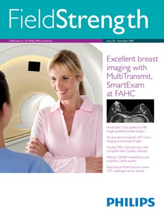
Excellent breast
imaging with
MultiTransmit,
SmartExam
at FAHC
4. 4
Philips MRI displays clinical and operational
excellence at RSNA 2009
Philips reinforces leadership role in 3.0T technology and advanced clinical solutions
At RSNA 2009, Philips exhibits a strong portfolio of MR products
that enhance clinical capabilities and simplify imaging workflow.
Achieva 3.0T TX fully equipped for breast imaging, biopsy
The Achieva 3.0T TX scanner with MultiTransmit technology has
demonstrated to be a successful product in clinical practice at many
sites. MultiTransmit addresses dielectric shading at the source
delivering improved image consistency and signal uniformity.
The Achieva 3.0T TX can be combined with the MammoTrak patient
support for superb, consistent high quality breast imaging and biopsy
with efficient workflow and patient friendliness. MultiTransmit now
also offers benefits for imaging applications in the brain.
Elite Clinical Solution for prostate
The Elite Prostate Clinical Solution brings together a full range
of imaging and spectroscopy capabilities, coil combinations, data
analysis and biopsy planning software and a new MR compatible
biopsy device for targeted MR image guided biopsies.
Further enhancements in Elite Clinical Solutions
The 32-channel SENSE Head coil offers enhanced 3.0T brain imaging
as well as fMRI, DTI and spectroscopic imaging. Increased signal-to-
noise ratio and multidirectional parallel imaging enable improved
imaging quality and high SENSE acceleration factors. MultiTransmit
parallel RF transmission in combination with this coil provides an
outstanding combination for excellent image quality and fast scanning.
The Cardiac Explorer and Vessel Explorer packages for automated,
task-guided analysis of CMR and MRA data strengthen the Elite
Cardiac and Elite Vascular Clinical Solutions.
5. Dear friends,
Sometimes, forces or ideas that appear to be in opposition
actually work together to bring better solutions for you
and your patients. This issue of FieldStrength will portray
how great clinical solutions and quality can still be
economically responsible and sustainable.
Achieva 3.0T TX, the MRI scanner that adjusts to each
patient through MultiTransmit technology, continues to
demonstrate clinical excellence with shorter scan times.
The article on breast imaging at Fletcher Allen shows this
again. Being the first to market with this true MultiTransmit
system, Philips has taken another lead in MRI knowledge.
Our Pediatric MRI experience brings more effective
outcomes (quality and economic) by recognizing these
patients’ unique needs. This issue offers two articles
building the awareness and understanding that Pediatric
MRI may require different approaches than adult imaging.
Philips is recognized as a leader in Sustainability. The
Achieva 1.5T SE has earned the Philips’ Green Flagship
designation for its PowerSave operating mode, smaller
footprint in the equipment room and lower operating
cost. Philips also offers FieldStrength readers an opportunity
to be more environmentally friendly by changing to an
electronic-only subscription, see page 13.
We look forward to sharing these and more developments
in Clinical and Operational Excellence at the RSNA 2009.
Conrad H. Smits
Chief Executive Officer Magnetic Resonance Imaging
Philips Healthcare
Five successful years confirm efficacy of Panorama HFO
Already five years ago Philips was the first vendor to introduce
a high field open system. Since then Panorama HFO has established
its reputation of combining high field image quality with patient
comfort offered by its wide open design. Last October a successful
international symposium on high field open MRI was held in Berlin,
attracting more than 80 participants from more than 10 countries.
The symposium covered the wide open clinical versatility of the
system – from breast imaging and biopsy to dynamic scanning of
joints, from fetal imaging to interventional therapy. Participants also
had a chance to experience hands-on how easy interventional MRI
procedures could be. Panorama highlights at RSNA are new 8-channel
coils for body and breast imaging.
More innovation proofpoints
The Philips booth also highlights SmartExam one-click reproducible
scanning for brain, spine, breast, shoulder, knee and the “rampable”
Achieva XR, the MR system that is easily upgraded from 1.5T to 3.0T
without a magnet swap. Also featured is Achieva 1.5T SE, providing
the best gradients in its class, and PowerSave, which smartly reduces
energy consumption.
For more information visit www.philips.com/RSNA
Investigational MR-HIFU system showcased
MR imaging is an optimal modality for guiding and monitoring High
Intensity Focused Ultrasound (HIFU) ablation procedures because of
its excellent soft tissue contrast, 3D imaging capabilities and its ability
to non-invasively measure temperature. Philips is showing its MR-
HIFU system, a tabletop design with an integrated transducer and 5D
positioning system is currenty under clinical investigation.
6. FieldStrength – Issue 39 – December 20096
Excellent breast imaging at FAHC
with MultiTransmit and SmartExam
MultiTransmit and SmartExam Breast address workflow and image quality challenges in 3.0T breast imaging
at Fletcher Allen Health Care
Sally Herschorn, MD
Fletcher Allen Health Care is Vermont's academic medical center,
based in Burlington.
Fletcher Allen clinicians employ MRI mostly when examining
high-risk patients and patients with a new diagnosis. MRI also is
used in occasional problem-solving cases, such as evaluating for
disease recurrence at a lumpectomy site and resolving one-view
mammographic densities. Frontline diagnostic modalities include
digital mammography and ultrasound – 30,000 studies per year
and 4,000 studies per year, respectively.
Transfer to 3.0T made impact, but challenges remained
Fletcher Allen shifted its breast studies from an old 1.5T system
to the Achieva 3.0T in October 2005, enabling clinicians to obtain
bilateral axial images with much higher SNR and resolution.
Dr. Herschorn and her colleagues have been using an advanced
ExamCard consisting of a T1-weighted non-fat saturated sequence,
a T2-weighted sequence with SPAIR fat saturation, followed by a
dynamic fat saturated THRIVE series. A high resolution THRIVE
is positioned between the second and third dynamic series.
In the beginning, results were inconsistent, Dr. Herschorn observes.
“In some patients, we obtained excellent images, while in others
there was poor or inhomogeneous fat saturation and every degree
in between.”
The high spatial resolution enabled by 3.0T is a clear benefit in breast imaging,
but obtaining uniform fat saturation can be more challenging at this field strength.
MultiTransmit and SmartExam Breast innovations in the new Achieva 3.0T TX are
designed to neutralize inhomogeneities – resulting in higher image quality and consistent
fat saturation – and make scan planning much easier and faster. Fletcher Allen’s
Dr. Sally Herschorn and her colleagues have been evaluating both solutions in
an Achieva 3.0T X-series system and recently on their Achieva 3.0T TX.
7. FieldStrength 7
This variability was due to a wide range of patient factors, and to
a lesser extent, technologist skill and experience, Dr. Herschorn
notes. At 3.0T, inhomogeneities of the main magnetic field (B0
) are
likely to occur in patients with large breasts, asymmetric breasts,
extensive breast folding and those who have had prior surgery
and have one or two implants. If these inhomogeneities are not
compensated for – either by manual or automatic shimming or by
repositioning the patient – the result will be poor fat suppression
and image quality.
Manual placement of shim boxes can be fairly tricky and time-
consuming, though, she says. Patients undergoing MRI are anxious
about a new cancer or about their risk for cancer to begin with.
It is critical for technologists to be confident that the sequences
they program will work right the first time. Technologist frustration
and insecurity are easily transmitted to patients, making them more
anxious. Therefore, it is vital that technologists work quickly
and expertly.
“SmartExam Breast has eliminated delays in
implementing fat saturation, resulting in better
workflow and increased patient comfort.”
SmartExam Breast improves workflow and image quality
“Workflow without SmartExam Breast was not ideal,”
Dr. Herschorn observes. “It was time-consuming to plan the volume
shim box and check the offset frequencies needed. This required
trial-and-error. Often manual central frequency determination was
also needed to improve fat suppression quality. And even after these
steps, outcomes were inconsistent. SmartExam Breast completely
eliminates this problem and has made achieving acceptable fat
saturation a non-issue.”
SmartExam Breast is the newest SmartExam application. It provides
intelligent assistance in planning, scanning and processing.
It automatically detects the breasts and chest wall to efficiently
plan the scan. Perhaps more importantly, it automatically optimizes
localized shimming. The automatic image-based higher order shim
results in better image quality and better fat suppression.
With SmartExam Breast, workflow is tremendously simplified.
And the result is excellent, reproducible image quality.
“With MultiTransmit, we are seeing
a significant uniformity improvement
in the non-fat saturated images.”
Standard THRIVE MultiTransmit and SmartExam
MultiTransmit and SmartExam Breast
47-year-old patient. Fat suppression
with SmartExam Breast image-based
shimming and MultiTransmit is
significantly better than in standard
THRIVE (arrows).
8. FieldStrength – Issue 39 – December 20098
According to Supervisor of MRI, Gretchen White, one of every
two 3.0T breast MRI examinations present shimming challenges.
“There are so few women whose breasts are exactly alike,” White
observes. “Even a little asymmetry can cause problems, particularly
in achieving good fat saturation at 3.0T, so manual shimming was a
monumental task. With SmartExam Breast, it was as if someone had
sprinkled magic dust on our magnet. Shimming was automated and
homogeneity was optimized for every patient. SmartExam Breast is
absolutely wonderful.”
“SmartExam Breast has made technologists feel confident that they
can get a good image on the first try. Patients can easily sense stress
or confidence in their technologist and feel more comfortable in their
exam when the technologist is confident. This leads to less patient
motion and better quality exams – not to mention happier patients
and in turn, happier referring physicians,” says Dr. Herschorn.
Standard MultiTransmit and SmartExam
“It was as if someone had sprinkled magic dust on our
magnet: shimming was automated and homogeneity
was optimized for every patient. SmartExam Breast
is absolutely wonderful.”
In 2008, Dr. Herschorn’s team used SmartExam Breast in 3.0T
breast MRI exams on nearly 400 patients (1-2 patients per day).
Previously, in difficult cases, the manual shimming step would add
an average of 15 minutes or more to each breast MRI examination,
which takes 30 minutes under optimal conditions. In especially
difficult cases, as the length of time it took to obtain decent fat
saturation increased, technologist and patient anxiety increased.
The longer it took, the worse fat saturation would get. Often, these
cases resulted in images of unacceptable quality. “SmartExam Breast
has eliminated delays in implementing fat saturation, resulting in
better workflow and increased patient comfort,” Dr. Herschorn says.
Patient-adaptive MultiTransmit achieves consistent
image uniformity at 3.0T
The dielectric effect causes RF field (B1
) non-uniformity and is
exacerbated at 3.0T due to a larger RF wavelength at this field
T1W TSE
THRIVE
42-year-old high-risk patient
Signal inhomogeneities are
noted in standard T1W TSE (red
arrows). Using MultiTransmit
the image is significantly more
uniform. While fat suppression
is inhomogeneous in standard
THRIVE (arrows), it is excellent
with MultiTransmit and
SmartExam Breast.
9. FieldStrength 9
strength. The result is non-uniform signal – or shading – in the
image, which can obscure anatomy and pathology. MultiTransmit
solves this problem by adding an extra RF transmit source to
provide simultaneous parallel RF transmission. The dielectric effect
and shading are nullified, resulting in enhanced signal uniformity
and consistency.
Dr. Herschorn and FAHC-based Philips clinical scientist Trevor
Andrews, PhD, have used MultiTransmit on about 20 women so far.
They currently are beginning a project to assess the impact of both
MultiTransmit and SmartExam Breast on image quality.
“We are seeing a significant uniformity improvement in the non-
fat saturated images,” she reports. “Not all patients present these
shading issues, but for the ones who do, the signal uniformity
is enhanced through MultiTransmit. Fat saturation also is more
uniform, which enables improved visualization of the axilla.
This is particularly important in patients with cancer.”
Standard MultiTransmit and SmartExam
“MultiTransmit helps us to achieve
a high level of image quality and
image uniformity in all of our
patients, which in turn makes
image interpretation easier.”
Dr. Herschorn says with 3.0T imaging, there often is a right
breast/left breast shading difference. “However,” she continues,
“MultiTransmit will automatically adapt the RF signal to each patient’s
unique anatomy. The result in every case so far has been neutralized
shading. This is where the true benefit of MultiTransmit lies: it helps
us achieve a high level of image quality and image uniformity in all of
our patients, which in turn makes image interpretation easier.”
Achieva 3.0T TX: premium breast imaging system
With its high spatial resolution enabled by the 3.0T field strength and its
powerful tools for enhanced workflow and consistently optimized image
uniformity, Achieva 3.0T TX with the Elite Breast Clinical Solution seems
destined to become the option of choice for breast MRI imaging.
“We have only just started breast imaging on Achieva 3.0T TX,”
Dr. Herschorn says. “But the nice thing is that SmartExam Breast
solves B0 inhomogeneities and MultiTransmit solves RF shading,
so you’re able to address both issues, enabling us to fully leverage
the benefits of 3.0T breast imaging.”
Patient with DCIS
50-year-old patient with DCIS and family
history of breast cancer. The standard
THRIVE images shows non-uniform
fat suppression (yellow arrows) and
parenchymal tissue signal is inhomogeneous
comparing right to left breast (red arrows).
In the THRIVE image with MultiTransmit
and SmartExam Breast, fat suppression is
uniform throughout the entire field of view.
10. FieldStrength – Issue 39 – December 200910
Hirslanden Clinic performs
MR image guided prostate biopsy
Elite Prostate Clinical Solution makes prostate biopsies easier and faster at Hirslanden Clinic
Beat Porcellini, MD
The Hirslanden Clinic (Zurich, Switzerland) is using the Philips Elite Prostate
Clinical Solution for faster, more controlled prostate biopsy procedures.
With a new biopsy device and DynaCAD for Prostate, MR image guided
prostate biopsies are vastly simplified.
The most common method for prostate biopsy involves a transrectal
procedure with tactile finger guidance performed by a urologist or
an ultrasound-guided process. Clinicians at the Hirslanden Clinic are
using their Achieva 3.0T with the Elite Prostate solution to perform
MR image guided biopsy in the prostate.
“With MR’s excellent soft tissue contrast, we can clearly visualize
where lesions are, and we can see how to angle and position the
biopsy needle. That’s the big advantage of MR image guided biopsy,”
says Beat Porcellini, MD, radiologist at Hirslanden Clinic.
He emphasizes that also ultrasound-guided biopsy is not comparable
to the MR image guided method. “A large number of lesions cannot
be detected by ultrasound and it may be difficult to reach the entire
organ, whereas it is easy to make biopsies of every region using MR
image guided biopsy.”
MR image guided biopsies help discover more lesions
About 50% to 70% of the time a patient’s first blind biopsy is
negative, says Dr. Porcellini. “Maybe you can reduce that by taking
more biopsy samples, but with ‘blind’ biopsies the problem remains
that even with many samples, a lesion can be missed if none of the
needles passes through it.”
They have thus far performed MR image guided prostate biopsy on
19 patients referred by urologists who wanted a second, MR image
guided biopsy after blind biopsy with negative results. “Out of these
first 19 patients, seven have had positive MR image guided biopsy
results,” says Dr. Porcellini. “In other words, seven patients had a
“In 19 patients with a
negative blind biopsy a
positive result was found
with MR image guided
biopsy. So these seven
patients had a carcinoma
that was missed by
previous blind biopsies.”
11. FieldStrength 11
T2-weighted ADC Biopsy
T2-weighted ADC Biopsy
Spectroscopy in different voxels
MR image guided biopsy of adenocarcinoma
66-year-old male with PSA rising from 5.1 to 7.2 in
two years. Previous TRUS biopsies in 2007 and 2008
were negative. T2-weighted image shows a suspicious
focus of 10 x 6 mm in the right side lobe. It has
restricted diffusion. The dynamic contrast series and
spectroscopy do not show any distinct abnormality.
No invasion of capsule, no enlarged lymph nodes or
mets. After MR image guided biopsy the lesion was
found to be adenocarcinoma Gleason 3 + 4 = 7. The
SENSE Cardiac coil and the endorectal coil were used
in dual-coil mode for the exam, a flexible coil was used
for the biopsy images.
MR image guided biopsy of adenocarcinoma
67-year-old male with PSA 33. Six
months ago PSA was 27 and TRUS
biopsy was negative. MR results show
a suspicious focal lesion of 25 x 30 mm
in the right middle lobe with strong
restriction of diffusion. Spectroscopy
shows an elevated cholin peak and
slightly reduced citrate in the lesion.
The dynamic contrast series is
unremarkable. No invasion of capsule,
no lymph nodes and/or mets. MR
image guided biopsy helped to identify
adenocarcinoma Gleason 3 + 4 = 7.
12. FieldStrength – Issue 39 – December 200912
carcinoma that was missed by the previous blind biopsies. That is the
dramatic part of MR image guided prostate biopsy: often still finding
a lesion in patients who went home after a negative blind biopsy.”
Urologists and radiologists work together for patient outcomes
“In Switzerland’s fee-for-service healthcare program urologists and
radiologists earn more doing ten biopsies instead of two biopsies,” says
Dr. Porcellini. “But clinicians are beginning to understand that the way
to a more efficient process and better diagnosis is with well-targeted
MR image guided biopsy. Our outlook is to bring more patients to both
urologists and radiologists by offering this method to their patients.”
Hirslanden Clinic urologists are convinced of the need for MR image
guided prostate biopsy. “But we also want to propagate and offer
this method to other urologists in Zurich,” Dr. Porcellini says.
“I think the fact that we are getting more patients will be a factor
in these referrals; urologists will profit, and consequently we as
radiologists also. And, of course, the patients benefit the most.
MR image guided biopsy helps us detect more prostate lesions
at an earlier stage.”
Dr. Porcellini hopes to expand the prostate program soon,
bringing in a larger group of urologists and staff.
Hirslanden Clinic
“Both urologists and radiologists
will benefit from implementing MR
image guided biopsy. And, of course,
the patients benefit the most.”
Elite Prostate: clinical solution for imaging and biopsy
T2w TSE
DynaLOC InterventionalDynaCAD for Prostate
(multi b-value) Diffusion
Dynamic T1w FFE
3D Spectroscopy
When a patient presents for MR image guided biopsy, first
dual-coil MR scanning can be performed to visualize suspicious
lesions in the prostate. Imaging techniques include high resolution
T2-weighted imaging, diffusion weighted imaging, dynamic T1 studies
and spectroscopy. DynaCAD for Prostate offers streamlined image
Imaging Techniques
SENSE Torso/Cardiac 32 Endo Coil (+ Cardiac coil) DynaTRIM Biopsy device
Supporting Coils Peripherals
Workflow Support Tools
analysis and biopsy guidance. For biopsy, the MR-compatible biopsy
device DynaTRIM is placed on the MR patient support, imaging
is performed with flexible surface coils. Planning of the biopsy
trajectory is an integrated part of the solution, that can be displayed
at the scanner’s console.
13. MR news
FieldStrength 13
FieldStrength going green: register for email version
FieldStrength now offers electronic-only subscriptions
in an effort to be more environmentally friendly. When
a new FieldStrength issue is available, subscribers
receive an email alert with an overview of the issue’s
contents and links to the articles online. We encourage
existing readers to switch to an email subscription, and
new readers to register for an email subscription.
Philips’ commitment to sustainability
Philips Healthcare is dedicated to being a world-class
sustainable development company by conducting
our business in an environmentally and socially
responsible manner.
Reducing our carbon footprint – Achieva 1.5T SE
Since 1994, Philips has been increasing the energy
efficiency of its products along with other environmental
improvements in manufacturing since 1984. We
seek new solutions for responsible energy practices,
and focus on the energy efficiency of our products
and production processes. Our continued focus on
sustainability has resulted in the PowerSave feature for
our MRI systems. The Achieva 1.5T Green Product line
reduces energy consumption by 28%. Green products
score at least 10% lower on environmental impact on
one of the Green focal areas, measured during the
entire life cycle of the product. So, Philips MRI systems
now help hospitals to reduce their carbon footprint.
Please consider the environment
before printing any document.
Most popular NetForum contributions
in third quarter of 2009
1. Web Seminar: Clinical experience with MultiTransmit technology at 3.0T – University of Bonn
2. ExamCard: 1.5T Non-CE renal/mesenteric MRA – Scottsdale Healthcare
3. Application Tip: Optimization of fat suppression in the head and neck area
4. Application Tip: Tips for cardiac black blood imaging
6. ExamCard: 1.5T Brain spectroscopy – Soroka University Medical Center
Visit the NetForum User Community
for downloading ExamCards and
viewing application tips, clinical cases,
extended versions of FieldStrength
articles, and more.
NetForum
www.philips.com/netforum
Visit www.philips.com/fieldstrength
to manage your subscription or to
preview FieldStrength online.
14. FieldStrength – Issue 39 – December 200914
32-channel coil boosts 3.0T
neuro imaging at Kennedy Krieger
Kennedy Krieger Institute sees significantly better fMRI, DTI, spectroscopic imaging with the 32-channel SENSE Head coil
The F.M. Kirby Research Center for Functional Brain Imaging at the Kennedy Krieger
Institute (Baltimore, Maryland, USA) is a general resource center for neuro imaging.
It provides 3.0T and 7.0T imaging for Kennedy Krieger, for Johns Hopkins University
School of Medicine and for the University of Maryland.
The Center recently began using the 32-channel SENSE
Head coil in its neuro studies on the Achieva 3.0T
X-series. Because the 32-channel coil provides increased
SNR, clinicians can either scan faster or get higher
spatial resolution or temporal resolution, depending on
the particular study.
Peter van Zijl, PhD, is the Director of the F.M. Kirby
Research Center at Kennedy Krieger and Professor of
Radiology at the Johns Hopkins University School of
Medicine. He explains how the coil benefits studies done
at the Institute.
High SENSE factors become practical
“With the 8-channel coil we wouldn’t do parallel
imaging in multiple directions because noise was much
bigger,” says Dr. van Zijl. “The 32-channel coil now
enables using SENSE in multiple directions and with
higher SENSE acceleration factors. This is particularly an
advantage for 3D acquisition techniques.”
Dr. van Zijl emphasizes that the high SENSE acceleration
factors achieved with the coil do not compromise
image quality. “For anatomical imaging, we have applied
SENSE acceleration factors of 6, and the images are still
beautiful. So, for instance, we can easily do a 3D FFE at
0.7 x 0.7 x 0.7 mm in about 4 minutes, which is much
better than the 1 x 1 x 1 mm in about 6 minutes that we
used to do with the 8-channel coil.”
“In spectroscopy, we have gone as high as a SENSE
acceleration factor of 9. The quality of these
spectroscopic images is still better than with the
8-channel coil using lower acceleration factors, because
Peter van Zijl, PhD
“The quality of the 32-channel
coil spectroscopic images with
SENSE factor 9 is still better
than 8-channel coil images
using lower SENSE factors.
These are most impressive
spectroscopic images.”
15. FieldStrength 15
there is less lipid contamination. These are the most
impressive spectroscopic images that I have seen in my
whole life,” he says, “and I have seen a lot of them.”
“This coil’s design enables very good quality images over
the whole of the brain, even without the front section.
I have not seen that in any other coil ,” says Dr. van Zijl.
Significantly faster spectroscopy promotes
clinical acceptance
Peter B. Barker, D.Phil., Professor, Department of
Radiology at Johns Hopkins University School of
Medicine, is working on several neuroimaging projects
at the Institute. These include using MR Spectroscopy
Imaging (MRSI) to assess rare metabolic diseases in
children, such as leukodystrophies and Rett syndrome.
“There are two main advantages to this 32-channel coil,”
says Dr. Barker. “Improved SNR and improved SENSE
reconstruction performance. These are critical for
demanding experiments such as multi-voxel spectroscopy.
The coil is also very comfortable for the patient.”
With the conventional 8-channel coil, Dr. Barker did
not use SENSE factors higher than 2 or 3 for 2D-MRSI,
because of low SNR and artifacts becoming more
apparent. “With the new 32-channel coil, we routinely
do a SENSE factor of 6 for MRSI,” he says. “This reduces
a conventional 30-minute MRSI scan to just five minutes.
Recently, we acquired some 3-minute 2D-MRSI scans
with a SENSE factor of 9. The data were surprisingly good
for such a short scan time.”
Peter Barker, D.Phil.
32-channel coil
SF 2x1.5=3
32-channel coil
SF 2x3=6
8 channel coil
SF 2x1.5=3
8 channel coil
SF 2x3=6
High SENSE factors for MRSI
Detailed comparisons by Dr. Henry Zhu
with Dr. Barker show that the 32-channel
coil choline spectroscopic scan with
SENSE factor 6 has better quality and is
faster than the 8-channel coil scan with
SENSE factor 3.
SENSE-MRSI of high-grade tumor
The left frontoparietal lesion shows very
characteristic spectra of a high grade
glioma, with a high Cho signal and a
decrease in NAA. Note that the lesion
shows FLAIR/T2 hyperintensity in a
region which closely matches the NAA
reduction. Individual spectra are shown
from the normal right side (blue) and the
abnormal region (red). Achieva 3.0T with
32-channel SENSE Head coil.
16. FieldStrength – Issue 39 – December 200916
Susumu Mori, PhD, Professor at Johns Hopkins
University School of Medicine, is a world leader in using
DTI to characterize human white matter anatomy, and
its abnormalities in conditions such as multiple sclerosis,
stroke, brain tumors and cerebral palsy.
“While DTI is an extremely powerful method to
delineate white matter anatomy, it’s easily affected
by patient motion,” says Prof. Mori. “To minimize
this motion effect, it is essential to use a rapid imaging
technique such as single-shot echo-planar imaging
(EPI). DTI, therefore, inherits many issues related
to single-shot EPI, such as low spatial resolution
and image distortion.”
In the past 10 years, DTI has been drastically improved
by the introduction of SENSE parallel imaging.
SENSE enables shorter echo times for high SNR, less
distortion, and higher spatial resolution.
New coil enables higher resolution and less
distortion in DTI
“SENSE was literally a ‘dream’ technology for DTI,” says
Prof. Mori. “However, there is a limitation in increasing
the SENSE factor. Depending on the coil geometry,
a SENSE factor higher than 2 often leads to artifacts.
With the 32-channel SENSE Head coil, however, we
can now push the boundary in DTI. First, the new coil
allows a higher SENSE factor; a SENSE factor of
3 produces clean, artifact-free images. Second,
it provides higher SNR, which leads to shorter
scanning time and/or higher image resolution.”
“In the past, we spent at least 9 minutes for DTI
with 2.5 mm resolution, but now the scanning time is
reduced to about 4.5 minutes with 2.2 mm resolution,”
says Prof. Mori. “This time reduction is the key for DTI
scans to be accepted as a routine clinical scan, where
time efficiency is so crucial.”
Susumu Mori, PhD
SENSE 2 SENSE 3
“With the 32-channel
SENSE Head coil, we
can now push the
boundary in DTI.”
Parallel imaging has improved DTI
Higher SENSE factor reduces distortion
Images with different SENSE factors. The red outline from the SENSE 3 image is superimposed on
the SENSE 2 image, highlighting the improvements near the temporal lobes. This clearly shows that
distortion is significantly reduced with the higher SENSE factor.
High SENSE factors enable high resolution DTI
The 32-channel SENSE Head coil enables high resolution DTI, which was not possible several years ago.
These DTI images with 1.9 mm isotropic resolution, 66 slices were obtained in 5:21 min. scan time.
17. FieldStrength 17
High resolution improves BOLD-fMRI
This right-hand finger-tap block-design
experiment demonstrates better
localization of the activation and higher
t-values (arrows) when resolution is
increased from 3 x 3 x 3 mm to
2 x 2 x 2 mm using the 32-channel
head coil.
“If we had done the same fMRI
acquisition with the 8-channel coil,
we would have seen nothing.”
3 x 3 x 3 mm 2 x 2 x 2 mm
The applications of MRSI might be expanded as clinicians
become aware of its feasibility. “Widespread adoption
of clinical applications has been hampered by a lack of
commercially available optimized pulse sequences and
analysis tools,” says Dr. Barker. “Philips now provides
an outstanding package for both single and multi-voxel
spectroscopy.”
Faster scans, smaller voxels particularly
important in pediatric fMRI
In Kennedy Krieger’s functional MRI (fMRI) studies,
the new 32-channel head coil far outperforms the
previous coil. “In standard fMRI the new coil works very
well,” says Dr. van Zijl. “The SNR is much better, and
we can go to higher spatial resolution.”
James J. Pekar, PhD, is Research Scientist at Kennedy
Krieger and Associate Professor in the Russel H. Morgan
Department of Radiology and Radiological Science at Johns
Hopkins University School of Medicine. In his fMRI work,
Dr. Pekar often works with children who have ADHD,
autism, dyslexia and other brain-related disorders. James Pekar, PhD
High resolution DTI allows detailed fiber-tracking
The small pixel size in combination with high
SENSE factor allows detailed visualization of
complex anatomy in the mid-brain region. Five
fiber bundles are shown: cortico-spinal tract (cst,
blue), medial lemniscus (ml, green), superior-,
medial-, and inferior-cerebellar peduncles (scp,
pink; mcp, red; icp, orange).
18. Dr. van Zijl explains that the coil benefits other studies
as well. “One of our colleagues, Dr. Jinyuan Zhou, has
a project to look at the content of small proteins and
peptides in brain tumors using Amide Proton Transfer
imaging, or APT imaging. With the 32-channel head
coil, the images from the increased baseline protein
and peptide content are very beautiful.” APT imaging
is a variant of a method called Chemical Exchange
Saturation Transfer (CEST) imaging developed for
research purposes by Drs. Zhou and van Zijl at
Kennedy Krieger and Johns Hopkins.
References
1 Mori, S., Wakana, S., Poetscher, N. L. M, van Zijl, P. C. M.
MRI Atlas of Human White Matter, First Edition.
Elsevier Science. (2005).
2 Zhou J, Blakeley J, Hua J, Kim M, Laterra J, Pomper M, van Zijl PCM.
A practical data acquisition method for human brain amide proton
transfer (APT) imaging.
Magn Reson Med. 2008 Oct; 60(4):842-9.
There are special challenges in working with children.
Investigators design child-friendly paradigms with stimuli
and tasks that are age appropriate. Faster scan times are
of great importance in children, who often cannot stay
still for long periods of time. The higher SENSE factors
also produce smaller voxels for better localization.
“We’re currently optimizing fMRI acquisitions using the
new 32-channel coil,” says Dr. Pekar. “Previously, our
default fMRI exam used 3 mm isotropic resolution. Our
initial scans with the new coil show better results using
2 mm isotropic voxels.”
“The new coil provides higher temporal SNR with 3 mm
isotropic voxels,” he says, “But the 2 mm isotropic
“The 32-channel coil
interfaces seamlessly with
our 3.0T system and is very
comfortable for the patient.”
Coil yields beautiful results in APT research
on brain tumors
voxels show higher t-values in a block-design activation
paradigm, which is due to reduced partial volume effects
when using smaller voxels.”
Increasing to more than 32 coil elements will add
less benefit
“Adding even more coil elements will not have a similar
impact as moving from 8 to 32,” says Dr. van Zijl. “There’s
a point of diminishing gain per added coil element as this
follows an exponential curve. Changing from 1 to 8 will
give more than a doubling in SNR, increasing from 8 to
16 may provide 50% more, and from 16 to 32 probably
another 10%. Even if you would go from 32 to 64,
you would probably gain only 10% in SNR. So, I think
32 channels is about right.”
Tumor core
Peritumoral
edema
APT of oligodendro-astrocytoma
Amide Proton Transfer (APT) imaging contrast is based on increased
endogenous cytosolic proteins and peptides in tumor core.
FieldStrength – Issue 39 – December 200918
19. FieldStrength 19
Cardiac MR is fast and easy with
complete Elite Cardiac Solution
32-channel cardiac coil and new Cardiac Explorer software smooth out the CMR process
at St. Luke’s Episcopal Hospital (SLEH) and German Heart Institute Berlin
Philips’ Elite Cardiac Clinical Solution now includes the 32-channel SENSE Torso/Cardiac coil
for 1.5T and 3.0T systems, and Cardiac Explorer and Vessel Explorer software for automated,
task-guided analysis of CMR data. These make Cardiac Magnetic Resonance (CMR) fast and
easy, which clinicians have indicated as an essential need for the growth of CMR.
The SENSE Torso/Cardiac coil is designed to allow
greater coverage of anatomy and improved signal-to-
noise ratio. The increased SENSE acceleration factors
help to either improve image spatial resolution, or
reduce breath holding duration during cardiac studies.
32-channel coil boosts cardiac studies at SLEH
St. Luke’s Episcopal Hospital, home of the Texas Heart®
Institute is
a tertiary referral center performing about 80 to 100 cardiovascular
MR exams in an average month. Benjamin Cheong, MD, cardiologist
at St. Luke’s, routinely uses the SENSE Torso/Cardiac coil for
cardiovascular MR.
“All of our cardiovascular studies, ranging from functional assessment
to aortic studies, benefit from the 32-channel coil,” he says. “With
the increase in imaging speed, the staff can complete an image
sequence and finish the examination much more quickly than before.
That helps our throughput, and our patients obviously appreciate a
faster examination and less time in the scanner.”
Multiple breath holds are often unnecessary with the 32-channel
coil, explains Dr. Cheong. “Cardiac MR has already established itself
as the gold standard for functional and morphological assessment
of the left and right ventricles with the traditional multislice cine
imaging that covers both ventricles in 10 to 12 slices. But with the
new 32-channel cardiac coil, knowing that we can increase the
Cardiac Explorer integrates image viewing and analysis,
with viewing formats selected from a predefined
list linked to the ExamCard. Many analyses can be
completed in less than 20 minutes, thanks to a high
degree of automation. Cardiac Explorer and Vessel
Explorer are available as optional packages on the
Extended MR WorkSpace (EWS) workstation.
Benjamin Cheong, MD
20. FieldStrength – Issue 39 – December 200920
Two-chamber view
Left ventricular apical non-compaction
58-year-old male with congestive
cardiac failure of unknown etiology.
CMR with SENSE Torso/Cardiac coil,
SENSE factor 2, voxels 1.8 x 1.8 x 8 mm,
breath hold 6 sec. Cine SSFP of the
two-chamber view shows increased
trabeculation confined to the left
ventricular apex (arrowhead) consistent
with left ventricular non-compaction.
imaging speed or the spatial resolution, we can actually
acquire multislice images of the left ventricle in a single
breath hold without the risk of slice misregistration.”
Coil enables improved speed and resolution
With the traditional 5-channel cardiac coil, typically
a SENSE factor of 2-3 is used. Higher acceleration
factors may result in decreased signal-to-noise ratio or
image artifacts. The high number of coil elements in the
32-channel coil enables use of higher SENSE acceleration
factors and multidimensional parallel imaging accelerations.
Dr. Cheong says in the past, using the 5-channel cardiac
coil with parallel imaging, a coronary whole-heart MRA
typically required an imaging time of 8 to 12 minutes,
even with reasonable respiratory gating efficiency.
“However, I was the first to try out the 32-channel coil,”
says Dr. Cheong. “The whole-heart coronary MRA of
myself was acquired in only four minutes, with a gating
efficiency close to 70% and an isotropic resolution of
1 x 1 x 1 mm. I had never seen such quality images
coming from a 1.5T scanner. All major epicardial
coronary arteries were well visualized.”
“I had never seen such quality images
coming from a 1.5T scanner.”
“The higher spatial and temporal resolution of the images
is impressive. One of the applications where MR really
shines is in the assessment of the arrhythmogenic right
ventricular dysplasia/cardiomyopathy (ARVD/C),” says Dr.
Cheong. “The right ventricle (RV) is a thin wall structure
of about 3-4 mm in thickness. The 32-channel coil allows
us to achieve a higher spatial resolution in order to image
the RV free wall, without a prolonged breath hold. That
is of paramount importance in the assessment of fatty
infiltration and fibrous replacement of the right ventricle
in patients suspected to have ARVD/C. Our image quality,
and therefore our diagnostic capability, is much higher.”
St. Luke’s senior physicist, Raja Muthupillai, PhD, recently
completed a study using the 32-channel coil to achieve
very high temporal resolution (3-6 ms) in a cine sequence
for evaluation of the left ventricular diastolic function.
As certain diastolic indices, such as isovolumic relaxation
time, are about 70-80 ms in normal individuals, this
high temporal resolution is essential to help distinguish
between normal and abnormal heart function.
“For cardiac imaging, MR is a modality that is accurate,
well established, non-invasive, and does not expose the
patient to radiation,” says Dr. Cheong. “And using the
32-channel coil gives us a huge advantage.”
Left ventricular outflow tract Mid short axis
Pre-operative CMR of left ventricular aneurysm
68-year-old male with prior myocardial infarction and congestive cardiac failure
presented for pre-operative surgical planning. Echocardiography demonstrated left
ventricular aneurysm. Cardiac MR was performed with 32-channel SENSE Torso/
Cardiac coil, SENSE factor 2, voxels 1.8 x 1.8 x 8 mm, breath hold 6 sec. Cine SSFP
of the left ventricular outflow tract and mid short-axis demonstrate left ventricular
aneurysmal dilation present in the proximal half of the inferolateral wall (left,
arrowhead) extending into the inferior wall (right, arrowhead). LV = left ventricle;
RA = right atrium; Ao = aorta; RV = right ventricle.
21. FieldStrength 21
“With the increase in
imaging speed, the
staff can finish the
examination much
more quickly than
before.”
Cardiologist Ingo Paetsch, MD, Director of the CMR
imaging department at the German Heart Institute (Berlin,
Germany) has been involved in Cardiac MR for more than
10 years. He uses both Achieva 1.5T and 3.0T for cardiac
MR. In the past months, he has been one of the first to
use the Cardiac Explorer and Vessel Explorer packages.
Cardiac Explorer software facilitates rapid viewing of
cardiac CMR images. “It’s particularly useful in stress
CMR imaging when looking for myocardial ischemic
reactions in the presence of coronary artery stenoses,”
Dr Paetsch says. “For both cine imaging and non-cine
dynamic functional imaging in stress CMR Cardiac
Explorer is extremely helpful. Side-by-side viewing of
rest and multiple stress scans for comparative purposes
– which is the hallmark of pharmacological stress CMR
testing – is easily accomplished.”
In the Institute’s recently developed combined stress
CMR protocol using a single stressor agent2,3
, multiple
cine views are acquired at each step of the protocol.
“With just two mouse clicks Cardiac Explorer displays
multiple views at different stress levels together with
the non-cine dynamic images.”
“Cardiac Explorer mimics a physician’s line of thought,”
says Dr. Paetsch. “Incoming images are automatically
placed in predefined viewports and can be reviewed
almost immediately thus avoiding the tedious job of
manual sorting. The side-by-side comparison of multiple
cine views assists the cardiologist not only to rapidly
establish a diagnosis but also helps to continuously
monitor wall motion changes during the scan procedure,
which is important as early recognition of wall motion
abnormalities leads to termination of the test. Cardiac
Explorer represents the cardiologist’s “best friend”
during the stress CMR examination.”
More time saving is seen in the Cardiac Explorer module
for left ventricular functional analysis. Automatic endo-
and epicardial contour detection routinely enables
measurement of LV volumes and mass using the disk
summation method (Simpson´s rule) in each patient in
less than 30 seconds.
Cardiac and Vessel Explorer facilitate CMR viewing and postprocessing, save physicians time
Five-chamber” cine at mid-systole Short-axis cine near base of left ventricle
Gerbode defect with shunting from left ventricle to right atrium
38-year-old male with prior Ross procedure and repaired co-arctation of aorta
presented with pulmonic stenosis. In addition, surface echocardiography demonstrated
a small left-to-right shunt. Cardiac MR was performed with 32-channel SENSE Torso/
Cardiac coil, SENSE factor 2, voxels 1.8 x 1.8 x 8 mm, breath hold 6 sec. The mid-
systolic “five-chamber” SSFP cine image shows a jet arising from the basal septum
of the left ventricle into the right atrium, indicating a left ventricular-to-right atrial
communication (arrowhead). The short-axis SSFP cine image (right) also demonstrates
the left ventricular-to-right atrial communication (arrowhead). LV = left ventricle; RV
= right ventricle; LA = left atrium; RA = right atrium.
Ingo Paetsch, MD
22. FieldStrength – Issue 39 – December 200922
Dobutamine stress study
Synchronized display of
multiple cine short axis
views of a dobutamine CMR
stress study. From left to
right: increasing levels of
graded dobutamine dosage.
From top to bottom: basal,
equatorial and apical short
axis orientation. Missing wall
thickening of the inferolateral
segment in all three short
axis views under maximum
dobutamine stress can easily
be recognized to assist in
diagnosis.
Targeted RCA reconstruction
Vessel Explorer used for
reconstruction of a 3D
whole-heart coronary
MR angiography. Targeted
reconstruction of the
right coronary artery
(RCA) is exemplified
using maximum intensity
projection, multiplanar
curved reformatting and
corresponding cross-sectional
view of RCA lumen.
“With just two mouse clicks Cardiac
Explorer displays multiple views at
different stress levels together with
the non-cine dynamic images.”
Vessel Explorer helps evaluate coronary arteries
Vessel Explorer not only facilitates 3D reconstruction of
large thoracic or mid-sized peripheral arteries, but also
of the coronary arteries with 2 to 4 mm luminal diameter
range. “The benefits of Vessel Explorer are enormous,”
says Dr. Paetsch. “Interactive and simultaneous display
of 3D volume rendering, MIPs and MPRs ensures
easier anatomical identification of complex vessel
anatomy and speeds up path identification for additional
reconstructions highlighting a specific pathology.
Several vessel parameters are simultaneously provided
23. FieldStrength 23
“Cardiac Explorer mimics a
physician’s line of thought.”
References
1 R Krishnamurthy, A Pednekar, B Cheong, R Muthupillai
High-Temporal Resolution SSFP Cine MRI for Estimation of Left Ventricular
Diastolic Parameters.
Submitted
2 R Gebker, C Jahnke, R Manka, A Hamdan, B Schnackenburg,
E Fleck, I Paetsch
Additional Value of Myocardial Perfusion Imaging During Dobutamine Stress
Magnetic Resonance for the Assessment of Coronary Artery Disease.
Circ Cardiovasc Imaging 2008; 1:122-130.
3 I Paetsch, C Jahnke, A Wahl, R Gebker, M Neuss, E Fleck, E Nagel.
Comparison of dobutamine stress magnetic resonance, adenosine stress
magnetic resonance, and adenosine stress magnetic resonance perfusion.
Circulation 2004; 110:835-842.
(luminal diameter, percentage of stenosis, vessel length,
ostial area, etc). A single click on a vessel displays its
cross-section view, and curved planar reformats can be
obtained with just a few more clicks.
In CMR and electrophysiological interventions, Vessel
Explorer permits fast review of 3D angiographic
datasets before and during interventions
Packages speed processing, reporting and
enhance reproducibility
“Cardiac Explorer and Vessel Explorer increase our
reproducibility and make results less user-dependent,”
Dr. Paetsch says. “Especially with ExamCards, you
need to ‘teach’ Cardiac Explorer only once to sort and
display images. Despite the increased speed and ease
of use, both flexibility and customizability are very well
preserved in these two packages.”
“More than 20 CMR studies are conducted every day
at the Institute, and our policy is to finish the report
immediately after the examination with the patient
taking it directly back to the referring physician.
A CMR study delivers a huge amount of images for
3D venous angiogram reconstruction with Vessel Explorer
Vessel Explorer is used for reconstruction of a 3D left
atrial and pulmonary venous MR angiogram. Assessment of
ostial area and luminal diameter of pulmonary veins assists
electrophysiological procedure planning (top). Simultaneous
display of surface rendering improves anatomical orientation
and can directly be used for ostial area and luminal diameter
measurements (bottom).
every patient, consisting of multiple cine views as well as
dynamic images and scar images,” explains Dr. Paetsch.
“We are extremely happy with Cardiac Explorer to free
our mind of tedious image sorting and other manual
processing before actually reviewing the case – which is
a physician’s true task.”
24. FieldStrength – Issue 39 – December 200924
Pediatric SENSE Head/Spine coil
simplifies infant studies
Dedicated, lightweight, integrated coil enables one-step examinations with high SNR
in Karolinska University Hospital, Stockholm.
Bo Ehnmark
When applying “adult” coils for infants, the large coil elements and their distance from
the patient can yield suboptimal SNR. In head/spine examinations in infants, another
problem has been the risk of waking sleeping patients when swapping head and spine
coils. At Astrid Lindgren Children’s Hospital, radiographers led by Bo Ehnmark, have
evaluated Philips’ Pediatric SENSE Head/Spine coil for 12 patients. The coil’s small
elements, integrated design and light weight have provided an excellent solution.
Astrid Lindgren Children’s Hospital
• 2,700 MRI examinations per year
• Daily volume: 8-10
• Age range: newborn to 16-years-old
• 40% sedated, 60% awake or Feed-and-Sleep
• 3-5 patients per week with Pediatric SENSE
Head/Spine coil
Astrid Lindgren Children’s Hospital, part of Karolinska University
Hospital (Stockholm, Sweden), developed an innovative way to scan their
youngest patients, children ranging in age from hours-old to 12 weeks
old. The “Feed-and-Sleep” protocol entails bringing infant patients in 30
minutes before their examination, feeding them and then allowing the
babies to fall asleep. Because the children are brought in overdue for
a nap anyway, it is more likely that the patients will sleep motionlessly
(with ear protection) for at least the 30 minutes it takes to scan them.
For Achieva 1.5T brain studies in this patient group, the Children’s
Hospital has commonly used the 8-channel SENSE Head coil or the
SENSE Spine coil.
“For brain-only studies we have used the SENSE Head coil,”
Ehnmark says. “It’s a good coil with ample space on the inside for
headsets, but its size also is a disadvantage. It’s too big for infants,
so we don’t obtain optimal SNR because the coil elements are too
far away from the head.”
In addition, if clinicians wanted to investigate the spine as well,
technologists risked waking the sleeping baby when swapping the
head coil for the SENSE Spine coil, and SNR was not optimal.
“Swapping coils is too clumsy,” Ehnmark observes. “And the SNR in
the spine has not been sufficient in two- to three-kilogram babies.”
Sequential infant head/spine studies became easier when Astrid
Lindgren’s Children’s Hospital acquired the 16-channel SENSE
NeuroVascular coil about a year ago. When integrated with the
SENSE Spine coil, clinicians could look at the brain, possible
25. FieldStrength 25
problems in the upper cervical spine (e.g. Chiari malformation) and
lower cervical spine (e.g. tethered cord), all the way to the lumbar
spine and pelvis, without changing coils. Still, signal issues persist
when using the adult-designed spine coil for infants.
“Babies-only” Pediatric SENSE Head/Spine coil performs
well in test
Astrid Lindgren Children’s Hospital evaluated the Pediatric SENSE
Head/Spine coil on Philips’ Try Buy program, using the coil in
12 examinations over an eight-week period. The typical weight
range of the patients was 4.5 to 6.5 lbs. (2-3 kilograms). Ehnmark
believes one of their patients, a 17.6 lb. (8 kilograms) 10-month-old
represents the maximum feasible size for the coil. The Children’s
Hospital’s first case for the pediatric SENSE Head/Spine coil was at
the opposite end of the spectrum, a 6-hour old infant (see images).
“We received the coil around lunchtime and by one o’clock it was
on the table ready to scan this 6-hour old baby with suspected
hydrocephalus,” Ehnmark recalls. “We obtained really nice images. It
was very impressive that we could just plug the coil in, run our standard
ExamCard and get excellent images on this ~4 lb. (1.9 kg) infant. The
neurosurgeon was able to confirm the diagnosis of hydrocephalus
and could make the decision to shunt the ventricle. With this first
patient, I immediately realized the potential of this coil.”
For the Feed-and-Sleep patients who require an examination of
head and spine, the pediatric SENSE Head/Spine coil seems tailor-
made, he adds. “With Feed-and-Sleep, we have a 30-minute window
to perform the examination. It’s usually not more, because of the
increasing chance that the baby will wake up from his nap and start
to cry and move. Then it’s all over,” Ehnmark explains. “With our
traditional solution, the lower SNR required more signal averages,
which could increase the scan time past 30 minutes. Sometimes we
got lucky and the baby stayed asleep, but most times we had to end
the study because the infant woke up.”
Clear visualization of congenital anomalies
Ehmark has found that the Pediatric SENSE Head/Spine coil
has provided excellent visualization of a tethered spinal cord, a
neurological disorder caused by tissue attachments that limit spinal
cord movement. The resultant stretching of the spinal cord as the
baby grows interferes with nerve function. “In infant spine imaging,
you need high resolution, which has been very tricky with a
standard spine coil,” he says.
“We received the coil around
lunchtime and by one o’clock
it was on the table ready to
scan this 6-hour old baby with
suspected hydrocephalus.”
T2W TSE Ax 4mm T1W 3D FFE T2W TSE DRIVE sag HR
Hydrocephalus in 6-hour-old infant
A 6-hour-old infant with suspected hydrocephalus was
examined with the Pediatric SENSE Head/Spine coil. Clinical
symptoms included bulging of anterior and posterior
fontanelles and wider than normal sutures between adjacent
skull bones. The examination confirmed the diagnosis of
hydrocephalus; the cerebral aqueduct was blocked and CSF
had accumulated within the ventricles. Hours after the MRI
scan, the neurosurgeon placed a cerebral shunt.
Pediatric SENSE Head/Spine coil
26. FieldStrength – Issue 39 – December 200926
“But with the dedicated pediatric coil, we can see this clearly in
babies.” Among the 12 patients that Astrid Lindgren’s Children’s
Hospital scanned using the pediatric coil, several had brain tumors,
which Ehnmark reports were visualized at least as well as they
had been with the NeuroVascular coil. The design of the Pediatric
SENSE Head/Spine coil, however, enabled better accommodation of
the tubing and wires of ancillary monitoring equipment.
From the start, he was surprised that technologists could use
the department’s existing ExamCards with the pediatric coil.
“We obtained very good results,” he says. “In the long run, with
more experience with this coil, I think we could still optimize the
protocols and get even better images, but I was amazed that we
acquired such good images with the standard sequences.”
A special feature of Achieva systems that contributes to these
results is the ability to adjust the FOV while maintaining the same
voxel size.
“That means we can use the same ExamCard whether the baby is
two kilograms or five kilograms,” Ehnmark observes. “With an older
system or with other vendor’s machines, when changing the FOV,
you also have to manually adapt the voxel size, increasing it for larger
patients and decreasing if for smaller patients. We don’t need to do
that – our Achieva is a very pediatric-friendly system.”
Astrid Lindgren Children’s Hospital neuroradiologist, Chen Wang,
MD, PhD, also reports favorable first impressions of the pediatric
T2W TSE Sag 3 mm T1W TSE Sag 4mm
T2W TSE Ax 4mm T1W SE post contrast
Treated metastatic lesion in 14-month-old
Follow-up MRI exam of a 14-month-old male
patient who was treated for a metastatic
lesion in the brain. This was the first use of
the pediatric SENSE Head/Spine coil with the
patient under general anesthesia. Note that
there is ample space in the coil for anesthesia
equipment. The high quality images clearly
show the lesion and surrounding brain
anatomy.
27. FieldStrength 27
Intraspinal extension, tethered cord in 2-week-old
A two-week-old infant was referred for MRI
investigation of a skin lesion in the midline of
the lower back. Such a lesion can be suspicious
of a meningomyelocele. Often covered by skin,
a meningomyelocele can result in a tethered
cord, a neurological disorder caused by tissue
attachments that limit spinal cord movement.
The resultant stretching of the spinal cord as
the baby grows interferes with nerve function.
In children, symptoms may include lesions and
hairy patches, in addition to leg weakness, lower
back pain, incontinence and loss of bowel control.
If the disorder is not addressed, symptoms will
progress. The patient was examined using the
Feed-and-Sleep protocol, which requires a rapid
study not exceeding 30 minutes (to ensure a
reasonable likelihood that the patient will remain
asleep). The images confirmed the question
of dermal sinus with intraspinal extension and
tethered cord.
SENSE Head/Spine coil. “Our preliminary test results are very
promising, with high quality images that to our eyes are superior, or
at least equal to, the images obtained with a standard coil,” he says.
“Controlled comparative studies may be needed before we make
firm conclusions, but we definitely are looking forward to further
evaluations of this RF coil.”
Lightweight coil makes technologists lives easier
The size and weight of the pediatric SENSE Head/Spine coil makes
it very easy to use, according to Ehnmark. “The pediatric coil seems
to weigh almost nothing,” he says. “Some of the other coils are
noticeably heavier.”
The Pediatric SENSE Head/Spine coil comes with a special mattress
that enables precise positioning of the coil centered on the patient
table. The mattress’s thickness brings it flush to the part of the coil
on which the patient lays, allowing technologists to wrap the baby in a
blanket outside the coil and gently pull him into position inside the coil.
Coil fulfills a real need
Scanning these infant patients is a growing application at the medical
center, Ehnmark reports. “We’re seeing a big increase in demand for
studies of these congenital problems, for which the pediatricians can
appreciate the importance of MRI,” he says. “And, unfortunately,
we also are seeing more emergency cases. We will put the Pediatric
SENSE Head/Spine coil to good use.”
Chen Wang, MD, PhD
“In infant spine imaging, you need
high resolution, which has been
very tricky with a standard spine
coil. But with the dedicated
pediatric coil, we can see this
clearly in babies.”
28. FieldStrength – Issue 39 – December 200928
Achieva XR offers Dritter Orden
Hospital easy conversion to 3.0T
Munich center enjoys 3.0T gradient power in its rampable 1.5T MRI system
Dritter Orden Hospital (DOH) had operated its Intera 1.5T system for just six years
before officials decided that MRI technological development since 2002 offered an
opportunity to upgrade imaging capabilities. In selecting the rampable Achieva XR,
Dritter Orden Hospital not only improved its 1.5T scanning, but also prepared for
an easy upgrade to 3.0T when new construction is complete in 2013.
Although DOH could not accommodate a 3.0T
system immediately – due to the configuration and
location of its current 1.5T scan room – hospital
administrators and radiology department clinicians
were well aware of 3.0T’s potential and were eager
to get their “foot in the door,” says Prof. Hermann
Helmberger, MD, Head of the Department of
Radiology and Nuclear Medicine at DOH, a 574-
bed teaching hospital in Munich, Germany.
“Acquiring the Achieva XR system would
accomplish two goals at once,” he observes.
“First, we would obtain improvements in 1.5T
image quality, patient comfort and diagnostic
capabilities, and second, we could wait until future
hospital renovations made it possible to build a
scan room suitable for 3.0T – at which point we
could quickly ramp up.”
When Philips representatives explained that the
ramp up to 3.0T would require just four days of
downtime, Prof. Helmberger admits to some initial
skepticism.
“At first, this sounded a bit mystical,” he recalls.
“Then, I spoke with some technicians and others
at Philips who gave me additional technical
explanations, and I became quite optimistic that
it was feasible.”
The ramp-up involves bringing the 1.5T field to
0T, replacing the RF subsystem, integrating a new
RF coil, ramping up to 3.0T and shimming and
calibrating the system.
Dritter Orden Hospital experiences 1.5T
scanning with 3.0T gradients
The Achieva XR became operational on
September 29, 2008. It provided the hospital
with new possibilities that come with the latest
software release and technology. By virtue of the
Achieva XR system’s X-series magnet technology
and dual-mode 80 mT/m X-series gradients,
1.5T imaging performance in several areas has
increased at DOH, Prof. Helmberger reports.
In DWI, for example, better spatial resolution and
anatomical coding of functional information has
resulted in easier study implementation and better
visualization. Also, clinicians now can perform
DWI scans in the sagittal plane, offering enhanced
resolution in neuro imaging. “For instance, we are
achieving better depiction of spots in the cerebral
myelon in patients with multiple sclerosis – within
just one session,” he says.
High gradient power has enhanced peripheral
vessel analysis, specifically in estimation of
stenoses, Prof. Helmberger notes. With the
Achieva XR, in combination with the 16-channel
SENSE XL Torso coil, Dritter Orden clinicians
have been able to reduce the number of additional
catheter angiographies that need to be performed
to determine therapeutic alternatives.
The Achieva XR has helped improve imaging
of pathology in the female pelvis and enhanced
diagnostic confidence for staging prostate and
rectal tumor, including local lymph nodes.
Prof. Hermann Helmberger, MD
“We are
achieving better
visualization
of spots in the
cerebral myelon
in patients
with multiple
sclerosis.”
29. FieldStrength 29
“In patients with rectal cancer, it is essential to
differentiate stage T2 from stage T3, the latter involving
invasion of surrounding fatty tissue,” he says. “T2
patients normally are resected immediately, while T3
patients undergo neoadjuvant radio-chemotherapy.”
Better spatial resolution in breast MRI has yielded 1 mm
reconstructions, Prof. Helmberger adds. In addition,
higher spatial resolution has benefitted MRA and
imaging of the joints and the cerebrum.
The Achieva XR system’s more robust gradients also
have enabled DOH to scan faster, with a 20% increase
in patients per day from 10-15 patients to 12-18 patients
during normal daily scanning hours.
Finally, fat suppression has been exceptional, Prof.
Helmberger says, “which is not unusual, since that is one
of the stars in the entire Philips MRI portfolio.”
An investment in the future
Prof. Helmberger regards Achieva XR as the ideal
solution for medical centers that are strictly limited to a
single MRI system, but which also desire the flexibility to
evolve to higher level scanning.
“Achieva XR is simply the best 1.5T you can get now,
because it is a sort of 1.5T-3.0T ‘hybrid’ with its 3.0T
gradient system,” he says. “But it’s also an investment
in the future – leaving the door open for 3.0T when
you’re ready.”
“Achieva XR is simply the best
1.5T you can get now, with its
3.0T gradient system at 1.5T.
Coronal T2W 3D MRA Post-contrast T1W
Newborn with teratoma
One-day-old female newborn with sacrococcygeal teratoma.
The fat saturated post-contrast T1-weighted view delineates a
heterogeneous enhancement also due to the different tissues within
the mass. 3D MRA depicts areas of neovascularity in the right parts
of the mass, including arteriovenous shunting (arrow). The SENSE
Flex M coil was used.
Single shot diffusion Post contrast Post contrast T2W
Multiple sclerosis
A 14-year-old girl with symptoms of ataxia and dysfunction of the deep sensibility due
to multiple sclerosis. The diffusion-weighted image shows a high intensity spot dorsal
of the right ventricle (arrow), that enhances immediately after contrast administration.
Another lesion in the corpus callosum also enhances immediately. In the spine two
regions of demyelinization are seen in the thoracic myelon (arrows). The 16-element
SENSE NeuroVascular coil was used.
30. FieldStrength – Issue 38 – August 200930
More patients and high
clinical versatility
Panorama HFO accommodates broader patient group and provides exceptional
results at two different sites
The Panorama HFO provides a spacious 160 cm-wide patient aperture to ensure a
comfortable, relaxing MRI experience for anxious, elderly, obese or claustrophobic
patients. At Cuero Community Hospital (Cuero, Texas, USA), Solstice MRI Center at
the Florida Neurological Center (Ocala, Florida, USA) and Wadi El-Neel Hospital (Cairo,
Egypt) clinicians appreciate the Panorama for its exceptional image quality.
Cuero Community Hospital went clinical with a Panorama HFO
in June 2009. Since then, patient volume has grown from about 10
patients a week to nearly 35 per week, with imaging needs that
range from spine to extremities to angiography.
Brian Olsovsky, RT, CT, Director of Radiology at the hospital, says,
“Within two weeks I brought on a consulting technologist just to
keep up. We’ll be extending our hours soon, to accommodate 40 to
50 patients per week.”
Repeat scans are a thing of the past
When Cuero Hospital began its search for an open MR system, the
project was met with skepticism. “Open MRI units have gotten a
bad reputation in the past because of their weak signal, but Philips
has introduced a great product with the Panorama HFO.”
“Our patients have good quality images for the next level of care
if they need it,” says Olsovsky. “There is no need to repeat these
scans. If they were done here, they don’t need to be done again
because of quality issues. The images will not be any better than
these, regardless of the vendor.”
The hospital’s radiologists say that in many cases the quality of
Panorama images exceeds that of their 1.5T systems. Additionally,
Olsovsky says they appreciate the Panorama’s ability to center the
patient directly under the isocenter of the magnet.
“Our overall experience
with Philips and the
Panorama has been a
solid 10. I am amazed
how many people are
calling the hospital and
asking about it.”
Hospital serves community with high quality MR scans
31. FieldStrength 31
Ambient Experience calms patients
The hospital uses Ambient Experience to enhance the MR
experience with sounds and lights of the patient’s choice. The
Ambient Experience suite was offered to Cuero Hospital as
a turnkey installation. “It puts patients in charge of their MR
environment,” Olsovsky explains. “Patients can bring their own CDs
or MP3 players, and that’s very comforting. I have scanned many
patients who have never had an MRI comfortably, who go into this
MR system and fall asleep because of Ambient Experience.”
“Our overall experience with Philips and the Panorama has been a
solid 10,” he adds. “I am amazed how many people are calling the
hospital and asking about it. They’ve been going to larger cities for
high quality imaging, and now they can stay right here. It really is an
excellent system.”
Solstice MRI Center, a part of the Florida Neurological Center,
began imaging patients on a Panorama HFO in August 2008.
Lance Kim, MD, says he had been frustrated with open MR systems.
“I realized that the gold standard for MRI is the resolution from a
closed MRI; however, many patients cannot tolerate the closed tube.
As a result, they were getting a low field open MRI scan, which, in
my opinion, is totally unacceptable. The image quality was so poor
that the surgeons simply did not have enough information for clinical
decision-making.”
MR
CT MRCP
Choledocholithiasis
74-year-old male with nausea and vomiting with elevated LFT’s.
With 64-slice CT biliary dilatation and a small mass were seen,
but type of mass was inconclusive. The high detail of Panorama
HFO MRCP images (standard protocol, ST Body/Spine M coil)
helped to diagnose the mass as a stone rather than a tumor.
The patient then underwent endoscopic ultrasound with stone
retrieval and sphinchterectomy.
MRI center making patients more comfortable, one scan at a time
Solstice MRI Center
32. Post contrast T2-weighted
Post contrast FLAIR
Acute subdural hematoma
In a 64-year-old male violent involuntary movement due
to severe REM behavior sleep disorder resulted in acute
subdural hematoma. The FLAIR image shows a high signal
fluid collection overlying the left parietal and frontal lobes
consistent with acute subdural hematoma.
Then Dr. Kim attended an RSNA meeting and saw a long line of
people at the Philips booth. “When I saw the Panorama HFO, there
was an instant attraction. It had an incredibly beautiful design and a
huge patient space.” When Dr. Kim met with the Philips sales rep and
saw actual MR images from the Panorama, his decision was made.
Dr. Kim’s experience with the Panorama has been exceptional.
“It’s just as I expected. The image quality is superb, and the whole
community has accepted it with open arms; surgeons, specialists,
and especially patients.”
Panorama puts patient comfort first
He says the main advantage of the Panorama is that it minimizes his
patients’ discomfort. “We have many larger patients who could not
have an MRI scan, because they just did not fit in the scanner, or
were beyond the weight limitation of the scanner. We can care for
those patients now.”
Solstice MRI also has the Ambient Experience suite. “We have
patients with profound claustrophobia, where even an open MR
system isn’t enough to offset it. I’m able to image many of those
patients because of Ambient Experience.”
Florida patients have responded accordingly, he says. “We have
not yet changed our marketing, and yet the word has spread like
wildfire. We’re enjoying referrals from three surrounding counties.”
Dr. Kim feels that the Panorama serves the true purpose of what an
MR scan is all about. “It meets the needs of the physician without
compromising the comfort of the patient.”
“The word has spread like wildfire.
We’re enjoying referrals from three
surrounding counties.”
“It meets the needs of
the physician without
compromising the comfort
of the patient.”
FieldStrength – Issue 38 – August 200932
Lance Kim, MD
33. “The Panorama enables excellent image quality and a
short scan time; it’s ideal for all kinds of patients, and
covers modern advanced MRI procedures including
diffusion, perfusion and fiber tracking.”
Prof. Hassan El-Kiki, Head of MR Unit, Wadi El-Neel Hospital
Professor of Radiodiagnosis, Cairo University, Egypt.
Advanced techniques enable broad use of Cairo Panorama HFO
Fetal Chiari malformation
Fetal MR images acquired at 37
weeks of pregnancy show Chiari
malformation. Performed on
Panorama HFO in Wadi El-Neel
Hospital, Cairo.
FieldStrength 33
Fiber tracking in spine
MR images show a focal
cervical cord area of
malacia. Fiber tracking
demonstrates fiber
interruption in that area.
Performed on Panorama
HFO in Wadi El-Neel
Hospital, Cairo.
34. MR news
FieldStrength – Issue 39 – December 200934
Versailles clinic lightens up
its MRI room
Ambient Lighting creates a calming and relaxing atmosphere in Panorama HFO examination room
Philips Healthcare has installed the first Ambient Lighting MR suite in the Clinique Medicale
de la Porte Verte, Versailles, France. This private clinic, which is located west of Paris,
focuses on elderly patients and delivers acute care, re-education, and aftercare services.
To maintain its prestigious reputation, the
clinic decided to buy a Panorama HFO
MRI system. In addition, the clinic installed
the Ambient Lighting solution to further
enhance the patient-friendly experience
offered by the wide open Panorama HFO.
In close cooperation with the clinic, Philips
had directed preparatory discussions and
provided architectural recommendations
for the room. A local contractor smoothly
installed the Ambient Lighting MR solution
in just two days.
The clinic’s staff reports that patients are
enjoying the more relaxing atmosphere of
the Philips Panorama MRI system with
Ambient Lighting.
Ambient Lighting increases patient
comfort
Under standard examination room lighting,
patients may feel vulnerable, exposed and
anxious. Installation of Ambient Lighting
MR along the perimeter of the room,
creates a more soothing and relaxing
atmosphere. Walls are washed by warm
inviting hues of red, green, yellow and blue,
calming patients and providing a source of
positive distraction. Furthermore, patients
can actively participate in the creation
of their personal scanning experience by
choosing their favorite wall color before the
procedure. Empowering patients in this way
can greatly increase the patient’s comfort,
thereby resulting in an enhanced experience
for patients and staff.
Putting people first
Ambient Lighting is an excellent example of
what makes Philips Healthcare products so
special. By focusing on the needs of patients
and staff, and combining human insight
and clinical and technical expertise, Philips
Healthcare creates innovations that put
people first in the entire care cycle.
From left to right: Mrs. Spender, Mr. Bechet, Mr. François, Mr. Denis, Dr. Terjan, Mr. Reibell.
35. FieldStrength 35
MultiTransmit solves 3.0T body
imaging challenges at the source
MultiTransmit RF technology enhances image quality by nullifying dielectric shading and
speeds up scanning by reducing local SAR. Patient-adaptive MultiTransmit is available on
Philips’ Achieva 3.0T TX, the first 3.0T system with parallel RF transmission.
Educational
Higher field strength paradox
3.0T has rapidly become the preferred field strength for
neuro and musculoskeletal MRI because of the higher
SNR, spatial resolution and scan speed it offers. However,
anyone involved in body and body-related imaging on a
3.0T system has seen the impact of the stronger dielectric
effects that may sometimes cause darkened, shaded areas
in MR images.
RF source
RF source
Non-uniform Uniform image
Standing
wave
MultiTransmit: parallel RF transmission from two well-tuned RF sources results
in a uniform signal distribution.
The dielectric effect is non-uniform RF distribution – also
called B1 inhomogeneity – caused by changes of the RF
wave due to the tissue’s properties. The effect is more
pronounced at 3.0T than at lower field strengths, as the
RF wavelength at 3.0T (about 25 cm), approaches the
size of the body. Depending on the size, shape and tissue
characteristics of each individual patient, it can result in
standing waves.
In the Achieva 3.0T X-series, RF-SMART technology
reduces SAR and dielectric problems, avoiding the need
to use cumbersome dielectric pads. However, only
MultiTransmit is able to nullify the dielectric effect before
the fact, as it addresses dielectric shading at the source.
Conventional imaging using one RF transmission source
may create a non-uniform image due to a standing wave.
With MultiTransmit, parallel RF sources are tuned so that
a standing wave generated by one source is countered
by the second source, resulting in images with uniform
signal and contrast.
An enabling technology for growing MRI
applications
Philips’ MultiTransmit RF technology now addresses
these obstacles at their origins. With Achieva 3.0T TX
system’s MultiTransmit technology, dielectric shading
and local SAR challenges have been effectively addressed,
for consistent results and enhanced diagnostic
confidence. The way is now clear for exceptional 3.0T
breast, abdomen, pelvis, spine and heart studies.
MultiTransmit cancels out dielectric effect as soon as it appears
36. FieldStrength – Issue 39 – December 200936
Conventional single transmit MultiTransmit
Consistent image uniformity for enhanced diagnostic confidence
Conventional 3T image sometimes shows dark areas and brighter spots, but MultiTransmit images consistently provide enhanced
uniformity of signal and contrast, patient after patient.
MultiTransmit creates uniform images by adapting the
RF transmission to a patient’s size, shape and relative
amounts of fat and water. The system automatically
optimizes the power, amplitude, phase and waveform
of each RF source to tailor the RF transmission to
the patient’s anatomy. Optimizing RF using parallel RF
transmit is also called RF shimming or B1 shimming.
Using two completely independent RF sources greatly
patient 2patient 2
Image signal
RF send
RF receive
Conventional single transmit MultiTransmit
RF adapts to each patient’s unique anatomy
patient 1patient 1
Conventional single transmit 3T uses only
one single RF source, sometimes resulting
in dielectric shading, depending on the
size, shape and tissue characteristics of the
patient. MultiTransmit with multiple RF
sources enables adaption to each patient’s
unique anatomy resulting in uniform images.
increases the “degrees of freedom” that the MRI system
can use for B1 shimming.
Particularly in torso imaging, elimination or significant
lessening of dielectric shading with MultiTransmit’s
patient-adaptive RF technology has resulted in enhanced
signal and contrast uniformity and consistent results,
patient after patient.
MultiTransmit automatically adjusts to each patient’s unique anatomy
37. FieldStrength 37
T1w T2w T2w T1w T1w+GDTPA
Higher resolution in same scan time
The gain in scan speed by preventing high local SAR can be traded to obtain higher spatial resolution in the same scan time.
Up to 40% faster scanning
MultiTransmit avoids areas of high local SAR, so that up to 40% faster imaging can be achieved. A standard spine
examination decreases from 15:07 min. to 9:43 min. with MultiTransmit.
Conventional: 3:14 + 4:25 +
2:52 + 2:18 +
2:18 = 15:07 Min.
2:23 + 2:12 +
2:10 + 1:29 +
1:29 = 09:43 Min.
5:14 Min.
MultiTransmit:
Saved time
With completely independent RF sources, MultiTransmit can also
make local SAR distribution more uniform and avoids local high SAR
areas, when RF transmit is automatically tailored for each patient.
With MultiTransmit hardware (the full chain from RF source via RF
transmission to RF reception), it has become possible to optimize
RF management, thus better accommodating speed increases in all
RF intensive applications.
In this way MultiTransmit RF technology has helped clinicians to
reduce 3.0T scan times by up to 40%. Alternatively, this gain in
speed may be traded for higher resolution at the same scan time.
Local SAR
Conventional single transmit MultiTransmit
Color coding indicates SAR levels in a patient’s body (red is high, blue is low).
MultiTransmit helps to make the local SAR distribution more uniform and the
lowering of localized high SAR enables faster scanning.
Conventional 2:52 min. MultiTransmit Higher resolution, 2:50 min.
MultiTransmit reduces local SAR, lifting scan speed limit
38. FieldStrength – Issue 39 – December 200938
Application tips
Optimizing pediatric brain imaging
Pediatric brain MRI is a challenging specialty that requires users to adapt to continually
shifting imaging requirements, such as patient size (0.5 to 300 Kg), age, anatomical
maturation, pathology, condition, widely varying level of consciousness/cooperation.
Michael C. Pawlak, BPS, RT
Clinical Product Specialist, MRI
Philips Healthcare, USA
Young brains are undergoing myelination, the formation of the myelin
sheath around a nerve fiber.Therefore, images of pediatric brains,
especially of children under three-years-old, are markedly different from
adult brain images.After three years, signal characteristics are similar to
those in adult brains, but in smaller anatomy. In children of eight years
or older, adult methods are acceptable, as head size does no longer
increase appreciably with age.
With the basic strategies described here, high quality pediatric
neuro MRI exams are readily achievable. And simple, the way they
should be.
Tip 1: Reset the preset for spatial resolution
Preset procedures are designed for healthy, cooperative adult
volunteers and therefore usually are inadequate for imaging children.
Tip 2: Adjust contrast parameters for optimal T2W imaging
Because the structures of interest are smaller, use the 20%-30% rule
of thumb for spatial resolution: reducing voxel sizes by 20%-30% will
sufficiently scale adult presets for pediatric patients to optimize
spatial resolution.
Users who are new to pediatric MRI, often are challenged by the
spectrum of tissue signal characteristics and patient physical sizes
encountered among children. For example, unmyelinated white
matter in infants is hyperintense on T2-weighted images. This finding
is normal at this stage of development.
Optimizing imaging of infants requires adjustment of contrast
and resolution parameters. Increasing TR (4000-5000 ms minimum)
for T2-weighted scans typically improves contrast, and this alone
often is sufficient.
In addition, an increased TE can help exploit the minimal available
contrast in infants. As the inherent SNR of T2-weighted TSE is quite
high, these adjustments are easily made without adverse effects to
overall image quality.
T2 TSE scan of an infant brain shows an
example of hyperintense, unmyelinated white
matter indicated by the arrows.
39. Tip 3: Overcoming T1-weighted imaging challenges with SE
T1-weighted imaging in infants presents challenges related to the
myelination process. Don’t expect to see good T1 contrast in
infants – regardless of field strength – as there is not enough white
matter in this stage. However, T1-weighted images still can reveal
hypoxic ischemic changes well before abnormalities become visible
in T2-weighted images. Hypoxic injury results in T1 signal changes in
the basal ganglia first, followed by the cerebral hemispheres in more
severe injuries.
Among T1 imaging methods, spin echo (SE) is still the gold standard.
In infants, SE images tend to be somewhat noisy, but this is normal,
especially in neonates, whose brains have ample water but minimal
myelin. By the age of three, SE T1 contrast approximates that of adults.
Controlling flow artifacts in SE
Flow artifacts in the posterior fossa have plagued post-contrast T1 SE
ever since contrast agents were introduced. Hyperintense, contrast-
enhanced blood flowing through the transverse sinuses causes bright
ghosting artifacts in the cerebellum. While this is seen in all patients, it
can be more severe in pediatric patients due to their higher heart rates.
Do NOT add a parallel REST slab to the scan. REST not only is
ineffective at suppressing contrast-enhanced flowing blood, but it also
reduces T1 contrast through the increased TR needed and through
Magnetization Transfer Contrast (MTC) effects. Using a REST slab also
may lead to increased motion artifacts due to longer overall scan time.
The only way to reduce flow artifacts in post-contrast T1 SE scans
is to use shortest TE combined with a flow compensation gradient.
T1 SE with REST shows reduced gray
matter/white matter differentiation.
Improved T1 contrast in image with
shortest TE and flow compensation.
This example of aT1TSE scan shows
poor gray matter/white matter
differentiation due largely to MTC effects.
T1W SE in neonate. T1W SE in 3-year-old.
T1 SE with a parallel REST. Arrows
indicate flow artifacts.
T1 SE with shortest TE and flow
compensation. Flow artifacts are
reduced as much as achievable.
Use of T1 TSE is not recommended as inherent MTC effects reduce
T1 contrast, and the pediatric patient’s inherently low T1 contrast
makes it a poor choice.
FieldStrength 39
40. FieldStrength – Issue 39 – December 200940
Tip 4: Setting up a robust 3D T1-TFE scan
Tip 5: Use optimum inversion delay in FLAIR imaging
3D T1 TFE can be a good alternative in terms of resolution, high
SNR, and excellent T1 contrast. Additionally, the multiplanar
reformatting feature is excellent.
At 3.0T, it offers the best compromise between scan time and image
contrast. It provides better T1 contrast than SE, TSE, or T1 FFE,
and it’s faster than IR-TSE methods.
Watch TE and pre-pulse delay when modifying 3D T1 TFE, as these
can significantly affect the outcome of brain scans.
At 1.5T, fat and water are out of phase every 2.3 ms, while at 3.0T
this happens every 1.15 ms. For that reason, with “shortest” TE, little
modification may already result in a shortest TE that is out of phase,
especially at 3.0T. Setting TE to “in-phase” will avoid this problem.
Similarly, a user-defined pre-pulse delay of 800-1000 ms will guarantee
dark CSF and enhanced gray matter/white matter differentiation.
Leaving a pre-pulse delay set to “shortest” can have dramatic
consequences. Turning on SENSE (factor 2), for example, will halve
the shortest pre-pulse delay and CSF will no longer be nulled.
Locking in these two values allows users to freely modify
the sequence without negatively impacting image contrast.
FLAIR imaging can be challenging in pediatric cases
due to the lower SNR of inversion recovery techniques.
However, because the FLAIR sequence provides so
much useful information, accepting a longer scan time
can be worthwhile. As usual in MRI, users need to
balance scan time, SNR, and resolution.
Left image is a FLAIR adjusted for maximum SNR.
The right image is a FLAIR adjusted to high spatial
resolution at the expense of SNR. The middle image
represents a good compromise.
Another great benefit of TFE is flow artifact control, especially after
contrast administration.
The 3D T1 TFE images below are acquired after contrast
administration. The complete lack of flow artifacts makes diagnosis
in these challenging regions a bit easier. When acquiring a midline
sagittal as 2D T1 SE, it tends to suffer from flow artifacts arising
from the sagittal sinus. The reformatted images show the level of
the transverse sinuses, an area that also suffers from flow artifacts
when acquired with 2D T1 SE.
3DT1TFE in neonate 3DT1TFE in 7-year-old
Midline sagittal MPR MPR
Maximum SNR Good compromise High resolution
41. FieldStrength 41
Tip 6: Frequency offset test scans aid fat suppression
Fat suppression techniques are very important in pediatrics,
for instance when imaging optic nerves in a neurofibromatosis case,
or the goal is to find the extent of a lymphangioma.
Pediatric patients are smaller than adults, but conditions such
as hydrocephalus, or implants, dental braces or shunts can make
fat suppression difficult.
Fortunately, there are some superb tools on Philips MRI systems to
optimize fat suppression, for instance volume shim. Also extremely
useful are the frequency offset test scans in the “Orbits” folder.
These very fast (≈ 9 sec), low resolution scans help to fine tune fat
suppression. Just a 36-second investment helps to find the optimum
value for the most homogeneous fat suppression in particular
patient. Note that the optimum value will change from one patient
to the next.
A typical frequency offset test is shown on the right. In this example,
the most homogeneous fat suppression occurs with a frequency
offset of 80 Hz. This value can be plugged into all fat suppressed
scans performed on this patient to achieve the best results.
Although running a frequency offset test is an extra step, it is worth
it considering how much time could be wasted if a five-minute high
resolution scan would have to be repeated because of poor results.
Note the dramatic improvement between the image acquired with
a default orbit protocol and the image obtained with the exact same
protocol, after determining the best frequency offset via the test scans.
When optimizing FLAIR for pediatric patients, the optimum
inversion delay depends on the selected TR. As TR increases, also
the optimum value for the inversion delay will increase. When a
user-defined TR value is between the preset procedures values
(6000 and 11000), use the inversion delays of the preset procedures
as a guideline for setting the optimum inversion delay.
The high heart rates of small children lead to more flow artifacts
compared to adults. Note that the number of packages affects flow
artifacts in FLAIR. Dividing FLAIR scans into more packages reduces
flow sensitivity. It costs more scan time, but reduces the potential
for misinterpretation of images.
Default protocol After frequency offset test scans
2 packages, increased flow
sensitivity
4 packages, reduced flow
sensitivity
Offset 50
Offset 80, best choice
Offset 110
Offset 140
42. Education calendar 2010
FieldStrength – Issue 39 – December 200942
3.0T
AMIGENICS/NIC 3.0T courses
Las Vegas, Nevada, USA
Info: Colleen Perone, cperone@niclv.com,
Tel. (+1) 702-214-9741
Visiting Physician Fellowship Programs
Combination of didactic lectures and
interactive MRI case reading with experienced
3.0T MR radiologists.
Radiology Technologist Practicum
Hands-on experience and technical insights.
Breast MRI
Advanced Breast MRI Workshop
Cleveland, Ohio, USA
Date: t.b.d.
Two-day course for radiologists, technologists.
Participants have basic knowledge of MRI,
breast imaging. The course combines lectures
and the clinical practice of breast MR. Note
that class size for this course is limited.
Info: vicki.milligan@philips.com
The Chicago International Breast Course
Chicago, USA
Date: Sep. 30 - Oct. 3
Info: http://www.radiology.northwestern.edu
Soc. of Breast Imaging course
Dallas, Texas
Date: January 16-17
Info: www.sbi-online.org
Advances in Breast MRI
Las Vegas, USA
Date: October 14-16
Info: radiologycme.stanford.edu/dest
Musculoskeletal
Erasmus Course on Musculoskeletal MRI
Vienna, Austria
Date: February 8-12
Info: www.emricourse.org
MR Angiography
Contrast-enhanced MRA in clinical
practice
Maastricht, The Netherlands
Date: t.b.d.
For physicians and radiographers. Includes
teaching sessions and volunteer and patient
scanning.
Info: Tim Leiner, MD, PhD,
leiner@rad.unimaas.nl
Cardiac MR
Cardiac MR courses at CMR Academy
German Heart Institute, Berlin
All courses are for cardiologists and
radiologists. Some parts will be offered
in separate groups.
Info: www.cmr-academy.com,
info@cmr-academy.com,
Tel. +49-30-4502 6280
Complete course
Dates:
Feb. 8 - Mar. 19; Oct. 25 - Dec. 3
Intensive course including hands-on training at
the German Heart Institute, and reading and
partially quantifying over 250 cases
Compact course
Dates: February 8-12; October 25-29
CMR diagnostics in theory and practice,
including performing examinations and case
interpretation.
CVMRI Practicum: New Techniques and
Better Outcomes
St. Luke’s Episcopal Hospital,
Houston,Texas
Date: February 22-25; October 18-21
On principles and practical applications of
Cardiac MRI.
Info: trose@sleh.com
Tel. +1-832-355-4201, Fax: +1-832-355-4741
International Cardiac MR course
Leeds, England
Dates: June 15-19, Oct. 18-22
Deals with theoretical principles and practical
applications of Cardiac MRI. Daily practical
scanning and post-processing sessions in small
groups.
Info: www.leedscmr.org/cardiac_course,
Mgreen@leedscmr.org
Erasmus Course on Cardiovascular MRI
Leiden, Netherlands
Date: October 8-9
Focuses on clinical applications of cardiac MR.
Info: www.emricourse.org
Cardiac MRI Training
Washington Hospital
Center, Washington, DC, USA
Date: Three-month fellowship
Info: www.cvmri.com
Pamela Wilson
Tel. +1-202-877-6889
Cardiac MR Imaging in Clinical Practice
Leeds, England
Date: t.b.d.
Designed by cardiologists for cardiology
trainees and cardiologists. Includes the basics
of CMR methodology and its daily applications.
Lectures are presented with firmly clinical
focus in a case-based format.
Info: www.cmr.leeds.ac.uk,
j.c.beeton@leeds.ac.uk, medmop@leeds.ac.uk
Tel. +44-113-3922735
CMR case review
Leeds, England
Date: t.b.d.
50 cases in a day ― intensive course for
cardiology or radiology trainees or physicians.
Info: www.cmr.leeds.ac.uk,
j.c.beeton@leeds.ac.uk
Tel. +44-113-3922735
Cardiovascular MR training courses and
fellowships
St. Louis, Mo., USA
Date: Spring 2010
Lecture format (2.5 days) or lecture plus
hands-on (4 days). Also offered are hands-on
technologist training courses and three-month
fellowships.
Info: ctrain.wustl.edu,
cme@wustl.edu
Tel. +1-314-454-7459
MR Spectroscopy
MR Spectroscopy course (1.5T and 3.0T)
Zurich, Switzerland
Date: t.b.d.
Theory sessions and daily practical scanning
and post-processing sessions in small groups.
Info: www.biomed.ee.ethz.ch/education/
education-centre,
dmeier@ethz.ch
Advanced MR Spectroscopy
Cleveland, Ohio, USA
Dates: t.b.d.
MR engineers, research technologists,
physicians, and physicists of Philips MR sites,
interested in MR spectroscopy. Participants
require basic MR scanning experience. Note
that class size for this course is limited
Info: vicki.milligan@philips.com