1. As the largest cancer center in Northern
Europe, combining a hospital and a
research arm (the Institute for Cancer
Research), the Norwegian Radium
Hospital is leading the way with new
cancer treatments and ground-breaking
scientific advancements. With the
implementation of IMPAX Volume
Viewing, one of the latest clinical
applications available for IMPAX, the
hospital has applied that same approach
to the technology and processes that
support the work of its professional
teams.
As a cancer center, the hospital's
imaging requirements are complex.
Many diverse groups within the hospital
work with patient images; and there is
also a high volume of images to manage
– both those that come from outside,
from referring physicians across Norway,
and those from exams performed within
the hospital itself. "We have more exams
coming in from referring doctors than
those we do ourselves," says Dr. Cathrine
Saxhaug, Specialist in Oncologic
Radiology, whose primary focus is on
brain tumors and head and neck cancer.
Diagnostic imaging is used to diagnose
cancer, and also to assess responses to
treatment. In addition, many research
protocols are more image-intensive
than clinical imaging, and they can also
require more types of imaging.
Customer Case: NORWEGIAN RADIUM HOSPITAL, OSLO, NORWAY
Helping multi-disciplinary teams work
together more efficiently
‘Easier image interpretation’ and streamlined management of high image volumes
deliver more informed treatment plans at leading cancer center
Interviewee Dr. Cathrine Saxhaug, Specialist in Oncologic Radiology
Another difference as a cancer center
is the close cooperation between
the radiologists and the oncologists,
physicists and technicians who perform
radiation therapy. "We really work
together as a team," says Dr. Saxhaug.
Faster, easier image
interpretation and detection of
pathologies
The heaviest imaging demands come
from the large volumes generated
by MR. "We do a lot of 3D volume
imaging, so we need to have a system
that displays the volumes effectively,"
explains Dr. Saxhaug. The hospital uses
IMPAX Volume Viewing mainly for MR,
but also CT and PET CT. "In MR we
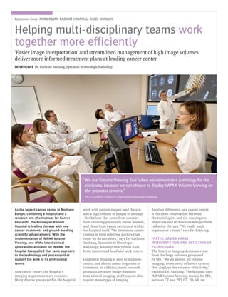
“We use Volume Viewing ‘live’ when we demonstrate pathology for the
clinicians, because we can choose to display IMPAX Volume Viewing on
the projector screens.”
Dr. Cathrine Saxhaug, Specialist in Oncologic Radiology
2. “The advanced functionality of
automatic registration and fusion
is extremely helpful and useful
for us.”
Dr. Cathrine Saxhaug,
Specialist in Oncologic Radiology
use it all the time," says Dr. Saxhaug.
"It gives us a better visualization of
the image volumes, and it is easier
to interpret the images and to find
pathologies. Another major use for
IMPAX Volume Viewing is to compare
two sets of images effectively by
using the registration tool: either a
comparison of two different sequences
from the same exam or a comparison of
the same sequence from two different
exams. This is an efficient way of
monitoring tumor development and
treatment response over time."
IMPAX Volume Viewing offers advanced
visualization tools including MIP/MPR,
volume rendering and segmentation,
and allows users to save reconstructed
series, static screenshots and video
captures. With IMPAX Volume Viewing
PLUS (an optional extension for IMPAX
Volume Viewing), users at the Norwegian
Radium Hospital can also view series in
fusion mode or subtraction mode, and
it offers automatic registration of both
volumes. "The advanced functionality
of automatic registration and fusion is
extremely helpful and useful for us," says
Dr. Saxhaug.
A tight integration with IMPAX PACS
makes IMPAX Volume Viewing highly
efficient. "It improves the workflow, and
you don't waste time loading all the
images," comments Dr. Saxhaug. "It is
seamless, easy to use, fast, offers a high
level of automation and it has a familiar
user interface."
Radiology-led MEETINGS benefit
with better view of patient
information
The radiologists at the hospital
hold daily and weekly meetings in
the radiology department, where
they demonstrate pathology for the
hospital's clinicians. "In Norway,
these meetings are a central way
for the radiologists to provide
information to clinicians," says Dr.
Saxhaug.
The daily meetings focus on cancer
patients who will be operated on, or
receive radiation treatment, that day.
Clinicians attend in groups, according
to their specialty area. For example
the oncologists who are working with
brain tumors come as a group, or the
surgeons who are working with breast
cancer. "We use Volume Viewing 'live'
when we demonstrate pathology for
the clinicians, because we can choose
to display IMPAX Volume Viewing on
the projector screens. It gives them
a better picture of the pathology. For
the surgeons, for example, it makes
it easier to visualize the tumors in
reference to neighboring structures."
The radiologists also host weekly
multi-disciplinary team meetings,
which are attended by surgeons,
oncologists, pathologists and nurses.
These meetings include discussions
on cancer treatment options and next
steps, and here also the participants
refer extensively to patient images.