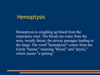
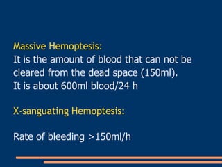
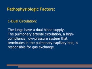
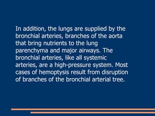
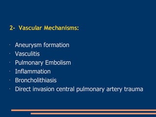
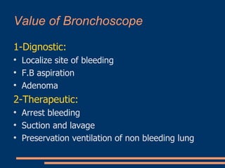
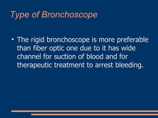
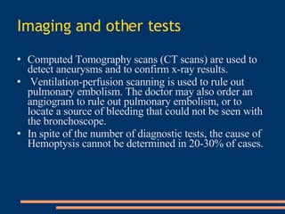
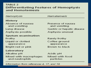
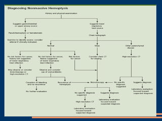
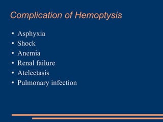
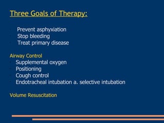
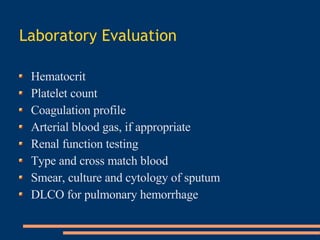
H e m o p t y s i s Prepared by  Dr. Tahany Mahmoud Banha Chest Department Banha University
References   .  Stedman TL. Stedman's Medical dictionary. 27th ed. Philidelphia: Lipincott Williams & Wilkins, 2000 . .  Thompson AB, Teschler H, Rennard SI. Pathogenesis, evaluation, and therapy for massive hemoptysis. Clin Chest Med 1992;13:69-82 . .  Knott-Craig CJ, Oostuizen JG, Rossouw G, Joubert JR, Barnard PM. Management and prognosis of massive hemoptysis. Recent experience with 120 patients. J Thorac Cardiovasc Surg 1993;105:394-7 . .  Cahill BC, Ingbar DH. Massive hemoptysis. Assessment and management. Clin Chest Med 1994;15:147-67 . .  Harrison TR, Braunwald E. Hemoptysis. In: Harrison's Principles of internal medicine. 15th ed. New York: McGraw-Hill
Reisz G, Stevens D, Boutwell C, Nair V. The causes of hemoptysis revisited. A review of the etiologies of hemoptysis between 1986 and 1995. Mo Med 1997;94:633-5 . Bond D, Vyas H. Viral pneumonia and hemoptysis. Crit Care Med 2001;29:2040-1 . Nelson JE, Forman M. Hemoptysis in HIV-infected patients. Chest 1996;110:737-43 . Santiago S, Tobias J, Williams AJ. A reappraisal of the causes of hemoptysis. Arch Intern Med 1991;151: 2449-51 . Hirshberg B, Biran I, Glazer M, Kramer MR. Hemoptysis: etiology, evaluation, and outcome in a tertiary referral hospital. Chest 1997;112:440-4 . Anish EJ, Mayewski RJ. Pulmonary embolism. In: Black ER, ed. Diagnostic strategies for common medical problems. Philadelphia: American College of Physicians, 1999:325-37 .
Hemoptysis Hemoptysis is coughing up blood from the respiratory tract. The blood can come from the nose, mouth, throat, the airway passages leading to the lungs. The word "hemoptysis" comes from the Greek "haima," meaning "blood," and "ptysis," which means "a spitting"
Pseudohemoptesis Causes: Blood not from lower respiratory tract it come from above the vocal cords: Mouth Pharynx Nose Aspiration of hematemsis Colonization by grame negative bacteria (seratia blue)‏
Massive Hemoptesis: It is the amount of blood that can not be  cleared from the dead space (150ml). It is about 600ml blood/24 h X-sanguating Hemoptesis: Rate of bleeding >150ml/h
Pathophysiologic Factors:  1-Dual Circulation: The lungs have a dual blood supply.  The pulmonary arterial circulation, a high-compliance, low-pressure system that terminates in the pulmonary capillary bed, is responsible for gas exchange.
In addition, the lungs are supplied by the bronchial arteries, branches of the aorta that bring nutrients to the lung parenchyma and major airways. The bronchial arteries, like all systemic arteries, are a high-pressure system. Most cases of hemoptysis result from disruption of branches of the bronchial arterial tree.
2-  Vascular Mechanisms :   Aneurysm formation   Vasculitis   Pulmonary Embolism   Inflammation   Broncholithiasis   Direct invasion central pulmonary artery trauma
Causes Blood-tinged mucus in a healthy nonsmoker usually indicates a mild infection and is generally no cause for concern. Indeed, the most common cause of Hemoptysis is the least serious – a ruptured small blood vessel caused by coughing and/or a bronchitic infection In patients with a history of smoking or who are otherwise at risk for lung disease, however, Hemoptysis is often a sign of serious illness, including cancer
Infectious: Chronic bronchitis Bronchiectasis Tuberculosis Nontuberculous mycobacteria Lung abscess Necrotizing pneumonia Mycetoma Cystic fibrosis  Common Causes of Hemoptysis
Neoplastic Lung cancer Bronchial adenoma Metastatic disease (osteogenic sarcoma, choriocarcinoma)  Vasculitic   Wegener's granulomatosis  Systemic lupus erythematosus  Churge Strauss syndrome
Cardiovascular Severe left ventricular heart failure Mitral stenosis Pulmonary embolism or infarction Septic pulmonary embolism or right-sided endocarditis Aortic aneurysm or bronchovascular fistula
Miscellaneous Idiopathic Pulmonary Hemosiderosis Aspirated foreign body Pulmonary contusion or trauma Posttransthoracic needle biopsy or transbronchial lung biopsy  Factitious hemoptysis
Bleeding Diathesis   Anticoagulant therapy   Deficiency of vitamin K–dependent factors: prothrombin (II), Stuart factor (X), factor VII, Christmas factor (IX)‏ Disseminated intravascular coaulation Fibrinolytic therapy: urokinase ,  streptokinase.
Evaluation of Hemoptysis 1-Types of Hemoptesis: Blood tinged sputum Blood streaked Red current jully Frank Hemoptesis Rusty sputum 2-Degree of Hemoptysis: Massive Non massive
Causes of Blood streaked Sputum :   Upper respiratory inflammation   Nose or Nasopharynx   Gums   Larynx   Severe coughing paroxysms   Trauma
Causes of Pink Sputum   : Blood and secretions mix in alveoli, small bronchioles   Conditions associated with pink   Sputum   Pneumonia   Pulmonary edema
Causes of Heavy Bleeding into Respiratory tree   : Pulmonary   Tuberculosis   Lung Abscess   Bronchiectasis   Pulmonary infarction   Pulmonary Embolism   Bronchogenic Carcinoma   Broncholithiasis   Mitral   Stenosis   Actinomycosis   Lung Abscess   Blastomycosis   Lung Abscess
Diagnosis   History: To differentiate between source of Hemoptysis: Respiratory Gastrointestinal Anticoagulant therapy Mitral valve disease
Physical Examination The doctor will examine the patient's nose, throat, mouth, and chest for bleeding from these areas and for signs of chest trauma. The doctor also listens to the patient's breathing and heart beat for indications of heart abnormalities or lung disease .
Laboratory Tests Laboratory tests include blood tests to rule out clotting disorders, and to look for food particles or other evidence of blood from the stomach. Sputum can be tested for fungi, bacteria, or parasites .
X - Ray and Bronchoscopy Chest x rays and bronchoscopy are the most important studies for evaluating hemoptysis. They are used to evaluate the cause, location, and extent of the bleeding.
Value of Bronchoscope 1-Dignostic: Localize site of bleeding F.B aspiration Adenoma 2-Therapeutic: Arrest bleeding Suction and lavage Preservation ventilation of non bleeding lung
Type of Bronchoscope  The rigid bronchoscope is more preferable than fiber optic one due to it has wide channel for suction of blood and for therapeutic treatment to arrest bleeding.
Imaging and other tests Computed Tomography scans (CT scans) are used to detect aneurysms and to confirm x-ray results. Ventilation-perfusion scanning is used to rule out pulmonary embolism. The doctor may also order an angiogram to rule out pulmonary embolism, or to locate a source of bleeding that could not be seen with the bronchoscope . In spite of the number of diagnostic tests, the cause of Hemoptysis cannot be determined in 20-30% of cases .
 
 
Complication of Hemoptysis  Asphyxia Shock Anemia Renal failure Atelectasis Pulmonary infection
Management
Three Goals of Therapy : Prevent asphyxiation   Stop bleeding   Treat primary disease   Airway Control   Supplemental oxygen   Positioning   Cough control   Endotracheal intubation a. selective intubation   Volume Resuscitation
Laboratory Evaluation   Hematocrit   Platelet count   Coagulation profile   Arterial blood gas, if appropriate   Renal function testing   Type and cross match blood   Smear, culture and cytology of sputum   DLCO for pulmonary hemorrhage
Consultations   Surgical   Definitive: resection   Medical   Antibiotics if indicated   Medical management if nonlocalized bleeding, severely impaired pulmonary function, disseminated terminal carcinoma, advanced bilateral pulmonary disease, vasculitis
Modalities   Bronchial artery Embolization   Balloon Tamponade   Intracavitary Anti-fungal therapy   Mortality   Medically managed patients with massive Hemoptysis: 75%   Surgically managed patients with massive Hemoptysis: 23%    
Role of Surgery in Hemoptysis Surgery is indicated in the following situations: Leaky thoracic aneurysm Chest trauma A-V fistula Localized bronchiectasis Chronic lung abscess

Hemoptysis

  • 1.
    H e mo p t y s i s Prepared by Dr. Tahany Mahmoud Banha Chest Department Banha University
  • 2.
    References . Stedman TL. Stedman's Medical dictionary. 27th ed. Philidelphia: Lipincott Williams & Wilkins, 2000 . . Thompson AB, Teschler H, Rennard SI. Pathogenesis, evaluation, and therapy for massive hemoptysis. Clin Chest Med 1992;13:69-82 . . Knott-Craig CJ, Oostuizen JG, Rossouw G, Joubert JR, Barnard PM. Management and prognosis of massive hemoptysis. Recent experience with 120 patients. J Thorac Cardiovasc Surg 1993;105:394-7 . . Cahill BC, Ingbar DH. Massive hemoptysis. Assessment and management. Clin Chest Med 1994;15:147-67 . . Harrison TR, Braunwald E. Hemoptysis. In: Harrison's Principles of internal medicine. 15th ed. New York: McGraw-Hill
  • 3.
    Reisz G, StevensD, Boutwell C, Nair V. The causes of hemoptysis revisited. A review of the etiologies of hemoptysis between 1986 and 1995. Mo Med 1997;94:633-5 . Bond D, Vyas H. Viral pneumonia and hemoptysis. Crit Care Med 2001;29:2040-1 . Nelson JE, Forman M. Hemoptysis in HIV-infected patients. Chest 1996;110:737-43 . Santiago S, Tobias J, Williams AJ. A reappraisal of the causes of hemoptysis. Arch Intern Med 1991;151: 2449-51 . Hirshberg B, Biran I, Glazer M, Kramer MR. Hemoptysis: etiology, evaluation, and outcome in a tertiary referral hospital. Chest 1997;112:440-4 . Anish EJ, Mayewski RJ. Pulmonary embolism. In: Black ER, ed. Diagnostic strategies for common medical problems. Philadelphia: American College of Physicians, 1999:325-37 .
  • 4.
    Hemoptysis Hemoptysis iscoughing up blood from the respiratory tract. The blood can come from the nose, mouth, throat, the airway passages leading to the lungs. The word "hemoptysis" comes from the Greek "haima," meaning "blood," and "ptysis," which means "a spitting"
  • 5.
    Pseudohemoptesis Causes: Bloodnot from lower respiratory tract it come from above the vocal cords: Mouth Pharynx Nose Aspiration of hematemsis Colonization by grame negative bacteria (seratia blue)‏
  • 6.
    Massive Hemoptesis: Itis the amount of blood that can not be cleared from the dead space (150ml). It is about 600ml blood/24 h X-sanguating Hemoptesis: Rate of bleeding >150ml/h
  • 7.
    Pathophysiologic Factors: 1-Dual Circulation: The lungs have a dual blood supply. The pulmonary arterial circulation, a high-compliance, low-pressure system that terminates in the pulmonary capillary bed, is responsible for gas exchange.
  • 8.
    In addition, thelungs are supplied by the bronchial arteries, branches of the aorta that bring nutrients to the lung parenchyma and major airways. The bronchial arteries, like all systemic arteries, are a high-pressure system. Most cases of hemoptysis result from disruption of branches of the bronchial arterial tree.
  • 9.
    2- VascularMechanisms : Aneurysm formation Vasculitis Pulmonary Embolism Inflammation Broncholithiasis Direct invasion central pulmonary artery trauma
  • 10.
    Causes Blood-tinged mucusin a healthy nonsmoker usually indicates a mild infection and is generally no cause for concern. Indeed, the most common cause of Hemoptysis is the least serious – a ruptured small blood vessel caused by coughing and/or a bronchitic infection In patients with a history of smoking or who are otherwise at risk for lung disease, however, Hemoptysis is often a sign of serious illness, including cancer
  • 11.
    Infectious: Chronic bronchitisBronchiectasis Tuberculosis Nontuberculous mycobacteria Lung abscess Necrotizing pneumonia Mycetoma Cystic fibrosis Common Causes of Hemoptysis
  • 12.
    Neoplastic Lung cancerBronchial adenoma Metastatic disease (osteogenic sarcoma, choriocarcinoma) Vasculitic Wegener's granulomatosis Systemic lupus erythematosus Churge Strauss syndrome
  • 13.
    Cardiovascular Severe leftventricular heart failure Mitral stenosis Pulmonary embolism or infarction Septic pulmonary embolism or right-sided endocarditis Aortic aneurysm or bronchovascular fistula
  • 14.
    Miscellaneous Idiopathic PulmonaryHemosiderosis Aspirated foreign body Pulmonary contusion or trauma Posttransthoracic needle biopsy or transbronchial lung biopsy Factitious hemoptysis
  • 15.
    Bleeding Diathesis Anticoagulant therapy Deficiency of vitamin K–dependent factors: prothrombin (II), Stuart factor (X), factor VII, Christmas factor (IX)‏ Disseminated intravascular coaulation Fibrinolytic therapy: urokinase , streptokinase.
  • 16.
    Evaluation of Hemoptysis1-Types of Hemoptesis: Blood tinged sputum Blood streaked Red current jully Frank Hemoptesis Rusty sputum 2-Degree of Hemoptysis: Massive Non massive
  • 17.
    Causes of Bloodstreaked Sputum : Upper respiratory inflammation Nose or Nasopharynx Gums Larynx Severe coughing paroxysms Trauma
  • 18.
    Causes of PinkSputum : Blood and secretions mix in alveoli, small bronchioles Conditions associated with pink Sputum Pneumonia Pulmonary edema
  • 19.
    Causes of HeavyBleeding into Respiratory tree : Pulmonary Tuberculosis Lung Abscess Bronchiectasis Pulmonary infarction Pulmonary Embolism Bronchogenic Carcinoma Broncholithiasis Mitral Stenosis Actinomycosis Lung Abscess Blastomycosis Lung Abscess
  • 20.
    Diagnosis History: To differentiate between source of Hemoptysis: Respiratory Gastrointestinal Anticoagulant therapy Mitral valve disease
  • 21.
    Physical Examination Thedoctor will examine the patient's nose, throat, mouth, and chest for bleeding from these areas and for signs of chest trauma. The doctor also listens to the patient's breathing and heart beat for indications of heart abnormalities or lung disease .
  • 22.
    Laboratory Tests Laboratorytests include blood tests to rule out clotting disorders, and to look for food particles or other evidence of blood from the stomach. Sputum can be tested for fungi, bacteria, or parasites .
  • 23.
    X - Rayand Bronchoscopy Chest x rays and bronchoscopy are the most important studies for evaluating hemoptysis. They are used to evaluate the cause, location, and extent of the bleeding.
  • 24.
    Value of Bronchoscope1-Dignostic: Localize site of bleeding F.B aspiration Adenoma 2-Therapeutic: Arrest bleeding Suction and lavage Preservation ventilation of non bleeding lung
  • 25.
    Type of Bronchoscope The rigid bronchoscope is more preferable than fiber optic one due to it has wide channel for suction of blood and for therapeutic treatment to arrest bleeding.
  • 26.
    Imaging and othertests Computed Tomography scans (CT scans) are used to detect aneurysms and to confirm x-ray results. Ventilation-perfusion scanning is used to rule out pulmonary embolism. The doctor may also order an angiogram to rule out pulmonary embolism, or to locate a source of bleeding that could not be seen with the bronchoscope . In spite of the number of diagnostic tests, the cause of Hemoptysis cannot be determined in 20-30% of cases .
  • 27.
  • 28.
  • 29.
    Complication of Hemoptysis Asphyxia Shock Anemia Renal failure Atelectasis Pulmonary infection
  • 30.
  • 31.
    Three Goals ofTherapy : Prevent asphyxiation Stop bleeding Treat primary disease Airway Control Supplemental oxygen Positioning Cough control Endotracheal intubation a. selective intubation Volume Resuscitation
  • 32.
    Laboratory Evaluation Hematocrit Platelet count Coagulation profile Arterial blood gas, if appropriate Renal function testing Type and cross match blood Smear, culture and cytology of sputum DLCO for pulmonary hemorrhage
  • 33.
    Consultations Surgical Definitive: resection Medical Antibiotics if indicated Medical management if nonlocalized bleeding, severely impaired pulmonary function, disseminated terminal carcinoma, advanced bilateral pulmonary disease, vasculitis
  • 34.
    Modalities Bronchial artery Embolization Balloon Tamponade Intracavitary Anti-fungal therapy Mortality Medically managed patients with massive Hemoptysis: 75% Surgically managed patients with massive Hemoptysis: 23%  
  • 35.
    Role of Surgeryin Hemoptysis Surgery is indicated in the following situations: Leaky thoracic aneurysm Chest trauma A-V fistula Localized bronchiectasis Chronic lung abscess