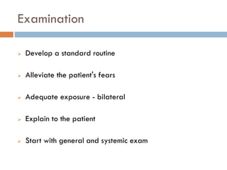
SPINE EXAMINATION
Dr. Fahad Al Mulhim
Faculty Member
Orthopaedic Surgery
Spine Conditions
Complains
Pain
Site Time and mode of onset
Severity or Intensity Character or Nature
Progression Referred pain
Aggravating factors Relieving factors
Any diurnal variation Any seasonal variation
History
Pain
Referred pain
More in the back
Doesn’t go beyond the knees
Radiating pain
Mainly in the LL , often associated with paresthesia
 Neural compression (Sciatica)
History
Pain
Claudication pain
Neurogenic VS Vascular
Q: How to differentiate between vascular and
neurogenic claudication?
History
Pain
Red Flags in Back Pain
 Age of Onset less than 20 or more than 55
 Violent Trauma
 Thoracic spine pain
 Constant progressive non-mechanical pain
 Weight loss or systemic unwell
 Widespread neurology
 Structural deformity
Stiffness
Deformity
Site
Associated Symptoms
• Neurological
• Vascular
• Articular
Amount of
disability
Time of Onset
• Congenital
• Developmental
• Acquired
Correctability
• Completely correctable
• Partially correctable
• Incorrectable
Weakness
Site
Generalised
Localised
Type
Pure Motor
Sensorimotor
Muscular
Mixed
Duration
Acute
Chronic
Onset
Sudden
Gradual
Progression
Progressive
Static
Regressive
Paresthesia
Aetiology Mode of
onset Duration
Site and
Pattern Progression
Aggravating
and Relieving
Factors
Examination
 Develop a standard routine
 Alleviate the patient's fears
 Adequate exposure - bilateral
 Explain to the patient
 Start with general and systemic exam
Local Examination
• InspectionLOOK
• PalpationFEEL
• Active/Passive movement
• Strength Testing
• Range of Motion
MOVE
• Dermatomes & Myotomes
• ReflexesNeurologic
• Depends upon specific region in considerationSPECIAL TESTS
Cervical
Lumbar
Inspection
Inspection
Standing
Look from front, back, and side
Spine alignment
Lower limb alignment and length
Shoulder and pelvic balance
Trunk shift
Walking
Gait pattern
Palpation
Setting
 Occipital protuberance and its muscular attachments
 Cervical spinous processes
 Paravertebral muscles
Standing
 Thoracic and lumbar spine spinous processes ( assess
for any deformity )
 Deep palpation by tapping
 Sacroiliac joint
Palpation
Prone
 Thoracic and lumbar spine
 Paravertebral muscles
 Ischeal tuberosity
 Sciatic nerve pathway
Range of Motion
Cervical:
Range of Motion
Thoracolumbar:
Neurological Examination- Dermatomes
 C5- Area over Deltoid
 C6- Thumb
 C7- Middle finger
 C8- Little finger
 T1- Medial forearm
 T4- Nipple
 T8- Xiphisternum
 T10- Umbilicus
 T12- Symphysis pubis
 L1- Anterior proximal thigh
 L2- Anterior middle thigh
 L3- Medial knee
 L4- Medial malleolus
 L5- 1st web space
 S1- Foot lateral border
 S3- Ischial tuberosity
 S4,5- Perianal region
Neurological Examination- Myotomes
Neurological Examination- Reflexes
Superficial
Abdominal Reflex
Use a blunt object such as a
key or tongue blade.
Stroke the abdomen lightly
on each side in an inward and
downward direction.
 Note the contraction of the
abdominal muscles and
deviation of the umbilicus
towards the stimulus.
Neurological Examination- Reflexes
Superficial
Plantar Reflex
 Stroke the lateral aspect
of the sole of each foot
with end of a reflex
hammer or key
 Observe for plantar
flexion of the foot
Neurological Examination- Reflexes
Deep Tendon Reflexes
Neurological Examination- Reflexes
Deep Tendon Reflexes
Biceps C5-6
 The patient's arm should be
partially flexed at the elbow
with the palm down.
 Place your thumb or finger
firmly on the biceps tendon.
 Strike your finger with the
reflex hammer.
 Look for contraction of the
biceps muscle and slight
flexion of the forearm.
Neurological Examination- Reflexes
Deep Tendon Reflexes
Triceps C6-7
 Support the upper arm and
let the patient's forearm hang
free
 Strike the triceps tendon
above the elbow with the
broad side of the hammer.
 Observe contraction of the
triceps muscle with extension
of the lower arm.
Neurological Examination- Reflexes
Deep Tendon Reflexes
Brachioradialis C5-6
 Have the patient rest the
forearm on the
abdomen or lap.
 Strike the radius about
1-2 inches above wrist.
 Watch for flexion and
supination of forearm
Neurological Examination- Reflexes
Deep Tendon Reflexes
Knee Reflex L3-4
 Have the patient sit with
knee flexed.
 Strike the patellar
tendon just below the
patella.
 Note contraction of the
quadriceps muscle and
extension of the knee
Neurological Examination- Reflexes
Deep Tendon Reflexes
Ankle Reflex S1-2
 Dorsiflex the foot at the
ankle.
 Strike the Achilles tendon.
 Watch and feel for
plantar flexion at the
ankle.
Special Tests
Lasegue’s Test for The Arm
The arm is 90 degrees abducted and extended
Ask the patient to tilt the head to the non affected
side
Positive test if the patient feels pain radiating distal
to the elbow and tingling in specific dermatome
Special Tests
Spurling’s Test
Ask the patient to extend the neck and to laterally
flex to the affected side
Positive if there is pain radiating distal to the elbow
and tingling in a specific dermatome
Additional pressure applied by the examiner on the
head sometimes needed
Special Tests
Straight Leg Raising (SLR) test
Ask the patient to raise the leg without flexing the
knee (actively) and assess for radiating pain from the
back to the leg or the foot
Repeat it passively to confirm
SLR considered positive only if the pain is distal to
the knees
Indicates disc prolapse or foramen stenosis in sciatic
nerve roots
Special Tests
Straight Leg Raising (SLR) test
Special Tests
Bragard’s Test
After SLR Lower the leg for 5 degrees (from the
degree that shows positive SLR)
Dorsiflex the ankle joint
Positive if similar finding to SLR
Special Tests
Femoral Nerve Stretch Test
Hip extension in prone position
Positive when the patient complains of pain at the front
of the thigh
Indicates tension at femoral nerve roots (L2,3,4)
Special Tests
Femoral Nerve Stretch Test
PS SESSION BACK EXAMINATION

PS SESSION BACK EXAMINATION