Case Presentation #54: 60 year old female failed minimally invasive scoliosis surgery
•
2 likes•982 views

A 60 year old woman presented after a failed minimally invasive XLIF surgery for Adult Idiopathic Scoliosis. The patient had multiple complications, and required extensive revision surgery.

Recommended
Recommended
More Related Content
What's hot
What's hot (20)
Viewers also liked
Viewers also liked (17)
Similar to Case Presentation #54: 60 year old female failed minimally invasive scoliosis surgery
Similar to Case Presentation #54: 60 year old female failed minimally invasive scoliosis surgery (17)
More from Robert Pashman
More from Robert Pashman (12)
Recently uploaded
Recently uploaded (20)
Case Presentation #54: 60 year old female failed minimally invasive scoliosis surgery
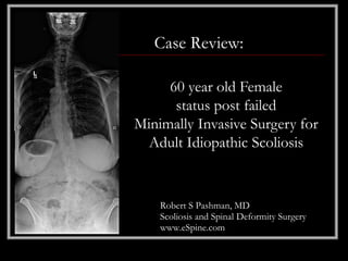
- 1. Case Review: 60 year old Female status post failed Minimally Invasive Surgery for Adult Idiopathic Scoliosis Robert S Pashman, MD Scoliosis and Spinal Deformity Surgery www.eSpine.com
- 2. Patient History • 60-year-old female • Status post an attempt at scoliosis correction with multiple level XLIF and a percutaneous transfascial para lumbar percutaneous pin fixation that was ultimately removed. • The patient did not do well. She has an iatrogenic flat back with collapse kyphosis probable pseudarthrosis 2 to L3. She had significant sagittal and coronal displacement and the patient is completely disabled from her symptoms.
- 3. Pre-op X-rays The patient will need pedicle subtraction osteotomy to reduce sagittal plane balance and she knows that this a significant morbid operation because of the redo nature.
- 4. Indications for Surgery 1. Status post "minimally invasive scoliosis correction with XLIF and percutaneous instrumentation lumbar spine." 2. Complete failure of minimally invasive scoliosis correction with removal of implants and collapse. 3. Iatrogenic kyphosis lumbar spine. 4. Multiple level pseudoarthrosis lumbar spine status post "minimally invasive scoliosis correction adult idiopathic scoliosis." 5. Severe flat back with low back plane in forward decompensation. 6. Failed conservative therapy. 7. Status post removal of a paraspinous boil.
- 5. Surgical Strategy • Segment 1, segmental spinal instrumentation thoracic 10 to pelvis using quarter-inch stainless steel rod screw construct. • Pedicle subtraction osteotomy in lumbar 3 with complete kyphectomy, vertebrectomy L3 under the microscope. • Laminectomy L2-L4 with spinal canal decompression under the microscope. • Posterior spinal fusion T10 to sacral pelvis using locally harvested autogenous bone and Rh BMP. • Intraoperative operating room neuro navigation. • Intraoperative SSEP motor-evoked potential.
- 6. Post-op Films The patient did very well post- operatively, and was very grateful for her results.