SlideShare a Scribd company logo
1 of 68
Copyright © John Wiley & Sons, Inc. All rights reserved.
Chapter 28
The Reproductive Systems
Copyright © John Wiley & Sons, Inc. All rights reserved.
 Sexual Reproduction is the process in which organisms
produce offspring by means of uniting gametes (sperm
and egg)
o Male reproductive organs secrete androgen hormones,
produce gametes (sperm), and facilitate fertilization
o Female reproductive organs secrete female hormones,
produce gametes (ova), facilitate fertilization and
sustain growth of the embryo and fetus
Reproduction Overview
Copyright © John Wiley & Sons, Inc. All rights reserved.
 Urology is the medical specialty that treats disorders and
diseases of the male reproductive system
 Gynecology is the medical specialty that treats disorders
and diseases of the female reproductive system
o Obstetrics focuses on the care of women during
pregnancy
Reproduction Overview
Copyright © John Wiley & Sons, Inc. All rights reserved.
 The genitals are all the structures of reproduction
o The gonads (the testes in males and ovaries in females)
are the site for gamete production and hormone
secretion
o Various ducts store and transport gametes
o Accessory sex glands produce secretions to protect and
support the gametes
o Supporting structures deliver and/or assist in joining
gametes (penis in male, vagina and uterus in female)
Male ReproductiveAnatomy
Copyright © John Wiley & Sons, Inc. All rights reserved.
 The male gonads are the testes (singular: testis)
o The ducts of the male reproductive system are the:
 vas deferens (ductus deferens)
 ejaculatory ducts
 urethra
o Male reproductive
glands are the:
 seminal vesicles (2)
 Prostate (1)
 bulbourethral glands (2)
Male ReproductiveAnatomy
Copyright © John Wiley & Sons, Inc. All rights reserved.
 The scrotum is a supporting structure for the testes
o It consists of a sac of loose skin and superficial fascia
that hangs from the root of the penis
o The location and contraction
of muscle fibers (dartos and
cremaster muscles) regulates
the testicular temp to that
required for sperm production
(2-3o
below the core temp)
Male ReproductiveAnatomy
Copyright © John Wiley & Sons, Inc. All rights reserved.
 The spermatic cord is a supportive structure that
ascends “out of” the scrotum, and consists of:
o The vas deferens
o The testicular artery (of course this
is going into the scrotum)
o Veins and lymphatics that
drain the testes and carry
testosterone to the body
o Autonomic nerves
Male ReproductiveAnatomy
Copyright © John Wiley & Sons, Inc. All rights reserved.
 The spermatic cord and ilioinguinal nerve pass through
the inguinal canal, a passageway in the aponeuroses of
the abdominal muscles (transversus abdominus, internal
oblique, and external
oblique muscles)
o In women, the round
ligament of the uterus and
the ilioinguinal nerve pass
through a very small
inguinal canal
Male ReproductiveAnatomy
Copyright © John Wiley & Sons, Inc. All rights reserved.
 The tunica albuginea (collagen) forms septa that divide
each testis into compartments
called lobules
o Each lobule contains 1-3
seminiferous tubules
where sperm are
produced
Male ReproductiveAnatomy
Copyright © John Wiley & Sons, Inc. All rights reserved.
 Prenatal secretion of testosterone assists testicular
descent and development of male external genital
 Secretion of testosterone at puberty leads to
development of male secondary sexual characteristics
 stimulation of anabolism (musculoskeletal and
protein growth)
 hair growth patterns
 lowering of the voice
 development of libido (sexual drive)
Male Reproductive Physiology
Copyright © John Wiley & Sons, Inc. All rights reserved.
 Spermatozoa are produced in the seminiferous tubules
by sperm stem cells called spermatogonia
 At the beginning of puberty, the
anterior pituitary increases secretion
of the gonadotrophs LH and FSH
o Follicle-stimulating hormone
(FSH) stimulates Sertoli cells
and increases the rate of
spermatogenesis
Spermatogenesis
Copyright © John Wiley & Sons, Inc. All rights reserved.
 LH stimulates Leydig cells, which are located between
seminiferous tubules, to secrete the hormone
testosterone (synthesized from cholesterol)
 Once the degree of spermatogenesis (sperm formation)
required for male reproductive functions
has been achieved,
Sertoli cells release
inhibin, a hormone
that inhibits FSH
Spermatogenesis
Copyright © John Wiley & Sons, Inc. All rights reserved.
Spermatogenesis
Copyright © John Wiley & Sons, Inc. All rights reserved.
 Each day about 300 million sperm complete the process
of spermatogenesis. A sperm contains several structures
that are highly adapted for reaching and penetrating a
secondary oocyte
o The major parts of a sperm are the head and the tail
 the nucleus contains 23 highly condensed
chromosomes (half the normal number)
 covering the anterior two-thirds of the nucleus is
the acrosome
Spermatozoa
Copyright © John Wiley & Sons, Inc. All rights reserved.
 The acrosome is a cap-like vesicle filled with enzymes
(hyaluronidase and proteases)
that help a sperm to penetrate
a secondary oocyte to bring about
fertilization
 The middle piece contains many
mitochondria which provide
the energy (ATP) for locomotion
Spermatozoa
Copyright © John Wiley & Sons, Inc. All rights reserved.
 Before ejaculation, sperm travel via the following route:
o Seminiferous tubules
o Rete testis (network)
o Efferent ducts
o Ductus epididymis
o Vas (ductus)
deferens…
Spermatozoa
Copyright © John Wiley & Sons, Inc. All rights reserved.
 Sperm travelogue continued:
o Vas (ductus) deferens …
o Ejaculatory duct (within
the prostate gland)
o Urethra, which has 3
portions to it:
prostatic
membranous
penile
Spermatozoa
Copyright © John Wiley & Sons, Inc. All rights reserved.
 The accessory glands contribute greatly to the
constituents of the ejaculate
o Seminal vesicles secrete a viscous, alkaline fluid
(mainly during ejaculation) which makes up 60% of
the total volume. It contains fructose (for energy),
prostaglandins (to stimulate smooth muscle
contractions), and clotting proteins (fibrinogen)
 the alkalinity neutralizes the acidity of the male
urethra and the female reproductive tract
Accessory Glands
Copyright © John Wiley & Sons, Inc. All rights reserved.
 The prostate is a chestnut-sized, donut-shaped gland
that secretes about 25% of ejaculate volume. Prostatic
fluid is a milky, slightly acidic solution containing citric
acid (for energy), acid phosphatase, and proteolytic
enzymes (PSA and hyaluronidase)
 The bulbourethral (Cowper’s) gland is a pea-sized gland
inferior to the prostate. It secretes a protective alkaline
mucus that decreases sperm damage in the urethra
Accessory Glands
Copyright © John Wiley & Sons, Inc. All rights reserved.
Accessory Glands
Copyright © John Wiley & Sons, Inc. All rights reserved.
 Semen is a mixture of sperm and seminal fluid, a liquid
that consists of the secretions of the seminiferous
tubules, seminal vesicles, prostate, and bulbourethral
glands
o The volume of semen in a typical ejaculation is 2.5–5
milliliters (mL), with 50–150 million sperm per mL
 when the number falls below 20 million/mL, the
male is likely to be infertile
Semen
Copyright © John Wiley & Sons, Inc. All rights reserved.
 The penis contains the urethra and is a passageway for
the ejaculation of semen and the excretion of urine
o It is cylindrical in shape and consists
of a body, glans penis, and a root
 The body of the penis is composed
of three cylindrical masses of
tissue, each surrounded by
fibrous tissue called the
tunica albuginea
The Penis
Copyright © John Wiley & Sons, Inc. All rights reserved.
 The two dorsolateral masses are the corpora cavernosa
penis, and the smaller midventral mass is the corpus
spongiosum penis (contains
the spongy urethra and
keeps it open during
Ejaculation)
The Penis
Copyright © John Wiley & Sons, Inc. All rights reserved.
 Upon sexual stimulation (visual, tactile, auditory,
olfactory, or imagined), sacral parasympathetic fibers
initiate and maintain an erection
o Under the influence of nitric oxide released from
parasympathetic neurons (“neurogenic NO”), arteries
that supply the penis dilate and blood enters penile
sinuses in the erectile tissue; NO also causes the
smooth muscle within the erectile tissue to relax,
resulting in widening of the blood sinuses
The Male Sexual Response
Copyright © John Wiley & Sons, Inc. All rights reserved.
 After an erection, sympathetic stimulation is necessary
for the rest of the sexual response, including ejaculation
o The smooth muscle sphincter at the base of the urinary
bladder must close, followed by semen being propelled
into the penile portion of the urethra (emission)
o Powerful peristaltic contractions culminate in the
release of semen from
the urethra to
the exterior
The Male Sexual Response
Copyright © John Wiley & Sons, Inc. All rights reserved.
 The organs of the female reproductive system include
the ovaries (female gonads);
the uterine tubes
(fallopian tubes); the
uterus; the vagina;
and the external
organs (collectively
called the vulva,
or pudendum)
Female ReproductiveAnatomy
Copyright © John Wiley & Sons, Inc. All rights reserved.
 The histology of the ovaries reveals the following parts:
o The germinal epithelium covers the surface of the
ovary, but does not give rise to ova – those cells arise
from the yolk sac and migrate to the ovaries
o The ovarian cortex contains the ovarian follicles
o The ovarian medulla
contains blood vessels,
lymphatic vessels
and nerves
Female ReproductiveAnatomy
Copyright © John Wiley & Sons, Inc. All rights reserved.
 The primary role of the ovaries are to produce mature
2o
oocytes (female gametes) and release one (ovulation)
during each monthly ovarian cycle
o another important
function of the ovaries
are to secrete the female
hormones estrogen,
progesterone, inhibin,
and relaxin
Female ReproductiveAnatomy
Copyright © John Wiley & Sons, Inc. All rights reserved.
 After receiving the 2o
oocyte at the infundibulum the
uterine tubes provide a site for fertilization, and then
transport for
the ovum if
fertilization occurs
o The uterine
tubes also have
an ampulla and
an isthmus
Female ReproductiveAnatomy
Copyright © John Wiley & Sons, Inc. All rights reserved.
 The main anchors for the ovaries are the suspensory
ligaments of the ovary (for pelvic wall attachment), and
the ovarian
ligament
(provides an
attachment to
the side wall of
the uterus)
Female ReproductiveAnatomy
Copyright © John Wiley & Sons, Inc. All rights reserved.
 The broad ligament is a
major support for the
uterus (provides side-to
-side and rotation
support)
oOther supportive
ligaments of the uterus
are depicted in these
graphics
Female ReproductiveAnatomy
Copyright © John Wiley & Sons, Inc. All rights reserved.
 The uterus is a pear shaped organ situated between the
urinary bladder and the rectum
o It serves as part of the pathway for sperm deposited
in the vagina to reach the uterine tubes
o It is also the site of implantation of a fertilized ovum,
development of the fetus during pregnancy, and
labor
o During reproductive cycles when implantation does
not occur, the uterus is the source of menstrual flow
Female ReproductiveAnatomy
Copyright © John Wiley & Sons, Inc. All rights reserved.
 Anatomical subdivisions of the uterus include:
o A dome-shaped superior portion called the fundus
o A central portion called the body, that tapers to a
narrow isthmus
o the inferior-most
cervix opens into
the vagina through
the cervical
canal
Female ReproductiveAnatomy
Copyright © John Wiley & Sons, Inc. All rights reserved.
 The interior of the body of the uterus is called the
uterine cavity
 The cervical canal
has an internal os
and an external os
that opens into
the uterine
cavity and the
vagina, respectively
Female ReproductiveAnatomy
Copyright © John Wiley & Sons, Inc. All rights reserved.
 The vagina is a fibromuscular canal lined with mucous
membrane that extends from the exterior of the body to
the uterine cervix. It is composed of both longitudinal
and circular muscle, and has 3 basic functions:
o Serve as a passageway
for menstrual flow
o Receive sperm
o Form the lower
birth canal
Female ReproductiveAnatomy
Copyright © John Wiley & Sons, Inc. All rights reserved.
 The vulva (female external genitalia) refers to the:
o Mons pubis (created by adipose tissue)
o Erectile tissue of the clitoris
o Labia majora (outer limits of
vulva) and labia minora
(covers the vestibule)
o Vestibule, the
area between the
labia minora
o Vaginal orifice
(opening)
Female ReproductiveAnatomy
Copyright © John Wiley & Sons, Inc. All rights reserved.
 Anterior to the vaginal orifice and posterior to the
clitoris is the opening of the external urethral orifice
o Mucus-secreting paraurethral glands flank the orifice
(homologous to the prostate gland in males)
 On either side of the vaginal orifice itself are the greater
vestibular (Bartholin’s) glands which open by ducts into
a groove between the hymen and labia minora. They
produce a small quantity of lubricating mucous during
sexual arousal
Female ReproductiveAnatomy
Copyright © John Wiley & Sons, Inc. All rights reserved.
 The perineum denotes the diamond-shaped area medial
to the thighs and buttocks of females (and males) – the
entire undersurface of the pelvis
o It contains the external
genitalia and
anus
Female ReproductiveAnatomy
Copyright © John Wiley & Sons, Inc. All rights reserved.
Female ReproductiveAnatomy
Copyright © John Wiley & Sons, Inc. All rights reserved.
 The breasts (mammary glands) are modified sudoriferous
glands that produce milk: Each contains 15–20 lobes
divided into lobules
o Each lobule is
composed of milk-
secreting glands called
alveoli. The nipple has a
pigmented area (areola)
and openings for the
lactiferous ducts
Female ReproductiveAnatomy
Copyright © John Wiley & Sons, Inc. All rights reserved.
Female ReproductiveAnatomy
Copyright © John Wiley & Sons, Inc. All rights reserved.
 During reproductive years, nonpregnant females
normally exhibit cyclical changes in the ovaries and
uterus
o Each cycle takes about a month and involves both
oogenesis (ovarian cycle) and preparation of the uterus
to receive a fertilized ovum with hormones secreted by
the hypothalamus, anterior pituitary, and ovaries
controling the main events
Female Reproductive Physiology
Copyright © John Wiley & Sons, Inc. All rights reserved.
 The hormones secreted in the brain constitute the part
of the cycle called the hypothalamic/pituitary cycle.
Those hormones are GnRH, FSH, and LH
oThe reproductive organs in the female pelvis respond
to the brain hormones by cycling at two “lower” levels
 the ovarian cycle occurs in the ovaries where 1o
, 2o
and 3o
follicles are formed
 the uterine cycle refers to the monthly cycling of
the endometrium when a woman is not pregnant
The Female Reproductive Cycle
Copyright © John Wiley & Sons, Inc. All rights reserved.
 The formation of gametes in the ovaries is termed
oogenesis. In contrast to spermatogenesis, which begins
in males at puberty, oogenesis is more complex and
begins in females before they are even born
o During early fetal development, primordial germ cells
migrate from the yolk sac to the ovaries where they
differentiate into oogonia
 Oogonia are diploid (2n) stem cells that divide
mitotically to produce millions of germ cells
The Ovarian Cycle
Copyright © John Wiley & Sons, Inc. All rights reserved.
 Even before birth, most oogonia degenerate, though a
few develop into larger cells called 1o
oocytes that enter
prophase of meiosis I (during fetal development) but do
not complete it until a fortunate few are called upon to
do so during the reproductive years
o During the interim (an arrested
stage of development), each 1o
oocyte is surrounded by follicular
cells in a primordial follicle
The Ovarian Cycle
Copyright © John Wiley & Sons, Inc. All rights reserved.
 At puberty, under the influence of LH and FSH (the brain
gonadotropins), several primordial follicles will be
stimulated each month, although only one will typically
reach the maturity needed for ovulation
o Maturing oocytes within maturing follicles undergo a
series of developmental stages which ultimately brings
one 2o
oocyte within a 3o
follicle to the point of
ovulation
 the ovulated 2o
oocyte will have completed meiosis I,
and so have the haploid number of chromosomes (1n)
The Ovarian Cycle
Copyright © John Wiley & Sons, Inc. All rights reserved.
 As the 1o
oocyte is prepared for ovulation, and receives
the signal to complete meiosis I, two haploid (1n) cells of
unequal size are produced – each with 23 chromosomes
o The smaller cell is called the first
polar body (essentially a packet
of discarded nuclear material)
The Ovarian Cycle
The ovarian cortex with many
follicles in different stages of
development.
o The larger 2o
oocyte inherits
most of the
cytoplasm
Copyright © John Wiley & Sons, Inc. All rights reserved.
 Be aware that the events depicted in these graphics
depict two separate, yet interrelated events: Oocyte
maturation vs. follicle maturation
o The follicle
has important
functions beyond
hosting an
oocyte!
The Ovarian Cycle
Copyright © John Wiley & Sons, Inc. All rights reserved.
The Ovarian Cycle
Copyright © John Wiley & Sons, Inc. All rights reserved.
High levels of
estrogens from
almost mature
follicle stimulate
release of more
GnRH and LH
Hypothalamus
Anterior pituitary
Ovary
Corpus hemorrhagicum
(ruptured follicle)
Almost mature
(graafian) follicle
LH
GnRH
1 High levels of
estrogens from
almost mature
follicle stimulate
release of more
GnRH and LH
Hypothalamus
Anterior pituitary
GnRH promotes
release of FSH
and more LH
Ovary
Corpus hemorrhagicum
(ruptured follicle)
Almost mature
(graafian) follicle
LH
GnRH
1
2
High levels of
estrogens from
almost mature
follicle stimulate
release of more
GnRH and LH
LH surge
brings about
ovulation
Ovulated
secondary
oocyte
Hypothalamus
Anterior pituitary
GnRH promotes
release of FSH
and more LH
Ovary
Corpus hemorrhagicum
(ruptured follicle)
Almost mature
(graafian) follicle
LH
GnRH
1
2
3
Ovulation
Copyright © John Wiley & Sons, Inc. All rights reserved.
 At ovulation the 3o
follicle (mature Graafian follicle)
ruptures to expel the 2o
oocyte into the pelvic cavity,
normally to be swept into the uterine tube - if not
fertilized, it degenerates
o if sperm are
present and
one penetrates
the 2o
oocyte,
meiosis II resumes
The Ovarian Cycle
Copyright © John Wiley & Sons, Inc. All rights reserved.
 The events of
oogenesis described
thus far are depicted
in this graphic
o The 2o
oocyte is
the cell that most
people call
“the egg”
The Ovarian Cycle
Copyright © John Wiley & Sons, Inc. All rights reserved.
 In the ovary, the mature Graafian follicle has become a
corpus luteum, a temporary structure essential for
establishing and maintaining pregnancy in females
o It secretes estrogens and progesterone, which are
responsible for the thickening of the endometrium and
its development and maintenance, respectively
o After approx. 14 days, if the 2o
oocyte is not fertilized,
the corpus luteum stops secreting progesterone and
degenerates into a corpus albicans (just a mass of
fibrous scar tissue)
The Ovarian Cycle
Copyright © John Wiley & Sons, Inc. All rights reserved.
 Without estrogen, and then with abrupt progesterone
withdrawal, the uterine lining cannot be maintained and
it sloughs (menses) – more on the uterine cycle shortly
 If, on the other hand, pregnancy occurs, the corpus
luteum is “rescued” from degeneration by an LH-like
hormone called human chorionic gonadotrophin (hCG –
produced by the developing embryo). With hCG support,
the corpus luteum goes on to produce hormones well into
the 1st trimester until the placenta can take over
The Ovarian Cycle
Copyright © John Wiley & Sons, Inc. All rights reserved.
 The window of opportunity for fertilization to happen
is approximately 2 days before ovulation to 1 day after
ovulation (the sperm can survive 48-72 hrs. in the
uterine tube)
 At the moment of conception, the successful sperm
penetrates the plasma membrane of the 2o
oocyte and
the nuclear material of the two cells unite to
reconstitute the normal number of chromosomes (2n)
o The new diploid cell is called a zygote
The Ovarian Cycle
Copyright © John Wiley & Sons, Inc. All rights reserved.
 The events of
oogenesis and
follicular
development
are summarized
here
The Ovarian Cycle
Copyright © John Wiley & Sons, Inc. All rights reserved.
 Estrogen, progesterone, relaxin, and
inhibin are all secreted by ovaries
(and the placenta during pregnancy)
o Estrogen is responsible for the
presence of secondary sex characteristics
(adipose tissue in the breasts, mons pubis, abdomen,
and hips, voice pitch, and broad pelvis)
 It also lowers blood cholesterol and assists with
fluid and electrolyte balance and protein anabolism
Ovarian Hormones
Copyright © John Wiley & Sons, Inc. All rights reserved.
 Progesterone is the principal hormone responsible for
maturation of the uterine endometrium, as well as an
important player in stimulating breast development
o It inhibits GnRH and LH through a negative feedback
loop
Ovarian Hormones
Copyright © John Wiley & Sons, Inc. All rights reserved.
 Relaxin is released by the corpus luteum; it relaxes the
myometrium and the pubic
symphysis at the
end of pregnancy
 Inhibin is released by
granulosa cells, and then in
large amount by the corpus
luteum; it inhibits FSH
and LH
Ovarian Hormones
Copyright © John Wiley & Sons, Inc. All rights reserved.
 In many ways the uterine or menstrual cycle closely
parallels the events happening in the ovaries
o Under the influence of the ovarian hormones, the
uterine lining undergoes cyclic events (4 phases)
every 28 days (on average)
 Menses marks the beginning of the cycle
 This is followed at day 5 by the pre-ovulatory phase
 Ovulation occurs on about day 14, after which the
post-ovulatory phase begins
The Uterine Cycle
Copyright © John Wiley & Sons, Inc. All rights reserved.
The Uterine Cycle
Copyright © John Wiley & Sons, Inc. All rights reserved.
 Menses manifests as a degeneration of the endometrium
when levels of progesterone become insufficient
o Prostaglandin released by the “unsupported”
endometrium causes constriction of supply arteries
causing a reduction in blood flow: Bloody endometrial
tissue eventually sloughs, and is passed out through
the two uterine os and into the vagina
 Menstruation lasts 1–7 days with 50–150 ml of
fluids and cells lost
The Uterine Cycle
Copyright © John Wiley & Sons, Inc. All rights reserved.
 Under the influence of estrogens released from the new
developing follicles the pre-ovulatory (proliferative
phase) begins, and the uterine epithelium is restored
 The post-ovulatory (secretory phase) begins around the
time of ovulation. Endometrial glands increase the
secretion of mucous-rich glycogen and the
endometrium reaches maximum thickness and maturity
under the influence of estrogens and progesterone
The Uterine Cycle
Copyright © John Wiley & Sons, Inc. All rights reserved.
 Equivalent  structures
during fetal development
 testes ≈ ovaries
 sperm ≈ ovum (2o
oocyte)
 scrotum ≈ labia majora
 spongy urethra and penile
skin ≈ labia minora
 glans penis ≈ clitoris
 prostate gland ≈
paraurethral glands
 bulbourethral glands ≈
vestibular glands
Homologous Structures
Copyright © John Wiley & Sons, Inc. All rights reserved.
 As in the male, the initiation of the sexual response
results in stimulation of sacral parasympathetic fibers
and nitric oxide dilation of the erectile tissues – in this
case, of the clitoris
o Orgasm occurs at the peak of the plateau phase of the
sexual response. Sexual climax culminates in a
sympathetic discharge, accompanied by quick cycles of
muscle contraction in the lower pelvic muscles and an
intense feeling of euphoria
The Female Sexual Response
Copyright © John Wiley & Sons, Inc. All rights reserved.
 The period after orgasm is known as the refractory
period. During this time, release of the neurohormones
oxytocin and prolactin produce a feeling of relaxation
o Release of these hormones (and others, like
vasopressin) also serves as a reward mechanism that
regulates pair-bonding and sexual imprinting
between the partners
The Female Sexual Response
Copyright © John Wiley & Sons, Inc. All rights reserved.
 Benign prostatic hypertrophy is an enlargement of the
prostate gland in the absence of cancer. It is a very
common affliction as men age, resulting in obstruction of
urine flow and inability to completely empty the bladder
 Impotence is the inability to maintain erection long
enough for sexual intercourse
 Primary infertility describes couples who have never
been able to become pregnant after at least 1 year of
unprotected sex
Disruptions of Homeostasis
Copyright © John Wiley & Sons, Inc. All rights reserved.
 Menstrual Abnormalities
o Amenorrhea is the absence of menstruation
o Dysmenorrhea describes unusual menstrual discomfort
(usually indicates an excess of prostaglandin secretion)
o Disfunctional uterine bleeding (DUB) is abnormal
uterine bleeding in the absence of organic disease
(usually an estrogen/progesterone imbalance)
o PMS (premenstrual syndrome) indicates a mild distress
near end of postovulatory (luteal) phase
Disruptions of Homeostasis

More Related Content

What's hot

histology of male reproductive system
histology of male reproductive systemhistology of male reproductive system
histology of male reproductive systemSubhajit Hajra
 
Anatomy & Physiology of the Male Reproductive System
Anatomy & Physiology of the Male Reproductive SystemAnatomy & Physiology of the Male Reproductive System
Anatomy & Physiology of the Male Reproductive SystemDr. Sarita Sharma
 
Male reproductive system
Male reproductive systemMale reproductive system
Male reproductive systemJegan Nadar
 
2 Spermatogenesis & Oogenesis
2  Spermatogenesis & Oogenesis2  Spermatogenesis & Oogenesis
2 Spermatogenesis & Oogenesismsu
 
Female reproductive system
Female  reproductive systemFemale  reproductive system
Female reproductive systemAPARNA C LAKSHMI
 
Anatomy and physiology of male reproductive system
Anatomy and physiology of male reproductive systemAnatomy and physiology of male reproductive system
Anatomy and physiology of male reproductive systemPallavi Lokhande
 
Anatomy of reproductive system
Anatomy of reproductive systemAnatomy of reproductive system
Anatomy of reproductive systemArturo Decano
 
The Male Reproductive System
The Male Reproductive SystemThe Male Reproductive System
The Male Reproductive SystemAlok Kumar
 
Spermatogenesis
SpermatogenesisSpermatogenesis
Spermatogenesisopkholwad
 
Human Reproductive system: Anatomy and physiology
Human Reproductive system: Anatomy and physiologyHuman Reproductive system: Anatomy and physiology
Human Reproductive system: Anatomy and physiologyA M O L D E O R E
 
Male reproductive system
Male reproductive systemMale reproductive system
Male reproductive systemAnnabel Base
 
Biology form 5 process of oogenesis
Biology form 5 process of oogenesisBiology form 5 process of oogenesis
Biology form 5 process of oogenesissahieyra
 
Spermatogenesis vs. oogenesis_-_germinario
Spermatogenesis vs. oogenesis_-_germinarioSpermatogenesis vs. oogenesis_-_germinario
Spermatogenesis vs. oogenesis_-_germinarioPakLiam
 
Malereproductivesystem
MalereproductivesystemMalereproductivesystem
MalereproductivesystemSoneeshah
 
28 [chapter 28 the reproductive system]
28 [chapter 28 the reproductive system]28 [chapter 28 the reproductive system]
28 [chapter 28 the reproductive system]Sompoch Thanachaikan
 
Spermatogenesis -PART 4
Spermatogenesis -PART 4Spermatogenesis -PART 4
Spermatogenesis -PART 4AnuSebastian18
 

What's hot (20)

histology of male reproductive system
histology of male reproductive systemhistology of male reproductive system
histology of male reproductive system
 
Anatomy & Physiology of the Male Reproductive System
Anatomy & Physiology of the Male Reproductive SystemAnatomy & Physiology of the Male Reproductive System
Anatomy & Physiology of the Male Reproductive System
 
Male reproductive system
Male reproductive systemMale reproductive system
Male reproductive system
 
2 Spermatogenesis & Oogenesis
2  Spermatogenesis & Oogenesis2  Spermatogenesis & Oogenesis
2 Spermatogenesis & Oogenesis
 
Female reproductive system
Female  reproductive systemFemale  reproductive system
Female reproductive system
 
Anatomy and physiology of male reproductive system
Anatomy and physiology of male reproductive systemAnatomy and physiology of male reproductive system
Anatomy and physiology of male reproductive system
 
Anatomy of reproductive system
Anatomy of reproductive systemAnatomy of reproductive system
Anatomy of reproductive system
 
1. Male reproductive system
1. Male reproductive system1. Male reproductive system
1. Male reproductive system
 
The Male Reproductive System
The Male Reproductive SystemThe Male Reproductive System
The Male Reproductive System
 
Spermatogenesis
SpermatogenesisSpermatogenesis
Spermatogenesis
 
Spermatogenesis
SpermatogenesisSpermatogenesis
Spermatogenesis
 
Human Reproductive system: Anatomy and physiology
Human Reproductive system: Anatomy and physiologyHuman Reproductive system: Anatomy and physiology
Human Reproductive system: Anatomy and physiology
 
Male reproductive system
Male reproductive systemMale reproductive system
Male reproductive system
 
Biology form 5 process of oogenesis
Biology form 5 process of oogenesisBiology form 5 process of oogenesis
Biology form 5 process of oogenesis
 
Reproductive system Notes
Reproductive system NotesReproductive system Notes
Reproductive system Notes
 
Spermatogenesis vs. oogenesis_-_germinario
Spermatogenesis vs. oogenesis_-_germinarioSpermatogenesis vs. oogenesis_-_germinario
Spermatogenesis vs. oogenesis_-_germinario
 
Malereproductivesystem
MalereproductivesystemMalereproductivesystem
Malereproductivesystem
 
28 [chapter 28 the reproductive system]
28 [chapter 28 the reproductive system]28 [chapter 28 the reproductive system]
28 [chapter 28 the reproductive system]
 
Female Reproductive System I
Female Reproductive System IFemale Reproductive System I
Female Reproductive System I
 
Spermatogenesis -PART 4
Spermatogenesis -PART 4Spermatogenesis -PART 4
Spermatogenesis -PART 4
 

Similar to Chapter 28

The Reproductive System
The Reproductive SystemThe Reproductive System
The Reproductive Systemitutor
 
Male reproductive system slideshare.pptx
Male reproductive system slideshare.pptxMale reproductive system slideshare.pptx
Male reproductive system slideshare.pptxDr.pavithra Anandan
 
Reproduction.pptx
Reproduction.pptxReproduction.pptx
Reproduction.pptxRupaSingh83
 
3. human reproduction male reproductive system
3. human reproduction male reproductive system3. human reproduction male reproductive system
3. human reproduction male reproductive systemInderjit Singh
 
Overview of reproductive physiology and anatomy
Overview of reproductive physiology and anatomyOverview of reproductive physiology and anatomy
Overview of reproductive physiology and anatomymukemilawol
 
REPRODUCTIVE SYSTEM.pptx
REPRODUCTIVE SYSTEM.pptxREPRODUCTIVE SYSTEM.pptx
REPRODUCTIVE SYSTEM.pptxRupaSingh83
 
The reproductive system
The reproductive systemThe reproductive system
The reproductive systemVanja Drljevic
 
Reproductive System
Reproductive SystemReproductive System
Reproductive Systemlogan_xavi93
 
MALE REPRODUCTIVE SYSTEM.pptx
MALE REPRODUCTIVE SYSTEM.pptxMALE REPRODUCTIVE SYSTEM.pptx
MALE REPRODUCTIVE SYSTEM.pptxSwetaGaude1
 
Reproductive physiology
Reproductive physiologyReproductive physiology
Reproductive physiologyKern Rocke
 
REPRODUCTIVE SYSTEM PHYSIOLOGY pkl 2414437kptx
REPRODUCTIVE SYSTEM PHYSIOLOGY pkl 2414437kptxREPRODUCTIVE SYSTEM PHYSIOLOGY pkl 2414437kptx
REPRODUCTIVE SYSTEM PHYSIOLOGY pkl 2414437kptxMuliChristopherKimeu
 
Male reproductive system Cyril Skaria
Male reproductive system Cyril SkariaMale reproductive system Cyril Skaria
Male reproductive system Cyril SkariaCyril Skaria
 
Male reproductive system
Male reproductive systemMale reproductive system
Male reproductive systemwaheed Bahir
 
Reproductive System.
Reproductive System.Reproductive System.
Reproductive System.Audumbar Mali
 
Grade 10 Bio Male reproductive system
Grade 10 Bio Male reproductive systemGrade 10 Bio Male reproductive system
Grade 10 Bio Male reproductive systemSonakshi Yadav
 

Similar to Chapter 28 (20)

The Reproductive System
The Reproductive SystemThe Reproductive System
The Reproductive System
 
Male reproductive system slideshare.pptx
Male reproductive system slideshare.pptxMale reproductive system slideshare.pptx
Male reproductive system slideshare.pptx
 
Presentation on testes ppt
Presentation on testes pptPresentation on testes ppt
Presentation on testes ppt
 
Reproduction.pptx
Reproduction.pptxReproduction.pptx
Reproduction.pptx
 
3. human reproduction male reproductive system
3. human reproduction male reproductive system3. human reproduction male reproductive system
3. human reproduction male reproductive system
 
Overview of reproductive physiology and anatomy
Overview of reproductive physiology and anatomyOverview of reproductive physiology and anatomy
Overview of reproductive physiology and anatomy
 
Reproductive System.pptx
Reproductive System.pptxReproductive System.pptx
Reproductive System.pptx
 
REPRODUCTIVE SYSTEM.pptx
REPRODUCTIVE SYSTEM.pptxREPRODUCTIVE SYSTEM.pptx
REPRODUCTIVE SYSTEM.pptx
 
The reproductive system
The reproductive systemThe reproductive system
The reproductive system
 
Reproductive System
Reproductive SystemReproductive System
Reproductive System
 
MALE REPRODUCTIVE SYSTEM.pptx
MALE REPRODUCTIVE SYSTEM.pptxMALE REPRODUCTIVE SYSTEM.pptx
MALE REPRODUCTIVE SYSTEM.pptx
 
Reproductive physiology
Reproductive physiologyReproductive physiology
Reproductive physiology
 
REPRODUCTIVE SYSTEM PHYSIOLOGY pkl 2414437kptx
REPRODUCTIVE SYSTEM PHYSIOLOGY pkl 2414437kptxREPRODUCTIVE SYSTEM PHYSIOLOGY pkl 2414437kptx
REPRODUCTIVE SYSTEM PHYSIOLOGY pkl 2414437kptx
 
22 Reproductive
22 Reproductive22 Reproductive
22 Reproductive
 
Male reproductive system Cyril Skaria
Male reproductive system Cyril SkariaMale reproductive system Cyril Skaria
Male reproductive system Cyril Skaria
 
Human Rep Part 1
Human Rep Part 1Human Rep Part 1
Human Rep Part 1
 
Male reproductive system
Male reproductive systemMale reproductive system
Male reproductive system
 
Reproduction
ReproductionReproduction
Reproduction
 
Reproductive System.
Reproductive System.Reproductive System.
Reproductive System.
 
Grade 10 Bio Male reproductive system
Grade 10 Bio Male reproductive systemGrade 10 Bio Male reproductive system
Grade 10 Bio Male reproductive system
 

More from Yukti Sharma (20)
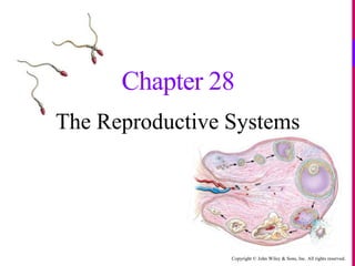
Chapter 1
Chapter 1Chapter 1
Chapter 1
 
Muscle funccellularlevel animal systems
Muscle funccellularlevel animal systemsMuscle funccellularlevel animal systems
Muscle funccellularlevel animal systems
 
Chapter 25
Chapter 25Chapter 25
Chapter 25
 
Chapter 24
Chapter 24Chapter 24
Chapter 24
 
Sensory receptors animal systems
Sensory receptors animal systemsSensory receptors animal systems
Sensory receptors animal systems
 
Action potentials animal systems
Action potentials  animal systemsAction potentials  animal systems
Action potentials animal systems
 
Chapter 27
Chapter 27Chapter 27
Chapter 27
 
Chapter 26
Chapter 26Chapter 26
Chapter 26
 
Chapter 18
Chapter 18Chapter 18
Chapter 18
 
Chapter 17
Chapter 17Chapter 17
Chapter 17
 
Chapter 16
Chapter 16Chapter 16
Chapter 16
 
Chapter 14
Chapter 14Chapter 14
Chapter 14
 
Chapter 13
Chapter 13Chapter 13
Chapter 13
 
Chapter 12
Chapter 12Chapter 12
Chapter 12
 
Chapter 25
Chapter 25Chapter 25
Chapter 25
 
Chapter 25
Chapter 25Chapter 25
Chapter 25
 
Chapter 24
Chapter 24Chapter 24
Chapter 24
 
Chapter 20
Chapter 20Chapter 20
Chapter 20
 
Chapter 3
Chapter 3Chapter 3
Chapter 3
 
Lecture 23
Lecture 23 Lecture 23
Lecture 23
 

Chapter 28

  • 1. Copyright © John Wiley & Sons, Inc. All rights reserved. Chapter 28 The Reproductive Systems
  • 2. Copyright © John Wiley & Sons, Inc. All rights reserved.  Sexual Reproduction is the process in which organisms produce offspring by means of uniting gametes (sperm and egg) o Male reproductive organs secrete androgen hormones, produce gametes (sperm), and facilitate fertilization o Female reproductive organs secrete female hormones, produce gametes (ova), facilitate fertilization and sustain growth of the embryo and fetus Reproduction Overview
  • 3. Copyright © John Wiley & Sons, Inc. All rights reserved.  Urology is the medical specialty that treats disorders and diseases of the male reproductive system  Gynecology is the medical specialty that treats disorders and diseases of the female reproductive system o Obstetrics focuses on the care of women during pregnancy Reproduction Overview
  • 4. Copyright © John Wiley & Sons, Inc. All rights reserved.  The genitals are all the structures of reproduction o The gonads (the testes in males and ovaries in females) are the site for gamete production and hormone secretion o Various ducts store and transport gametes o Accessory sex glands produce secretions to protect and support the gametes o Supporting structures deliver and/or assist in joining gametes (penis in male, vagina and uterus in female) Male ReproductiveAnatomy
  • 5. Copyright © John Wiley & Sons, Inc. All rights reserved.  The male gonads are the testes (singular: testis) o The ducts of the male reproductive system are the:  vas deferens (ductus deferens)  ejaculatory ducts  urethra o Male reproductive glands are the:  seminal vesicles (2)  Prostate (1)  bulbourethral glands (2) Male ReproductiveAnatomy
  • 6. Copyright © John Wiley & Sons, Inc. All rights reserved.  The scrotum is a supporting structure for the testes o It consists of a sac of loose skin and superficial fascia that hangs from the root of the penis o The location and contraction of muscle fibers (dartos and cremaster muscles) regulates the testicular temp to that required for sperm production (2-3o below the core temp) Male ReproductiveAnatomy
  • 7. Copyright © John Wiley & Sons, Inc. All rights reserved.  The spermatic cord is a supportive structure that ascends “out of” the scrotum, and consists of: o The vas deferens o The testicular artery (of course this is going into the scrotum) o Veins and lymphatics that drain the testes and carry testosterone to the body o Autonomic nerves Male ReproductiveAnatomy
  • 8. Copyright © John Wiley & Sons, Inc. All rights reserved.  The spermatic cord and ilioinguinal nerve pass through the inguinal canal, a passageway in the aponeuroses of the abdominal muscles (transversus abdominus, internal oblique, and external oblique muscles) o In women, the round ligament of the uterus and the ilioinguinal nerve pass through a very small inguinal canal Male ReproductiveAnatomy
  • 9. Copyright © John Wiley & Sons, Inc. All rights reserved.  The tunica albuginea (collagen) forms septa that divide each testis into compartments called lobules o Each lobule contains 1-3 seminiferous tubules where sperm are produced Male ReproductiveAnatomy
  • 10. Copyright © John Wiley & Sons, Inc. All rights reserved.  Prenatal secretion of testosterone assists testicular descent and development of male external genital  Secretion of testosterone at puberty leads to development of male secondary sexual characteristics  stimulation of anabolism (musculoskeletal and protein growth)  hair growth patterns  lowering of the voice  development of libido (sexual drive) Male Reproductive Physiology
  • 11. Copyright © John Wiley & Sons, Inc. All rights reserved.  Spermatozoa are produced in the seminiferous tubules by sperm stem cells called spermatogonia  At the beginning of puberty, the anterior pituitary increases secretion of the gonadotrophs LH and FSH o Follicle-stimulating hormone (FSH) stimulates Sertoli cells and increases the rate of spermatogenesis Spermatogenesis
  • 12. Copyright © John Wiley & Sons, Inc. All rights reserved.  LH stimulates Leydig cells, which are located between seminiferous tubules, to secrete the hormone testosterone (synthesized from cholesterol)  Once the degree of spermatogenesis (sperm formation) required for male reproductive functions has been achieved, Sertoli cells release inhibin, a hormone that inhibits FSH Spermatogenesis
  • 13. Copyright © John Wiley & Sons, Inc. All rights reserved. Spermatogenesis
  • 14. Copyright © John Wiley & Sons, Inc. All rights reserved.  Each day about 300 million sperm complete the process of spermatogenesis. A sperm contains several structures that are highly adapted for reaching and penetrating a secondary oocyte o The major parts of a sperm are the head and the tail  the nucleus contains 23 highly condensed chromosomes (half the normal number)  covering the anterior two-thirds of the nucleus is the acrosome Spermatozoa
  • 15. Copyright © John Wiley & Sons, Inc. All rights reserved.  The acrosome is a cap-like vesicle filled with enzymes (hyaluronidase and proteases) that help a sperm to penetrate a secondary oocyte to bring about fertilization  The middle piece contains many mitochondria which provide the energy (ATP) for locomotion Spermatozoa
  • 16. Copyright © John Wiley & Sons, Inc. All rights reserved.  Before ejaculation, sperm travel via the following route: o Seminiferous tubules o Rete testis (network) o Efferent ducts o Ductus epididymis o Vas (ductus) deferens… Spermatozoa
  • 17. Copyright © John Wiley & Sons, Inc. All rights reserved.  Sperm travelogue continued: o Vas (ductus) deferens … o Ejaculatory duct (within the prostate gland) o Urethra, which has 3 portions to it: prostatic membranous penile Spermatozoa
  • 18. Copyright © John Wiley & Sons, Inc. All rights reserved.  The accessory glands contribute greatly to the constituents of the ejaculate o Seminal vesicles secrete a viscous, alkaline fluid (mainly during ejaculation) which makes up 60% of the total volume. It contains fructose (for energy), prostaglandins (to stimulate smooth muscle contractions), and clotting proteins (fibrinogen)  the alkalinity neutralizes the acidity of the male urethra and the female reproductive tract Accessory Glands
  • 19. Copyright © John Wiley & Sons, Inc. All rights reserved.  The prostate is a chestnut-sized, donut-shaped gland that secretes about 25% of ejaculate volume. Prostatic fluid is a milky, slightly acidic solution containing citric acid (for energy), acid phosphatase, and proteolytic enzymes (PSA and hyaluronidase)  The bulbourethral (Cowper’s) gland is a pea-sized gland inferior to the prostate. It secretes a protective alkaline mucus that decreases sperm damage in the urethra Accessory Glands
  • 20. Copyright © John Wiley & Sons, Inc. All rights reserved. Accessory Glands
  • 21. Copyright © John Wiley & Sons, Inc. All rights reserved.  Semen is a mixture of sperm and seminal fluid, a liquid that consists of the secretions of the seminiferous tubules, seminal vesicles, prostate, and bulbourethral glands o The volume of semen in a typical ejaculation is 2.5–5 milliliters (mL), with 50–150 million sperm per mL  when the number falls below 20 million/mL, the male is likely to be infertile Semen
  • 22. Copyright © John Wiley & Sons, Inc. All rights reserved.  The penis contains the urethra and is a passageway for the ejaculation of semen and the excretion of urine o It is cylindrical in shape and consists of a body, glans penis, and a root  The body of the penis is composed of three cylindrical masses of tissue, each surrounded by fibrous tissue called the tunica albuginea The Penis
  • 23. Copyright © John Wiley & Sons, Inc. All rights reserved.  The two dorsolateral masses are the corpora cavernosa penis, and the smaller midventral mass is the corpus spongiosum penis (contains the spongy urethra and keeps it open during Ejaculation) The Penis
  • 24. Copyright © John Wiley & Sons, Inc. All rights reserved.  Upon sexual stimulation (visual, tactile, auditory, olfactory, or imagined), sacral parasympathetic fibers initiate and maintain an erection o Under the influence of nitric oxide released from parasympathetic neurons (“neurogenic NO”), arteries that supply the penis dilate and blood enters penile sinuses in the erectile tissue; NO also causes the smooth muscle within the erectile tissue to relax, resulting in widening of the blood sinuses The Male Sexual Response
  • 25. Copyright © John Wiley & Sons, Inc. All rights reserved.  After an erection, sympathetic stimulation is necessary for the rest of the sexual response, including ejaculation o The smooth muscle sphincter at the base of the urinary bladder must close, followed by semen being propelled into the penile portion of the urethra (emission) o Powerful peristaltic contractions culminate in the release of semen from the urethra to the exterior The Male Sexual Response
  • 26. Copyright © John Wiley & Sons, Inc. All rights reserved.  The organs of the female reproductive system include the ovaries (female gonads); the uterine tubes (fallopian tubes); the uterus; the vagina; and the external organs (collectively called the vulva, or pudendum) Female ReproductiveAnatomy
  • 27. Copyright © John Wiley & Sons, Inc. All rights reserved.  The histology of the ovaries reveals the following parts: o The germinal epithelium covers the surface of the ovary, but does not give rise to ova – those cells arise from the yolk sac and migrate to the ovaries o The ovarian cortex contains the ovarian follicles o The ovarian medulla contains blood vessels, lymphatic vessels and nerves Female ReproductiveAnatomy
  • 28. Copyright © John Wiley & Sons, Inc. All rights reserved.  The primary role of the ovaries are to produce mature 2o oocytes (female gametes) and release one (ovulation) during each monthly ovarian cycle o another important function of the ovaries are to secrete the female hormones estrogen, progesterone, inhibin, and relaxin Female ReproductiveAnatomy
  • 29. Copyright © John Wiley & Sons, Inc. All rights reserved.  After receiving the 2o oocyte at the infundibulum the uterine tubes provide a site for fertilization, and then transport for the ovum if fertilization occurs o The uterine tubes also have an ampulla and an isthmus Female ReproductiveAnatomy
  • 30. Copyright © John Wiley & Sons, Inc. All rights reserved.  The main anchors for the ovaries are the suspensory ligaments of the ovary (for pelvic wall attachment), and the ovarian ligament (provides an attachment to the side wall of the uterus) Female ReproductiveAnatomy
  • 31. Copyright © John Wiley & Sons, Inc. All rights reserved.  The broad ligament is a major support for the uterus (provides side-to -side and rotation support) oOther supportive ligaments of the uterus are depicted in these graphics Female ReproductiveAnatomy
  • 32. Copyright © John Wiley & Sons, Inc. All rights reserved.  The uterus is a pear shaped organ situated between the urinary bladder and the rectum o It serves as part of the pathway for sperm deposited in the vagina to reach the uterine tubes o It is also the site of implantation of a fertilized ovum, development of the fetus during pregnancy, and labor o During reproductive cycles when implantation does not occur, the uterus is the source of menstrual flow Female ReproductiveAnatomy
  • 33. Copyright © John Wiley & Sons, Inc. All rights reserved.  Anatomical subdivisions of the uterus include: o A dome-shaped superior portion called the fundus o A central portion called the body, that tapers to a narrow isthmus o the inferior-most cervix opens into the vagina through the cervical canal Female ReproductiveAnatomy
  • 34. Copyright © John Wiley & Sons, Inc. All rights reserved.  The interior of the body of the uterus is called the uterine cavity  The cervical canal has an internal os and an external os that opens into the uterine cavity and the vagina, respectively Female ReproductiveAnatomy
  • 35. Copyright © John Wiley & Sons, Inc. All rights reserved.  The vagina is a fibromuscular canal lined with mucous membrane that extends from the exterior of the body to the uterine cervix. It is composed of both longitudinal and circular muscle, and has 3 basic functions: o Serve as a passageway for menstrual flow o Receive sperm o Form the lower birth canal Female ReproductiveAnatomy
  • 36. Copyright © John Wiley & Sons, Inc. All rights reserved.  The vulva (female external genitalia) refers to the: o Mons pubis (created by adipose tissue) o Erectile tissue of the clitoris o Labia majora (outer limits of vulva) and labia minora (covers the vestibule) o Vestibule, the area between the labia minora o Vaginal orifice (opening) Female ReproductiveAnatomy
  • 37. Copyright © John Wiley & Sons, Inc. All rights reserved.  Anterior to the vaginal orifice and posterior to the clitoris is the opening of the external urethral orifice o Mucus-secreting paraurethral glands flank the orifice (homologous to the prostate gland in males)  On either side of the vaginal orifice itself are the greater vestibular (Bartholin’s) glands which open by ducts into a groove between the hymen and labia minora. They produce a small quantity of lubricating mucous during sexual arousal Female ReproductiveAnatomy
  • 38. Copyright © John Wiley & Sons, Inc. All rights reserved.  The perineum denotes the diamond-shaped area medial to the thighs and buttocks of females (and males) – the entire undersurface of the pelvis o It contains the external genitalia and anus Female ReproductiveAnatomy
  • 39. Copyright © John Wiley & Sons, Inc. All rights reserved. Female ReproductiveAnatomy
  • 40. Copyright © John Wiley & Sons, Inc. All rights reserved.  The breasts (mammary glands) are modified sudoriferous glands that produce milk: Each contains 15–20 lobes divided into lobules o Each lobule is composed of milk- secreting glands called alveoli. The nipple has a pigmented area (areola) and openings for the lactiferous ducts Female ReproductiveAnatomy
  • 41. Copyright © John Wiley & Sons, Inc. All rights reserved. Female ReproductiveAnatomy
  • 42. Copyright © John Wiley & Sons, Inc. All rights reserved.  During reproductive years, nonpregnant females normally exhibit cyclical changes in the ovaries and uterus o Each cycle takes about a month and involves both oogenesis (ovarian cycle) and preparation of the uterus to receive a fertilized ovum with hormones secreted by the hypothalamus, anterior pituitary, and ovaries controling the main events Female Reproductive Physiology
  • 43. Copyright © John Wiley & Sons, Inc. All rights reserved.  The hormones secreted in the brain constitute the part of the cycle called the hypothalamic/pituitary cycle. Those hormones are GnRH, FSH, and LH oThe reproductive organs in the female pelvis respond to the brain hormones by cycling at two “lower” levels  the ovarian cycle occurs in the ovaries where 1o , 2o and 3o follicles are formed  the uterine cycle refers to the monthly cycling of the endometrium when a woman is not pregnant The Female Reproductive Cycle
  • 44. Copyright © John Wiley & Sons, Inc. All rights reserved.  The formation of gametes in the ovaries is termed oogenesis. In contrast to spermatogenesis, which begins in males at puberty, oogenesis is more complex and begins in females before they are even born o During early fetal development, primordial germ cells migrate from the yolk sac to the ovaries where they differentiate into oogonia  Oogonia are diploid (2n) stem cells that divide mitotically to produce millions of germ cells The Ovarian Cycle
  • 45. Copyright © John Wiley & Sons, Inc. All rights reserved.  Even before birth, most oogonia degenerate, though a few develop into larger cells called 1o oocytes that enter prophase of meiosis I (during fetal development) but do not complete it until a fortunate few are called upon to do so during the reproductive years o During the interim (an arrested stage of development), each 1o oocyte is surrounded by follicular cells in a primordial follicle The Ovarian Cycle
  • 46. Copyright © John Wiley & Sons, Inc. All rights reserved.  At puberty, under the influence of LH and FSH (the brain gonadotropins), several primordial follicles will be stimulated each month, although only one will typically reach the maturity needed for ovulation o Maturing oocytes within maturing follicles undergo a series of developmental stages which ultimately brings one 2o oocyte within a 3o follicle to the point of ovulation  the ovulated 2o oocyte will have completed meiosis I, and so have the haploid number of chromosomes (1n) The Ovarian Cycle
  • 47. Copyright © John Wiley & Sons, Inc. All rights reserved.  As the 1o oocyte is prepared for ovulation, and receives the signal to complete meiosis I, two haploid (1n) cells of unequal size are produced – each with 23 chromosomes o The smaller cell is called the first polar body (essentially a packet of discarded nuclear material) The Ovarian Cycle The ovarian cortex with many follicles in different stages of development. o The larger 2o oocyte inherits most of the cytoplasm
  • 48. Copyright © John Wiley & Sons, Inc. All rights reserved.  Be aware that the events depicted in these graphics depict two separate, yet interrelated events: Oocyte maturation vs. follicle maturation o The follicle has important functions beyond hosting an oocyte! The Ovarian Cycle
  • 49. Copyright © John Wiley & Sons, Inc. All rights reserved. The Ovarian Cycle
  • 50. Copyright © John Wiley & Sons, Inc. All rights reserved. High levels of estrogens from almost mature follicle stimulate release of more GnRH and LH Hypothalamus Anterior pituitary Ovary Corpus hemorrhagicum (ruptured follicle) Almost mature (graafian) follicle LH GnRH 1 High levels of estrogens from almost mature follicle stimulate release of more GnRH and LH Hypothalamus Anterior pituitary GnRH promotes release of FSH and more LH Ovary Corpus hemorrhagicum (ruptured follicle) Almost mature (graafian) follicle LH GnRH 1 2 High levels of estrogens from almost mature follicle stimulate release of more GnRH and LH LH surge brings about ovulation Ovulated secondary oocyte Hypothalamus Anterior pituitary GnRH promotes release of FSH and more LH Ovary Corpus hemorrhagicum (ruptured follicle) Almost mature (graafian) follicle LH GnRH 1 2 3 Ovulation
  • 51. Copyright © John Wiley & Sons, Inc. All rights reserved.  At ovulation the 3o follicle (mature Graafian follicle) ruptures to expel the 2o oocyte into the pelvic cavity, normally to be swept into the uterine tube - if not fertilized, it degenerates o if sperm are present and one penetrates the 2o oocyte, meiosis II resumes The Ovarian Cycle
  • 52. Copyright © John Wiley & Sons, Inc. All rights reserved.  The events of oogenesis described thus far are depicted in this graphic o The 2o oocyte is the cell that most people call “the egg” The Ovarian Cycle
  • 53. Copyright © John Wiley & Sons, Inc. All rights reserved.  In the ovary, the mature Graafian follicle has become a corpus luteum, a temporary structure essential for establishing and maintaining pregnancy in females o It secretes estrogens and progesterone, which are responsible for the thickening of the endometrium and its development and maintenance, respectively o After approx. 14 days, if the 2o oocyte is not fertilized, the corpus luteum stops secreting progesterone and degenerates into a corpus albicans (just a mass of fibrous scar tissue) The Ovarian Cycle
  • 54. Copyright © John Wiley & Sons, Inc. All rights reserved.  Without estrogen, and then with abrupt progesterone withdrawal, the uterine lining cannot be maintained and it sloughs (menses) – more on the uterine cycle shortly  If, on the other hand, pregnancy occurs, the corpus luteum is “rescued” from degeneration by an LH-like hormone called human chorionic gonadotrophin (hCG – produced by the developing embryo). With hCG support, the corpus luteum goes on to produce hormones well into the 1st trimester until the placenta can take over The Ovarian Cycle
  • 55. Copyright © John Wiley & Sons, Inc. All rights reserved.  The window of opportunity for fertilization to happen is approximately 2 days before ovulation to 1 day after ovulation (the sperm can survive 48-72 hrs. in the uterine tube)  At the moment of conception, the successful sperm penetrates the plasma membrane of the 2o oocyte and the nuclear material of the two cells unite to reconstitute the normal number of chromosomes (2n) o The new diploid cell is called a zygote The Ovarian Cycle
  • 56. Copyright © John Wiley & Sons, Inc. All rights reserved.  The events of oogenesis and follicular development are summarized here The Ovarian Cycle
  • 57. Copyright © John Wiley & Sons, Inc. All rights reserved.  Estrogen, progesterone, relaxin, and inhibin are all secreted by ovaries (and the placenta during pregnancy) o Estrogen is responsible for the presence of secondary sex characteristics (adipose tissue in the breasts, mons pubis, abdomen, and hips, voice pitch, and broad pelvis)  It also lowers blood cholesterol and assists with fluid and electrolyte balance and protein anabolism Ovarian Hormones
  • 58. Copyright © John Wiley & Sons, Inc. All rights reserved.  Progesterone is the principal hormone responsible for maturation of the uterine endometrium, as well as an important player in stimulating breast development o It inhibits GnRH and LH through a negative feedback loop Ovarian Hormones
  • 59. Copyright © John Wiley & Sons, Inc. All rights reserved.  Relaxin is released by the corpus luteum; it relaxes the myometrium and the pubic symphysis at the end of pregnancy  Inhibin is released by granulosa cells, and then in large amount by the corpus luteum; it inhibits FSH and LH Ovarian Hormones
  • 60. Copyright © John Wiley & Sons, Inc. All rights reserved.  In many ways the uterine or menstrual cycle closely parallels the events happening in the ovaries o Under the influence of the ovarian hormones, the uterine lining undergoes cyclic events (4 phases) every 28 days (on average)  Menses marks the beginning of the cycle  This is followed at day 5 by the pre-ovulatory phase  Ovulation occurs on about day 14, after which the post-ovulatory phase begins The Uterine Cycle
  • 61. Copyright © John Wiley & Sons, Inc. All rights reserved. The Uterine Cycle
  • 62. Copyright © John Wiley & Sons, Inc. All rights reserved.  Menses manifests as a degeneration of the endometrium when levels of progesterone become insufficient o Prostaglandin released by the “unsupported” endometrium causes constriction of supply arteries causing a reduction in blood flow: Bloody endometrial tissue eventually sloughs, and is passed out through the two uterine os and into the vagina  Menstruation lasts 1–7 days with 50–150 ml of fluids and cells lost The Uterine Cycle
  • 63. Copyright © John Wiley & Sons, Inc. All rights reserved.  Under the influence of estrogens released from the new developing follicles the pre-ovulatory (proliferative phase) begins, and the uterine epithelium is restored  The post-ovulatory (secretory phase) begins around the time of ovulation. Endometrial glands increase the secretion of mucous-rich glycogen and the endometrium reaches maximum thickness and maturity under the influence of estrogens and progesterone The Uterine Cycle
  • 64. Copyright © John Wiley & Sons, Inc. All rights reserved.  Equivalent  structures during fetal development  testes ≈ ovaries  sperm ≈ ovum (2o oocyte)  scrotum ≈ labia majora  spongy urethra and penile skin ≈ labia minora  glans penis ≈ clitoris  prostate gland ≈ paraurethral glands  bulbourethral glands ≈ vestibular glands Homologous Structures
  • 65. Copyright © John Wiley & Sons, Inc. All rights reserved.  As in the male, the initiation of the sexual response results in stimulation of sacral parasympathetic fibers and nitric oxide dilation of the erectile tissues – in this case, of the clitoris o Orgasm occurs at the peak of the plateau phase of the sexual response. Sexual climax culminates in a sympathetic discharge, accompanied by quick cycles of muscle contraction in the lower pelvic muscles and an intense feeling of euphoria The Female Sexual Response
  • 66. Copyright © John Wiley & Sons, Inc. All rights reserved.  The period after orgasm is known as the refractory period. During this time, release of the neurohormones oxytocin and prolactin produce a feeling of relaxation o Release of these hormones (and others, like vasopressin) also serves as a reward mechanism that regulates pair-bonding and sexual imprinting between the partners The Female Sexual Response
  • 67. Copyright © John Wiley & Sons, Inc. All rights reserved.  Benign prostatic hypertrophy is an enlargement of the prostate gland in the absence of cancer. It is a very common affliction as men age, resulting in obstruction of urine flow and inability to completely empty the bladder  Impotence is the inability to maintain erection long enough for sexual intercourse  Primary infertility describes couples who have never been able to become pregnant after at least 1 year of unprotected sex Disruptions of Homeostasis
  • 68. Copyright © John Wiley & Sons, Inc. All rights reserved.  Menstrual Abnormalities o Amenorrhea is the absence of menstruation o Dysmenorrhea describes unusual menstrual discomfort (usually indicates an excess of prostaglandin secretion) o Disfunctional uterine bleeding (DUB) is abnormal uterine bleeding in the absence of organic disease (usually an estrogen/progesterone imbalance) o PMS (premenstrual syndrome) indicates a mild distress near end of postovulatory (luteal) phase Disruptions of Homeostasis