3. To produce the gametes ,sperms and ova
To transport and sustain these gametes
To nurture the developing offspring
To produce hormones
Functions of Male & Female reproductive system
5. The penis is the male organ of copulation (Sexual intercourse) and urination.
It consists of three parts: the root, the body (shaft), and the glans (tip).
The shaft of the penis contains erectile tissue, including the two corpus cavernosa
and the corpus spongiosum, which fill with blood during sexual arousal, leading to
an erection.
The glans of the penis is highly sensitive and contains the external opening of the
urethra, through which both urine and semen exit the body.
Penis
6. Erectile Tissues
Corpus spongiosum
Made up of spongy erectile tissue that contains a network of blood vessels, smooth muscle fibers,
and connective tissue.
It has a more open and porous structure compared to the denser corpus cavernosa.
The corpus spongiosum surrounds the urethra and extends from the base of the penis to the glans
(the tip of the penis).
Main roles :
Spongy tissue helps keep the urethra open during
different situations.
For example, when a guy gets an erection, the
corpus spongiosum helps keep the urethra open so
that urine and semen can pass without any
blockages.
7. Corpus cavernosa
Each corpus cavernosum is composed of spongy erectile tissue made up of a
network of blood vessels, smooth muscle fibers, and connective tissue.
The tissue has a honeycomb-like structure with large spaces that can fill with
blood during an erection.
8. • Eg : When a guy becomes aroused, these tubes fill up with blood, causing the penis to
become bigger, longer, and harder. This is what creates an erection.
The corpus cavernosa is responsible for the stiffness and firmness of the penis during
sexual arousal.
The corpus spongiosum works
in conjunction with the corpus
cavernosa to facilitate sexual
function.
9. The scrotum is a sac-like structure located behind the penis.
It contains the testicles (testes), which are the primary male reproductive organs
responsible for producing sperm and testosterone.
The scrotum helps regulate the temperature of the testicles, keeping them
slightly cooler than the body's core temperature. This temperature regulation is
crucial for maintaining proper sperm development and function.
Scrotum
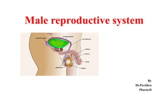
10. • The internal genital organs of the male reproductive system include the testes (singular:
testis), epididymis, vas deferens (ductus deferens), ejaculatory ducts, seminal vesicles,
prostate gland, and bulbourethral glands. These structures are responsible for producing,
storing, and delivering sperm and other components of semen.
Internal Genital organs
11. Testes (Testicles):
The testes are the primary male reproductive organs responsible
for producing sperm and testosterone.
Sperm are produced within the seminiferous tubules found in
the testes.
Epididymis:
The epididymis is a coiled tube located on the surface of each
testis.
It serves as a storage and maturation site for sperm.
Sperm produced in the testes mature and gain the ability to swim
and fertilize an egg as they pass through the epididymis.
12. Vas Deferens (Ductus Deferens):
The vas deferens is a muscular tube that
transports mature sperm from the epididymis to
the ejaculatory ducts.
It travels through the spermatic cord and
eventually joins with the duct of the seminal
vesicle to form the ejaculatory duct.
13. Ejaculatory Ducts:
The ejaculatory ducts are short tubes formed by the junction of the vas deferens
and the duct of the seminal vesicle.
They pass through the prostate gland and empty into the urethra within the
prostate
14. Seminal Vesicles:
The seminal vesicles are pouch-like structures located near the base of the
bladder.
They produce a significant portion of the fluid that makes up semen. This fluid
contains fructose (a sugar that provides energy to sperm) and various proteins.
15. Prostate Gland:
The prostate gland is a walnut-sized gland situated just below the bladder and
surrounds the urethra.
It produces a milky fluid that constitutes a significant portion of semen. This
fluid contains enzymes, citric acid (provides energy to sperm), and substances
that help activate sperm.
16. Bulbourethral Glands (Cowper's Glands):
The bulbourethral glands are small pea-sized glands located beneath the prostate
gland.
They produce a clear, slippery fluid that is released before ejaculation. This fluid
helps lubricate the urethra and neutralize any acidic urine remnants, creating a
favorable environment for sperm.
Collectively, these internal genital organs work
together to produce, store, and deliver sperm and
seminal fluid during ejaculation.
17. Anatomy of Spermatozoa
Each sperm is an intricate motile cell, rich in DNA,
with a head comprised
mostly of chromosomal material. Approximately 60
μm long and 1 μm wide. Each sperm is composed:
1) Head that contains:
Nucleus and densely packed chromosomes
2) Middle piece that contains
Mitochondria that produce the ATP needed
to move the tail
3) Tail : The only cell with flagellum in the
human body which enables the spermatozoa to
swim
18. Head:
The head is the anterior (front) portion of the spermatozoa and contains the genetic material
necessary for fertilization.
Nucleus: The head contains a condensed nucleus with the haploid set of chromosomes
(23 chromosomes in humans).
Acrosome:
The acrosome is a cap-like structure covering the anterior part of the nucleus.
It contains enzymes that are released during the acrosome reaction, which helps the sperm
penetrate the protective layers of the egg.
19. Midpiece (Neck):
The midpiece is a narrow region located between the head and the tail.
Mitochondria: The midpiece contains a high concentration of mitochondria, which
produce energy (in the form of adenosine triphosphate or ATP) required for the sperm's
movement.
Tail (Flagellum):
The tail is a long, whip-like structure that propels the sperm forward.
Axoneme: The core of the tail contains microtubules organized in a 9+2 arrangement,
forming the axoneme. This structure is responsible for the beating movement of the tail,
allowing the sperm to swim in a characteristic whip-like motion.
20. Cells of seminiferous tubule
The seminiferous tubules are the intricate structures found
within the testes where sperm production, known as
spermatogenesis, takes place.
These tubules are composed of several types of cells that
work together to produce and support the development of
spermatozoa (sperm cells).
21. Spermatogonia:
These are the stem cells located along the periphery of the seminiferous tubules.
They undergo mitotic divisions to produce spermatocytes, which then undergo meiosis to
form spermatids.
Sertoli Cells:
These are large, supportive cells that extend from the periphery to the lumen of the
seminiferous tubules.
They provide physical support, nourishment, and protection to developing sperm cells.
Sertoli cells also create a blood-testis barrier, which prevents immune cells from attacking
developing sperm.
22. Spermatocytes:
These are the cells resulting from the mitotic division of spermatogonia.
Primary spermatocytes undergo meiosis I to produce secondary spermatocytes.
Spermatids:
•These are the immediate precursors to mature spermatozoa (Mature Sperm cells )
•They undergo a process called spermiogenesis, during which they transform into mature
sperm cells.
Mature Spermatozoa:
These are the fully developed, motile sperm cells that are released into the lumen of the
seminiferous tubules.
They will eventually travel through the male reproductive system to be ejaculated during
sexual intercourse.
23. Hypothalamus-Pituitary-Testes axis – Male RS & Hormonal
Control
The hypothalamic-pituitary- gonadal axis is a key
regulatory system that governs the hormonal control of the
male reproductive system.
It involves the release of GnRH from the hypothalamus,
which stimulates the anterior pituitary to release LH and FSH.
LH acts on Leydig cells to promote testosterone secretion,
while FSH acts on Sertoli cells to support sperm development.
24. Hormones Secreted Functions
Gonadotropin-Releasing Hormone
(GnRH)
Produced by the hypothalamus Stimulates the release of luteinizing hormone
(LH) and follicle-stimulating hormone (FSH)
from the pituitary gland.
Luteinizing Hormone (LH) Produced by the anterior pituitary
gland
Acts on Leydig cells in the testes.
Stimulates the production and secretion of
testosterone by Leydig cells
Follicle-Stimulating Hormone (FSH) Produced by the anterior pituitary gland.
Acts on Sertoli cells in the seminiferous
tubules of the testes
Stimulates spermatogenesis and supports the
growth and maturation of sperm cells
25. Hormones Secreted Functions
Testosterone Produced by Leydig cells in the testes. Regulates various aspects of male reproductive function,
including the development of male secondary sexual
characteristics (e.g., facial hair, deep voice), sperm
production, and sex drive.
Inhibin Produced by Sertoli cells in the testes Inhibin inhibits the secretion of follicle-stimulating
hormone (FSH) from the pituitary gland.
Prolactin Produced by the anterior pituitary gland Prolactin levels increase after ejaculation, leading to a
temporary refractory period during which it's harder to
achieve another erection and ejaculation
26. How Sperm travels ?
Testes: Sperm are produced in the seminiferous tubules
within the testes.
Epididymis: Newly produced sperm move to the epididymis
for maturation and development.
Vas Deferens: Mature sperm travel through the vas deferens,
a muscular tube.
Ejaculatory Ducts: Vas deferens joins with seminal fluid-
producing structures to form ejaculatory ducts.
Ejaculation: Sperm and seminal fluid move through the
urethra and out of the penis during ejaculation.