SlideShare a Scribd company logo
1 of 39
Dr. Soran M Karim
COA 302
Required readings/reference
books
1. Clinical Anatomy for
Medical Students
[snell, richard] ;
chapter 12 pages;
432-446.
2. Netter's Clinical
Anatomy; chapter 8;
pages: 234- 247.
The palatine tonsil is an
ovoid (almond shaped)
mass of lymphoid tissue
located in the oropharynx
between the
anterior and posterior
pillars
It has a 2 surfaces – medial
and lateral and
2 poles – upper and lower
Introduction;
LYMPHOID TISSUE
1. Primary – Thymus
- Bone marrow
2. Secondary - lymph nodes,
lymphoid follicles in tonsils,
Peyer's patches,
spleen,
adenoids,
skin, etc.
3. Tertiary - Distributed groups
of lymphocytes
Generate lymphocytes from
Immature progenitor cells
Functions of tonsils
It has a protective function in that it prevents
entry of pathogens
through the nasal and oral route (Tonsils prevent
infection).
The crypts on the surface of the tonsil serve to
increase the
surface area and increase the efficiency of
protection against pathogens
Introduction;
• There are two palatine tonsils.
• Within tonsillar fossa (recess), which is situated in the lateral
wall of oropharynx between anterior and posterior faucial
pillars.
• They belong to MALT (Mucosa Associated Lymphoid Tissue).
• Size: Variable, 10-15 mm intransverse diameter and 20-25 mm
in vertical dimension (larger in children?)
• Surface marking (level): over the lower part of masseter
muscle, a little above and in front of the angle of mandible
marks the tonsil on the surface (C1 or C1-C2 VL).
Tonsils maintain its position:
1.Connected with the tongue by the
suspensory ligament of the tonsils.
2.The palatoglossus and palatopharyngeus
muscles attached to the fibrous capsules of
the tonsils
3.By the perivascular stalks
Boundaries and anatomical relations
Anterior- Anterior faucial pillar
(palatoglossal arch).
Posterior- Posterior faucial pillar
(palatopharyngeal arch).
Apex- Soft palate.
Base- Dorsal surface of posterior
1/3rd of tongue.
Lateral wall [Tonsillar Bed]-
Superior Constrictor Muscle
(mainly).
Tonsillar bed:
The mucosal layer between the palatoglossal
and palatopharyngeal arches that is filled with
the palatine tonsil
The tonsil is separated from its bed by loose
areolar tissue.
The structures forming the bed of the tonsil
are:
Superior constrictor muscle
Styloglossus muscle
The structures related to the bed of the tonsil;
1. styloid process !?(if enlarged)
2. Glossopharyngeal nerve
3. Facial artery
4. Submandibular salivary gland
5. Posterior belly of digastric
6. Medial pterygoid muscle
7. Angle of mandible
Parts
2 surfaces- Medial & Lateral
2 borders- Anterior & Posterior
2 Poles- Upper & Lower
Medial Surface-
• It bulges into oropharynx.
• It is covered by epithelium.
• It has crypts.
• There are ~ 12-15 crypts.
Crypta Magna-
• A very large and deep crypt
located near upper pole.
• It represents the remnant of 2nd
pharyngeal pouch.
Lateral Surface
• It is covered by fibrous
capsule.
Peritonsillar Space-
• A space between fibrous
capsule and tonsillar bed.
• It is filled with loose
areolar tissue.
• It is the site of collection
of pus in peritonsillar
abscess.
• During tonsillectomy,
tonsil is dissected in this
plane with ?
Internal Carotid Artery is
~2.5 cm posterolateral to
the tonsil
What is the tonsil capsule?
The tonsil capsule is a thin fibrous partition between
the tonsil and the underlying muscles of the throat. An
extracapsular tonsillectomy involves complete
removal of the tonsils and results in exposure of the
muscle bed and the blood vessels that lie within the
muscle.
Lateral Surface cont.…
 Superior constrictor separates
the lateral surface from
following structures:
1. Facial artery and its ascending
palatine and tonsillar branches.
2. Styloglossus muscle.
3. Glossopharyngeal nerve.
4. Angle of mandible.
5. Medial Pterygoid muscle.
6. Submandibular salivary gland.
Medial surface
The crypts are 12-15 in number
Secondary crypts arise from the primary crypts
and extend into the
substance of the tonsil
On of the crypts located in the upper part are
larger than
the rest – crypta magna
The crypts serve to increase the surface area of
the tonsil
The crypts may be filled witth cheesy material
– epithelial debris, food particles and
bacteria
Superiorly, the tonsil is
free and expands into the
soft palate where both
arches join.
Inferiorly, the suspensory
ligament, a band of
fibrous tissue, connects
the lower pole with the
posterior one third of the
tongue.
Poles
Blood supply
The tonsil is supplied by branches of external carotid
artery
(5 arteries) which are:
1. Tonsillar branch of facial artery (main supply)
2. Ascending palatine branch of facial artery
3. Ascending pharyngeal branch of external carotid artery
4. Dorsal linguae branch of lingual artery
5. Descending palatine branch of maxillary artery
Venous drainage:
1.Tonsillar vein →
Common facial vein
( drain into the internal
jugular vein, roughly at
the level of the hyoid
bone)
1.Para tonsillar vein →
Pharyngeal venous plexus
or Common facial vein →
Internal Jugular Vein
(IJV).
Lymphatic Drainage
• Upper deep cervical
lymph nodes [mainly
Jugulo-digastric
nodes].
• Jugulo-digastric nodes
are called ‘Tonsillar
Lymph Nodes’ (as
they ten to enlarge
with tonsillitis).
Nerve Supply
• Glossopharyngeal nerve (CN IX)
• Greater and lesser palatine nerve (CN V2)
Applied anatomy
1. Damage of paratonsillar vein during tonsillectomy
leads to excessive venous haemorhhage
2. Damage to glossopharygeal nerve leads to loss of
taste sensation
3. Infected tonsillar pain may be referred to middle
ear because of same nerve supply
4. Accumulation of pus in in the peritonsillar space in
chronic tonsillitis gives rise to peritonsillar
abscess or quinsy.
5. Jugulo-digastric lymph node is often enlarged in
tonsillitis.
Applied Aspects cont.…
Tonsillectomy-
• Surgical removal of tonsil.
• If paratonsillar vein gets damaged during
tonsillectomy, severe bleeding occurs
from tonsillar fossa.
• To check bleeding, blood clots should be
removed because they interfere with
retraction of walls of vein.
• Blood clots prevent the contraction of
surrounding muscles.
• After tonsillectomy, postoperative edema
of tonsillar bed can affect the
Glossopharyngeal nerve.
Tonsil Cancer
• Tonsil cancer is the most
common form of
oropharyngeal cancer.
• The condition is commonly
linked to HPV (human
papilloma virus) infection,
though it can also be caused
by heavy alcohol and
tobacco use.
• Tonsil cancer treatments
include surgery,
chemotherapy and radiation
therapy.
Quick review
1. The palatine tonsil are masses of lymphoid tissue
located in the oropharynx and part of the Waldeyr
tonsillar ring rests within within the tonsillar fossa on
the tonsillar bed with 2 poles, 2 surfaces and 2 borders.
2. A; 5 arteries all from ECA
3. V; Tonsillar an Para tonsillar veins
4. N; CN5B and CN 9
5. L; DCLN
6. Tonsils prevent infection but when these are infected,
acts as septic foci of the body which require surgical
removal.
7. Internal carotid artery, although only 1 inch (2.5cm)
behind the tonsil, is never injured in this operation
since it lies safely freed from the pharynx by fatty
tissue around the carotid sheath.
End
Questions!
Next lecture;
• Pharynx and Larynx!
Pop question!
1. Mention the compartments of of WTR!
2. Why (WTR) located at that particular
place(Specific! Region) !?

More Related Content

What's hot

SURGICAL ANATOMY OF DEEP NECK SPACES
SURGICAL ANATOMY OF DEEP NECK SPACESSURGICAL ANATOMY OF DEEP NECK SPACES
SURGICAL ANATOMY OF DEEP NECK SPACESAjay Manickam
 
Pterygopalatine ganglion 1
Pterygopalatine ganglion 1Pterygopalatine ganglion 1
Pterygopalatine ganglion 1Omar Eraky
 
Nasopharynx gross anatomy and applied anatomy in dental and medical aspects
Nasopharynx  gross anatomy and applied anatomy in dental and medical aspectsNasopharynx  gross anatomy and applied anatomy in dental and medical aspects
Nasopharynx gross anatomy and applied anatomy in dental and medical aspectsPratapMd
 
Pterygopalatine fossa and approaches by Dr.Ashwin Menon
Pterygopalatine fossa and approaches by Dr.Ashwin MenonPterygopalatine fossa and approaches by Dr.Ashwin Menon
Pterygopalatine fossa and approaches by Dr.Ashwin MenonDr.Ashwin Menon
 
Anatomy of tonsil and oropharynx
Anatomy of tonsil and oropharynxAnatomy of tonsil and oropharynx
Anatomy of tonsil and oropharynxsunheri2003
 
Pharynx Dr.N.Mugunthan.M.S.,
Pharynx  Dr.N.Mugunthan.M.S.,Pharynx  Dr.N.Mugunthan.M.S.,
Pharynx Dr.N.Mugunthan.M.S.,mgmcri1234
 
Anatomy of Soft palate
Anatomy of  Soft palateAnatomy of  Soft palate
Anatomy of Soft palateddert
 
Middle ear anatomy raju 1
Middle ear anatomy raju 1Middle ear anatomy raju 1
Middle ear anatomy raju 1Raju Mandal
 
Anatomy of nose and pns
Anatomy of nose and pnsAnatomy of nose and pns
Anatomy of nose and pnsManpreet Nanda
 
Anatomy of the Oral cavity Proper
Anatomy of the Oral cavity ProperAnatomy of the Oral cavity Proper
Anatomy of the Oral cavity ProperRahaf Sn
 

What's hot (20)

Pterygopalatine fossa
Pterygopalatine  fossaPterygopalatine  fossa
Pterygopalatine fossa
 
Anatomy of Pharynx
Anatomy of PharynxAnatomy of Pharynx
Anatomy of Pharynx
 
SURGICAL ANATOMY OF DEEP NECK SPACES
SURGICAL ANATOMY OF DEEP NECK SPACESSURGICAL ANATOMY OF DEEP NECK SPACES
SURGICAL ANATOMY OF DEEP NECK SPACES
 
Pterygopalatine ganglion 1
Pterygopalatine ganglion 1Pterygopalatine ganglion 1
Pterygopalatine ganglion 1
 
Deep cervical fascia
Deep cervical fasciaDeep cervical fascia
Deep cervical fascia
 
Nasopharynx gross anatomy and applied anatomy in dental and medical aspects
Nasopharynx  gross anatomy and applied anatomy in dental and medical aspectsNasopharynx  gross anatomy and applied anatomy in dental and medical aspects
Nasopharynx gross anatomy and applied anatomy in dental and medical aspects
 
Pterygopalatine fossa and approaches by Dr.Ashwin Menon
Pterygopalatine fossa and approaches by Dr.Ashwin MenonPterygopalatine fossa and approaches by Dr.Ashwin Menon
Pterygopalatine fossa and approaches by Dr.Ashwin Menon
 
Anatomy of tonsil and oropharynx
Anatomy of tonsil and oropharynxAnatomy of tonsil and oropharynx
Anatomy of tonsil and oropharynx
 
Cavernous sinus
Cavernous sinusCavernous sinus
Cavernous sinus
 
Lateral Wall of Nose
Lateral Wall of NoseLateral Wall of Nose
Lateral Wall of Nose
 
Anatomy of pharynx
Anatomy of pharynxAnatomy of pharynx
Anatomy of pharynx
 
Anatomy of infratemporal region
Anatomy of infratemporal regionAnatomy of infratemporal region
Anatomy of infratemporal region
 
Pharynx Dr.N.Mugunthan.M.S.,
Pharynx  Dr.N.Mugunthan.M.S.,Pharynx  Dr.N.Mugunthan.M.S.,
Pharynx Dr.N.Mugunthan.M.S.,
 
Anatomy of Pharynx
Anatomy of PharynxAnatomy of Pharynx
Anatomy of Pharynx
 
Muscles of tongue
Muscles of tongueMuscles of tongue
Muscles of tongue
 
Paranasal sinuses
Paranasal sinusesParanasal sinuses
Paranasal sinuses
 
Anatomy of Soft palate
Anatomy of  Soft palateAnatomy of  Soft palate
Anatomy of Soft palate
 
Middle ear anatomy raju 1
Middle ear anatomy raju 1Middle ear anatomy raju 1
Middle ear anatomy raju 1
 
Anatomy of nose and pns
Anatomy of nose and pnsAnatomy of nose and pns
Anatomy of nose and pns
 
Anatomy of the Oral cavity Proper
Anatomy of the Oral cavity ProperAnatomy of the Oral cavity Proper
Anatomy of the Oral cavity Proper
 

Similar to palatine_tonsils.pptx

adenoid and tonsilllll.pptx
adenoid and tonsilllll.pptxadenoid and tonsilllll.pptx
adenoid and tonsilllll.pptxSruthiNaren
 
Adeno tonsillitis dr.p.k arthikeyan, 11.07.16
Adeno tonsillitis dr.p.k arthikeyan, 11.07.16Adeno tonsillitis dr.p.k arthikeyan, 11.07.16
Adeno tonsillitis dr.p.k arthikeyan, 11.07.16ophthalmgmcri
 
Adeno tonsillitis dr.p.k arthikeyan, 11.07.16
Adeno tonsillitis dr.p.k arthikeyan, 11.07.16Adeno tonsillitis dr.p.k arthikeyan, 11.07.16
Adeno tonsillitis dr.p.k arthikeyan, 11.07.16ophthalmgmcri
 
palatine tonsil, its anatomy, diseases and their management
palatine tonsil, its anatomy, diseases and their managementpalatine tonsil, its anatomy, diseases and their management
palatine tonsil, its anatomy, diseases and their managementVaibhav Lahane
 
Tonsil anatomy and acute tonsil
Tonsil anatomy and acute tonsilTonsil anatomy and acute tonsil
Tonsil anatomy and acute tonsilSaurabh Ranjan
 
Anatomy and physiology of the palatine tonsil
Anatomy and physiology of the palatine tonsilAnatomy and physiology of the palatine tonsil
Anatomy and physiology of the palatine tonsilSalman Syed
 
Anatomy and physiology of oral cavity oropharynx waldeyer’s
Anatomy and physiology of oral cavity oropharynx waldeyer’sAnatomy and physiology of oral cavity oropharynx waldeyer’s
Anatomy and physiology of oral cavity oropharynx waldeyer’sENTDOST
 
Deep neck space infections
Deep neck space infectionsDeep neck space infections
Deep neck space infectionsAhlam Alzuway
 
TUNDIKERI -ACUTE AND CHRONIC TONSILITIS
TUNDIKERI -ACUTE AND CHRONIC TONSILITISTUNDIKERI -ACUTE AND CHRONIC TONSILITIS
TUNDIKERI -ACUTE AND CHRONIC TONSILITISDr. Raju
 
Cervical Viscera lecture delivered by Saad Datti
Cervical Viscera lecture delivered by Saad DattiCervical Viscera lecture delivered by Saad Datti
Cervical Viscera lecture delivered by Saad DattiSadiq787794
 
Anatomy of Waldeyers Ring .pptx
Anatomy of Waldeyers Ring .pptxAnatomy of Waldeyers Ring .pptx
Anatomy of Waldeyers Ring .pptxAleenaCJeeson
 
Anatomy of Waldeyers Ring .pptx
Anatomy of Waldeyers Ring .pptxAnatomy of Waldeyers Ring .pptx
Anatomy of Waldeyers Ring .pptxAleenaCJeeson
 
Anatomy of larynx & physiology, 29.08.16, dr.bakshi
Anatomy of larynx & physiology, 29.08.16, dr.bakshiAnatomy of larynx & physiology, 29.08.16, dr.bakshi
Anatomy of larynx & physiology, 29.08.16, dr.bakshiophthalmgmcri
 
Gross anatomy of the parotid gland part 1
Gross anatomy of the parotid gland part 1Gross anatomy of the parotid gland part 1
Gross anatomy of the parotid gland part 1obaje godwin sunday
 

Similar to palatine_tonsils.pptx (20)

adenoid and tonsilllll.pptx
adenoid and tonsilllll.pptxadenoid and tonsilllll.pptx
adenoid and tonsilllll.pptx
 
Adeno tonsillitis dr.p.k arthikeyan, 11.07.16
Adeno tonsillitis dr.p.k arthikeyan, 11.07.16Adeno tonsillitis dr.p.k arthikeyan, 11.07.16
Adeno tonsillitis dr.p.k arthikeyan, 11.07.16
 
Adeno tonsillitis dr.p.k arthikeyan, 11.07.16
Adeno tonsillitis dr.p.k arthikeyan, 11.07.16Adeno tonsillitis dr.p.k arthikeyan, 11.07.16
Adeno tonsillitis dr.p.k arthikeyan, 11.07.16
 
palatine tonsil, its anatomy, diseases and their management
palatine tonsil, its anatomy, diseases and their managementpalatine tonsil, its anatomy, diseases and their management
palatine tonsil, its anatomy, diseases and their management
 
Tonsil anatomy and acute tonsil
Tonsil anatomy and acute tonsilTonsil anatomy and acute tonsil
Tonsil anatomy and acute tonsil
 
Anatomy and physiology of the palatine tonsil
Anatomy and physiology of the palatine tonsilAnatomy and physiology of the palatine tonsil
Anatomy and physiology of the palatine tonsil
 
Tonsils ppt
Tonsils pptTonsils ppt
Tonsils ppt
 
Anatomy and physiology of oral cavity oropharynx waldeyer’s
Anatomy and physiology of oral cavity oropharynx waldeyer’sAnatomy and physiology of oral cavity oropharynx waldeyer’s
Anatomy and physiology of oral cavity oropharynx waldeyer’s
 
Acute & chronic tonsillitis and their management
Acute & chronic tonsillitis and their managementAcute & chronic tonsillitis and their management
Acute & chronic tonsillitis and their management
 
Deep neck space infections
Deep neck space infectionsDeep neck space infections
Deep neck space infections
 
TUNDIKERI -ACUTE AND CHRONIC TONSILITIS
TUNDIKERI -ACUTE AND CHRONIC TONSILITISTUNDIKERI -ACUTE AND CHRONIC TONSILITIS
TUNDIKERI -ACUTE AND CHRONIC TONSILITIS
 
Cervical Viscera lecture delivered by Saad Datti
Cervical Viscera lecture delivered by Saad DattiCervical Viscera lecture delivered by Saad Datti
Cervical Viscera lecture delivered by Saad Datti
 
Pharynx & Palate
Pharynx & PalatePharynx & Palate
Pharynx & Palate
 
Anatomy of Waldeyers Ring .pptx
Anatomy of Waldeyers Ring .pptxAnatomy of Waldeyers Ring .pptx
Anatomy of Waldeyers Ring .pptx
 
Anatomy of Waldeyers Ring .pptx
Anatomy of Waldeyers Ring .pptxAnatomy of Waldeyers Ring .pptx
Anatomy of Waldeyers Ring .pptx
 
Histology study guide
Histology study guideHistology study guide
Histology study guide
 
Anatomy of larynx & physiology, 29.08.16, dr.bakshi
Anatomy of larynx & physiology, 29.08.16, dr.bakshiAnatomy of larynx & physiology, 29.08.16, dr.bakshi
Anatomy of larynx & physiology, 29.08.16, dr.bakshi
 
23231.pptx
23231.pptx23231.pptx
23231.pptx
 
Gross anatomy of the parotid gland part 1
Gross anatomy of the parotid gland part 1Gross anatomy of the parotid gland part 1
Gross anatomy of the parotid gland part 1
 
Ct of the larynx
Ct of the larynxCt of the larynx
Ct of the larynx
 

Recently uploaded

Biopesticide (2).pptx .This slides helps to know the different types of biop...
Biopesticide (2).pptx  .This slides helps to know the different types of biop...Biopesticide (2).pptx  .This slides helps to know the different types of biop...
Biopesticide (2).pptx .This slides helps to know the different types of biop...RohitNehra6
 
Nightside clouds and disequilibrium chemistry on the hot Jupiter WASP-43b
Nightside clouds and disequilibrium chemistry on the hot Jupiter WASP-43bNightside clouds and disequilibrium chemistry on the hot Jupiter WASP-43b
Nightside clouds and disequilibrium chemistry on the hot Jupiter WASP-43bSérgio Sacani
 
Botany 4th semester file By Sumit Kumar yadav.pdf
Botany 4th semester file By Sumit Kumar yadav.pdfBotany 4th semester file By Sumit Kumar yadav.pdf
Botany 4th semester file By Sumit Kumar yadav.pdfSumit Kumar yadav
 
Hubble Asteroid Hunter III. Physical properties of newly found asteroids
Hubble Asteroid Hunter III. Physical properties of newly found asteroidsHubble Asteroid Hunter III. Physical properties of newly found asteroids
Hubble Asteroid Hunter III. Physical properties of newly found asteroidsSérgio Sacani
 
PossibleEoarcheanRecordsoftheGeomagneticFieldPreservedintheIsuaSupracrustalBe...
PossibleEoarcheanRecordsoftheGeomagneticFieldPreservedintheIsuaSupracrustalBe...PossibleEoarcheanRecordsoftheGeomagneticFieldPreservedintheIsuaSupracrustalBe...
PossibleEoarcheanRecordsoftheGeomagneticFieldPreservedintheIsuaSupracrustalBe...Sérgio Sacani
 
Natural Polymer Based Nanomaterials
Natural Polymer Based NanomaterialsNatural Polymer Based Nanomaterials
Natural Polymer Based NanomaterialsAArockiyaNisha
 
DIFFERENCE IN BACK CROSS AND TEST CROSS
DIFFERENCE IN  BACK CROSS AND TEST CROSSDIFFERENCE IN  BACK CROSS AND TEST CROSS
DIFFERENCE IN BACK CROSS AND TEST CROSSLeenakshiTyagi
 
Hire 💕 9907093804 Hooghly Call Girls Service Call Girls Agency
Hire 💕 9907093804 Hooghly Call Girls Service Call Girls AgencyHire 💕 9907093804 Hooghly Call Girls Service Call Girls Agency
Hire 💕 9907093804 Hooghly Call Girls Service Call Girls AgencySheetal Arora
 
Orientation, design and principles of polyhouse
Orientation, design and principles of polyhouseOrientation, design and principles of polyhouse
Orientation, design and principles of polyhousejana861314
 
Chromatin Structure | EUCHROMATIN | HETEROCHROMATIN
Chromatin Structure | EUCHROMATIN | HETEROCHROMATINChromatin Structure | EUCHROMATIN | HETEROCHROMATIN
Chromatin Structure | EUCHROMATIN | HETEROCHROMATINsankalpkumarsahoo174
 
Traditional Agroforestry System in India- Shifting Cultivation, Taungya, Home...
Traditional Agroforestry System in India- Shifting Cultivation, Taungya, Home...Traditional Agroforestry System in India- Shifting Cultivation, Taungya, Home...
Traditional Agroforestry System in India- Shifting Cultivation, Taungya, Home...jana861314
 
GBSN - Biochemistry (Unit 1)
GBSN - Biochemistry (Unit 1)GBSN - Biochemistry (Unit 1)
GBSN - Biochemistry (Unit 1)Areesha Ahmad
 
Recombination DNA Technology (Nucleic Acid Hybridization )
Recombination DNA Technology (Nucleic Acid Hybridization )Recombination DNA Technology (Nucleic Acid Hybridization )
Recombination DNA Technology (Nucleic Acid Hybridization )aarthirajkumar25
 
Botany 4th semester series (krishna).pdf
Botany 4th semester series (krishna).pdfBotany 4th semester series (krishna).pdf
Botany 4th semester series (krishna).pdfSumit Kumar yadav
 
TEST BANK For Radiologic Science for Technologists, 12th Edition by Stewart C...
TEST BANK For Radiologic Science for Technologists, 12th Edition by Stewart C...TEST BANK For Radiologic Science for Technologists, 12th Edition by Stewart C...
TEST BANK For Radiologic Science for Technologists, 12th Edition by Stewart C...ssifa0344
 
Zoology 4th semester series (krishna).pdf
Zoology 4th semester series (krishna).pdfZoology 4th semester series (krishna).pdf
Zoology 4th semester series (krishna).pdfSumit Kumar yadav
 
Pests of cotton_Borer_Pests_Binomics_Dr.UPR.pdf
Pests of cotton_Borer_Pests_Binomics_Dr.UPR.pdfPests of cotton_Borer_Pests_Binomics_Dr.UPR.pdf
Pests of cotton_Borer_Pests_Binomics_Dr.UPR.pdfPirithiRaju
 
9654467111 Call Girls In Raj Nagar Delhi Short 1500 Night 6000
9654467111 Call Girls In Raj Nagar Delhi Short 1500 Night 60009654467111 Call Girls In Raj Nagar Delhi Short 1500 Night 6000
9654467111 Call Girls In Raj Nagar Delhi Short 1500 Night 6000Sapana Sha
 

Recently uploaded (20)

Biopesticide (2).pptx .This slides helps to know the different types of biop...
Biopesticide (2).pptx  .This slides helps to know the different types of biop...Biopesticide (2).pptx  .This slides helps to know the different types of biop...
Biopesticide (2).pptx .This slides helps to know the different types of biop...
 
Nightside clouds and disequilibrium chemistry on the hot Jupiter WASP-43b
Nightside clouds and disequilibrium chemistry on the hot Jupiter WASP-43bNightside clouds and disequilibrium chemistry on the hot Jupiter WASP-43b
Nightside clouds and disequilibrium chemistry on the hot Jupiter WASP-43b
 
Botany 4th semester file By Sumit Kumar yadav.pdf
Botany 4th semester file By Sumit Kumar yadav.pdfBotany 4th semester file By Sumit Kumar yadav.pdf
Botany 4th semester file By Sumit Kumar yadav.pdf
 
Hubble Asteroid Hunter III. Physical properties of newly found asteroids
Hubble Asteroid Hunter III. Physical properties of newly found asteroidsHubble Asteroid Hunter III. Physical properties of newly found asteroids
Hubble Asteroid Hunter III. Physical properties of newly found asteroids
 
PossibleEoarcheanRecordsoftheGeomagneticFieldPreservedintheIsuaSupracrustalBe...
PossibleEoarcheanRecordsoftheGeomagneticFieldPreservedintheIsuaSupracrustalBe...PossibleEoarcheanRecordsoftheGeomagneticFieldPreservedintheIsuaSupracrustalBe...
PossibleEoarcheanRecordsoftheGeomagneticFieldPreservedintheIsuaSupracrustalBe...
 
Natural Polymer Based Nanomaterials
Natural Polymer Based NanomaterialsNatural Polymer Based Nanomaterials
Natural Polymer Based Nanomaterials
 
DIFFERENCE IN BACK CROSS AND TEST CROSS
DIFFERENCE IN  BACK CROSS AND TEST CROSSDIFFERENCE IN  BACK CROSS AND TEST CROSS
DIFFERENCE IN BACK CROSS AND TEST CROSS
 
Hire 💕 9907093804 Hooghly Call Girls Service Call Girls Agency
Hire 💕 9907093804 Hooghly Call Girls Service Call Girls AgencyHire 💕 9907093804 Hooghly Call Girls Service Call Girls Agency
Hire 💕 9907093804 Hooghly Call Girls Service Call Girls Agency
 
Orientation, design and principles of polyhouse
Orientation, design and principles of polyhouseOrientation, design and principles of polyhouse
Orientation, design and principles of polyhouse
 
CELL -Structural and Functional unit of life.pdf
CELL -Structural and Functional unit of life.pdfCELL -Structural and Functional unit of life.pdf
CELL -Structural and Functional unit of life.pdf
 
Chromatin Structure | EUCHROMATIN | HETEROCHROMATIN
Chromatin Structure | EUCHROMATIN | HETEROCHROMATINChromatin Structure | EUCHROMATIN | HETEROCHROMATIN
Chromatin Structure | EUCHROMATIN | HETEROCHROMATIN
 
Traditional Agroforestry System in India- Shifting Cultivation, Taungya, Home...
Traditional Agroforestry System in India- Shifting Cultivation, Taungya, Home...Traditional Agroforestry System in India- Shifting Cultivation, Taungya, Home...
Traditional Agroforestry System in India- Shifting Cultivation, Taungya, Home...
 
GBSN - Biochemistry (Unit 1)
GBSN - Biochemistry (Unit 1)GBSN - Biochemistry (Unit 1)
GBSN - Biochemistry (Unit 1)
 
Recombination DNA Technology (Nucleic Acid Hybridization )
Recombination DNA Technology (Nucleic Acid Hybridization )Recombination DNA Technology (Nucleic Acid Hybridization )
Recombination DNA Technology (Nucleic Acid Hybridization )
 
Botany 4th semester series (krishna).pdf
Botany 4th semester series (krishna).pdfBotany 4th semester series (krishna).pdf
Botany 4th semester series (krishna).pdf
 
Engler and Prantl system of classification in plant taxonomy
Engler and Prantl system of classification in plant taxonomyEngler and Prantl system of classification in plant taxonomy
Engler and Prantl system of classification in plant taxonomy
 
TEST BANK For Radiologic Science for Technologists, 12th Edition by Stewart C...
TEST BANK For Radiologic Science for Technologists, 12th Edition by Stewart C...TEST BANK For Radiologic Science for Technologists, 12th Edition by Stewart C...
TEST BANK For Radiologic Science for Technologists, 12th Edition by Stewart C...
 
Zoology 4th semester series (krishna).pdf
Zoology 4th semester series (krishna).pdfZoology 4th semester series (krishna).pdf
Zoology 4th semester series (krishna).pdf
 
Pests of cotton_Borer_Pests_Binomics_Dr.UPR.pdf
Pests of cotton_Borer_Pests_Binomics_Dr.UPR.pdfPests of cotton_Borer_Pests_Binomics_Dr.UPR.pdf
Pests of cotton_Borer_Pests_Binomics_Dr.UPR.pdf
 
9654467111 Call Girls In Raj Nagar Delhi Short 1500 Night 6000
9654467111 Call Girls In Raj Nagar Delhi Short 1500 Night 60009654467111 Call Girls In Raj Nagar Delhi Short 1500 Night 6000
9654467111 Call Girls In Raj Nagar Delhi Short 1500 Night 6000
 

palatine_tonsils.pptx

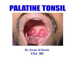
  • 1. Dr. Soran M Karim COA 302
  • 2. Required readings/reference books 1. Clinical Anatomy for Medical Students [snell, richard] ; chapter 12 pages; 432-446. 2. Netter's Clinical Anatomy; chapter 8; pages: 234- 247.
  • 3. The palatine tonsil is an ovoid (almond shaped) mass of lymphoid tissue located in the oropharynx between the anterior and posterior pillars It has a 2 surfaces – medial and lateral and 2 poles – upper and lower Introduction;
  • 4. LYMPHOID TISSUE 1. Primary – Thymus - Bone marrow 2. Secondary - lymph nodes, lymphoid follicles in tonsils, Peyer's patches, spleen, adenoids, skin, etc. 3. Tertiary - Distributed groups of lymphocytes Generate lymphocytes from Immature progenitor cells
  • 5.
  • 6. Functions of tonsils It has a protective function in that it prevents entry of pathogens through the nasal and oral route (Tonsils prevent infection). The crypts on the surface of the tonsil serve to increase the surface area and increase the efficiency of protection against pathogens
  • 7. Introduction; • There are two palatine tonsils. • Within tonsillar fossa (recess), which is situated in the lateral wall of oropharynx between anterior and posterior faucial pillars. • They belong to MALT (Mucosa Associated Lymphoid Tissue). • Size: Variable, 10-15 mm intransverse diameter and 20-25 mm in vertical dimension (larger in children?) • Surface marking (level): over the lower part of masseter muscle, a little above and in front of the angle of mandible marks the tonsil on the surface (C1 or C1-C2 VL).
  • 8.
  • 9. Tonsils maintain its position: 1.Connected with the tongue by the suspensory ligament of the tonsils. 2.The palatoglossus and palatopharyngeus muscles attached to the fibrous capsules of the tonsils 3.By the perivascular stalks
  • 10.
  • 11. Boundaries and anatomical relations Anterior- Anterior faucial pillar (palatoglossal arch). Posterior- Posterior faucial pillar (palatopharyngeal arch). Apex- Soft palate. Base- Dorsal surface of posterior 1/3rd of tongue. Lateral wall [Tonsillar Bed]- Superior Constrictor Muscle (mainly).
  • 12.
  • 13. Tonsillar bed: The mucosal layer between the palatoglossal and palatopharyngeal arches that is filled with the palatine tonsil The tonsil is separated from its bed by loose areolar tissue. The structures forming the bed of the tonsil are: Superior constrictor muscle Styloglossus muscle
  • 14. The structures related to the bed of the tonsil; 1. styloid process !?(if enlarged) 2. Glossopharyngeal nerve 3. Facial artery 4. Submandibular salivary gland 5. Posterior belly of digastric 6. Medial pterygoid muscle 7. Angle of mandible
  • 15.
  • 16. Parts 2 surfaces- Medial & Lateral 2 borders- Anterior & Posterior 2 Poles- Upper & Lower Medial Surface- • It bulges into oropharynx. • It is covered by epithelium. • It has crypts. • There are ~ 12-15 crypts. Crypta Magna- • A very large and deep crypt located near upper pole. • It represents the remnant of 2nd pharyngeal pouch.
  • 17.
  • 18. Lateral Surface • It is covered by fibrous capsule. Peritonsillar Space- • A space between fibrous capsule and tonsillar bed. • It is filled with loose areolar tissue. • It is the site of collection of pus in peritonsillar abscess. • During tonsillectomy, tonsil is dissected in this plane with ? Internal Carotid Artery is ~2.5 cm posterolateral to the tonsil
  • 19. What is the tonsil capsule? The tonsil capsule is a thin fibrous partition between the tonsil and the underlying muscles of the throat. An extracapsular tonsillectomy involves complete removal of the tonsils and results in exposure of the muscle bed and the blood vessels that lie within the muscle.
  • 20.
  • 21. Lateral Surface cont.…  Superior constrictor separates the lateral surface from following structures: 1. Facial artery and its ascending palatine and tonsillar branches. 2. Styloglossus muscle. 3. Glossopharyngeal nerve. 4. Angle of mandible. 5. Medial Pterygoid muscle. 6. Submandibular salivary gland.
  • 22. Medial surface The crypts are 12-15 in number Secondary crypts arise from the primary crypts and extend into the substance of the tonsil On of the crypts located in the upper part are larger than the rest – crypta magna The crypts serve to increase the surface area of the tonsil The crypts may be filled witth cheesy material – epithelial debris, food particles and bacteria
  • 23. Superiorly, the tonsil is free and expands into the soft palate where both arches join. Inferiorly, the suspensory ligament, a band of fibrous tissue, connects the lower pole with the posterior one third of the tongue. Poles
  • 24.
  • 25. Blood supply The tonsil is supplied by branches of external carotid artery (5 arteries) which are: 1. Tonsillar branch of facial artery (main supply) 2. Ascending palatine branch of facial artery 3. Ascending pharyngeal branch of external carotid artery 4. Dorsal linguae branch of lingual artery 5. Descending palatine branch of maxillary artery
  • 26.
  • 27.
  • 28. Venous drainage: 1.Tonsillar vein → Common facial vein ( drain into the internal jugular vein, roughly at the level of the hyoid bone) 1.Para tonsillar vein → Pharyngeal venous plexus or Common facial vein → Internal Jugular Vein (IJV).
  • 29. Lymphatic Drainage • Upper deep cervical lymph nodes [mainly Jugulo-digastric nodes]. • Jugulo-digastric nodes are called ‘Tonsillar Lymph Nodes’ (as they ten to enlarge with tonsillitis).
  • 30.
  • 31. Nerve Supply • Glossopharyngeal nerve (CN IX) • Greater and lesser palatine nerve (CN V2)
  • 32. Applied anatomy 1. Damage of paratonsillar vein during tonsillectomy leads to excessive venous haemorhhage 2. Damage to glossopharygeal nerve leads to loss of taste sensation 3. Infected tonsillar pain may be referred to middle ear because of same nerve supply 4. Accumulation of pus in in the peritonsillar space in chronic tonsillitis gives rise to peritonsillar abscess or quinsy. 5. Jugulo-digastric lymph node is often enlarged in tonsillitis.
  • 33.
  • 34. Applied Aspects cont.… Tonsillectomy- • Surgical removal of tonsil. • If paratonsillar vein gets damaged during tonsillectomy, severe bleeding occurs from tonsillar fossa. • To check bleeding, blood clots should be removed because they interfere with retraction of walls of vein. • Blood clots prevent the contraction of surrounding muscles. • After tonsillectomy, postoperative edema of tonsillar bed can affect the Glossopharyngeal nerve.
  • 35.
  • 36. Tonsil Cancer • Tonsil cancer is the most common form of oropharyngeal cancer. • The condition is commonly linked to HPV (human papilloma virus) infection, though it can also be caused by heavy alcohol and tobacco use. • Tonsil cancer treatments include surgery, chemotherapy and radiation therapy.
  • 37. Quick review 1. The palatine tonsil are masses of lymphoid tissue located in the oropharynx and part of the Waldeyr tonsillar ring rests within within the tonsillar fossa on the tonsillar bed with 2 poles, 2 surfaces and 2 borders. 2. A; 5 arteries all from ECA 3. V; Tonsillar an Para tonsillar veins 4. N; CN5B and CN 9 5. L; DCLN 6. Tonsils prevent infection but when these are infected, acts as septic foci of the body which require surgical removal. 7. Internal carotid artery, although only 1 inch (2.5cm) behind the tonsil, is never injured in this operation since it lies safely freed from the pharynx by fatty tissue around the carotid sheath.
  • 39. Pop question! 1. Mention the compartments of of WTR! 2. Why (WTR) located at that particular place(Specific! Region) !?

Editor's Notes

  1. Tonsils are large lymphoid tissue situated in the lateral wall of the oropharynx. They form lateral part of the Waldeyer's ring. Tonsil occupies the tonsillar fossa between diverging palato-pharyngeal and palatoglossal folds Tonsils are lymphoid tissue aggregates situated near the entrance of the digestive and respiratory tracts and play a key role in our immune system. They act as a front-line defense forming the initial immunological response to inhaled or ingested pathogens. The lymphatic tissues located in the oropharynx are composed of a circumferential tonsillar ring, known as the Waldeyer's ring which consists of the palatine tonsils (faucial tonsils), adenoid (nasopharyngeal tonsil), lingual tonsil, and tubal tonsils. When patients and doctors discuss tonsils, they are often referring to the palatine tonsils located at the back of the throat between the two palatine arches (pillars). 
  2. Secondary or peripheral lymphoid organs maintain mature naive lymphocytes and initiate an adaptive immune response Theses are the sites of lymphocyte activation by antigen Activation leads to clonal expansion and affinity maturation Mature Lymphocytes recirculate between the blood and the peripheral lymphoid organs until they encounter their specific antigen GALT Gut associated lymphoid tissue MALT mucosa-associated lymphatic tissue; lymphoid tissue associated with the mucosa of the female reproductive tract, respiratory tract, etc SALT Skin associated - dermis of the skin Lymph "Means clear water and it is basically the fluid and protein that has been squeezed out of the blood (i.e. blood plasma). "Unlike the cardiovascular system, the lymphatic system is not closed and has no central pump."  "Lymph movement occurs despite low pressure due to peristalsis – smooth muscle and skeletal activity (everyday activity and motion of the body).
  3. The Waldeyer's ring is made up of the tonsils, adenoids, and other lymphoid tissue. It contains lymphocytes (a type of immune cell) that help the body fight infection and disease.
  4. The activity of this lymphatic organ is especially pronounced during childhood, when immunologic challenges from the environment induce hyperplasia of the palatine tonsils. Following this “active phase” of immune initiation, which lasts until about 8–10 years of age, the lymphatic tonsillar tissue becomes less important as an immune organ, and there is a corresponding decline in the density of lymphocytes in all regions of the tonsils. While the tonsils become less important immunologically with ageing, the tonsillar tissue continues to perform immune functions even at an advanced age, although this should not alter the decision to remove the tonsils if a valid indication for tonsillectomy exists. Tonsils receive the afferent supply from the tonsillar plexus, with contributions from the trigeminal nerve (CN 5) via the lesser palatine nerves, as well as the glossopharyngeal nerve (CN IX). CN IX continues distally to the tonsils supplying general sensory and taste sensation to the posterior one-third of the tongue. CN IX is the nerve most likely to be damaged during a tonsillectomy.
  5. Almond shaped, ovoid mass of lymphoid tissue situated bilaterally in the lateral wall of oropharynx within the tonsillar recess or sinus bounded by palatoglossal fold anteriorly i.e. anterior pillar and palatopharyngeal fold posteriorly i.e. posterior pillar. The palatine tonsils are two large, conspicuous almond-shaped mass of the lymphoid tissue forming the lower lateral aspect of the ring and localized in a triangular tonsillar fossa along the anterolateral border of the oropharynx on each side. The dimensions of the tonsils are about 10–15 mm in width and 20–25 mm in length in adults, but increase in children. 
  6. Anchored by Perivascular spaces are small fluid-filled structures that surround blood vessels Inferiorly, the suspensory ligament, a band of fibrous tissue, connects the lower pole with the posterior one third of the tongue. Most of carcinomas develop in the tonsillolingual sulcus which separates the tonsil from tongue anteroinferiorly By the suspensory ligament of the tonsils it is connected with the tongue The palatoglossus and palatopharyngeus muscles attached to the fibrous capsules of the tonsils By the perivascular stalks
  7. Relations of tonsillar bed: Arteries: Facial artery and it’s ascending palatine branch Ascending pharyngeal artery Internal carotid artery (lies about 25 mm behind and lateral to the tonsil) Styloid process (if enlarged) Submandibular salivary gland Medial pterygoid muscle Angle of mandible
  8. Following structures form the tonsillar bed ( from inside outwards): Pharyngobasilar fascia. Superior Constrictor muscle. Buccopharyngeal fascia.
  9. Anterior Border- Passes underneath the palatoglossal arch. Posterior Border- Passes underneath the palatopharyngeal arch. Upper Pole- extends up into the soft palate. Lower Pole- It is attached to the tongue by a band of fibrous tissue called suspensory ligament of tonsil. What is the Crypta Magna of the tonsil? Mucosa invaginates into the substance of tonsil to form 12-15 tonsillar crypts. Largest crypt known as crypta magna or intratonsillar cleft opens near the upper part (represents ventral part of 2nd pharyngeal pouch) Crypts increase the surface area of tonsil.
  10. Poles Superiorly, the tonsil is free and expands into the soft palate where both arches join. Inferiorly, the suspensory ligament, a band of fibrous tissue, connects the lower pole with the posterior one third of the tongue. Most of carcinomas develop in the tonsillolingual sulcus which separates the tonsil from tongue anteroinferiorly  Pilica triangularis; The mucosal folds and arches of the palatine tonsil
  11. Lateral surface It is covered by the fibrous capsule of the tonsil The tonsillar bed is separated from the capsule by loose areolar tissue This makes it is easy to dissect the tonsil from its bed during tonsillectomy It is the site of collection of pus in peritonsillar abscess (quinsy) Some fibers of palatoglossus and palatopharyngeus gets attached to capsule of tonsil What is the relationship between the internal carotid artery and the tonsil? The internal carotid artery (ICA) is about 2.5 cm away from the tonsils. It has no branches in the cervical portion. Lateral surface is a base of the tonsil that is covered by well-defined fibrous capsule at the lateral wall of the tonsillar fossa, which is composed of five layers from within outward
  12. From the pharyngeal side, the palatine tonsils are covered with a stratified squamous epithelium (the tonsillar capsule), whereas a fibrous capsule links them to the wall of the pharynx. Through Tonsillar capsule, a thin false or surgical sheet, covers the tonsillar fossa as an appendage of the pharyngobasilar fascia. The upper part of this condensed capsule is even and loosely fixed, whereas the lower part is irregular and intermingled with the pharyngeal muscle fibers and is firmly attached anteroinferiorly by the suspensory ligament to the posterior one third of the tongue. The tonsillar artery enters near this ligament. So, the surgical removal of the upper part of the capsule up to the lower third is very easy, but the lower part requires cautious resection the capsule pass trabecules that contain small blood vessels, nerves and lymphatic vessels.
  13. Lateral surface extends deep to surrounding boundaries. It is coated with a fibrous sheet, an extension of pharyngobasilar fascia called capsule of the tonsil. The capsule is loosely attached to the muscular wall but antero-inferiorly it is attached firmly to the side of the tongue just in front of insertion of palatoglossus and palatopharyngeus muscles
  14. Tonsil has two surfaces, medial and lateral; two borders anterior and posterior; two poles upper and lower; two developmental folds plica triangulris and plica semilumris; and one cleft intratonsillar cleft. Medial surface is covered by squamous epithelium and presents 15-20 crypts usually plugged with epithelial and bacterial debris It is lined by stratified squamous non keratinising epithelium which dips into the crypts
  15. Most of carcinomas develop in the tonsillolingual sulcus which separates the tonsil from tongue anteroinferiorly It is attached to the tongue A triangular fold of mucous membrane extends from the anterior tonsillar pillar to the lower pole. It encloses a space – anterior tonsillar space. The lower pole is separated from the tongue by the tonsillo-lingual sulcus This sulcus may harbour carcinoma!
  16. Facial Artery- Tonsillar branch. Ascending Palatine branch. Lingual Artery- Dorsalis Linguae branches. Ascending Pharyngeal Artery. Maxillary Artery- Greater Palatine Branch.
  17. Anterior tonsillar artery (1): from dorsal lingual branches of lingual artery Posterior tonsillar artery (2): from ascending palatine branch of facial, and ascending pharyngeal arteries Superior tonsillar artery (1): from greater palatine branch of maxillary artery Inferior tonsillar artery (1; main artery): from facial artery
  18. Venous drainage:  Tonsillar vein → Common facial vein Paratonsillar vein → Pharyngeal venous plexus or Common facial vein → Internal Jugular Vein (IJV)
  19. The pharyngeal plexus (venous) is a network of veins beginning on the outer surface of the pharynx, and, after receiving some posterior meningeal veins and the vein of the pterygoid canal, end in the internal jugular. Lymphatics from the tonsil pierce the superior constrictor and drain into the upper cervical lymph nodes especially jugulodigastric (tonsillar) lymph node Enlarged non tender jugulodigastric lymph node is a sign of chronic tonsillitis
  20. This is a ring of lymphatic tissue at the beginning of food and air passages (in the nasopharynx and oropharynx). The ring is composed of MALT which is characterized by the close approximation of epithelium and lymphatic tissue. The components are: Palatine tonsils (largest) on either side on lateral wall of oropharynx Nasopharyngeal tonsil (adenoids) on the posterior wall of nasopharynx Tubal tonsils on lateral walls of nasopharynx Lingual tonsils on the dorsum of posterior 1/3 of tongue in the floor of oropharynx
  21. The innervation of the palatine tonsils is provided by the lesser palatine nerve which arises from the maxillary division of the trigeminal nerve and the tonsillar branches of the glossopharyngeal nerve. The innervation of the palatine tonsils is provided by the lesser palatine nerve which arises from the maxillary division of the trigeminal nerve and the tonsillar branches of the glossopharyngeal nerve. Lesser palatine branch of sphenopalatine ganglion The glossopharyngeal nerve leaves the brainstem in a form of the several rootlets from the posterior lateral sulcus of the medulla oblongata, superiorly to the place of the exit of the vagus nerve. Tonsillar nerves : These nerves provide sensory innervation to the palatine tonsils and to the mucosa of the isthmus of the fauces. Nerve supply: Glossopharyngeal nerve (CN IX) Greater and lesser palatine nerve (CN V)
  22. Ligature of the tonsillar arteries specially inferior tonsillar artery is important during tonsillectomy. 9. During tonsillectomy, paratonsillar vein may be damaged causing excessive hemorrhage. 10. Referred pain from the tonsils sometimes may radiate into the middle ear due to common nerve supply (CN IX). 11. After tonsillectomy, glossopharyngeal nerve may be affected which causes loss of taste sensation. Glossopharyngeal nerve lesions produce difficulty swallowing; impairment of taste over the posterior one-third of the tongue and palate; impaired sensation over the posterior one-third of the tongue, palate, and pharynx; an absent gag reflex; and dysfunction of the parotid gland.
  23. Tonsils prevent infection but when these are infected, acts as septic foci of the body which require surgical removal. Peritonsillar space is the plane of dissection during tonsillectomy. In tonsillectomy, the tonsil is dissected out along with its capsule from its bed. Tonsils are surgically removed by dissecting between the tonsillar capsule and the superior constrictor muscle using either the “hot” or “cold” technique.  In the “hot” tonsillectomy technique, electrocautery is employed to dissect and coagulate simultaneously. In the “cold” tonsillectomy technique, a superior incision is made through the mucosal layers, and blunt dissection is used to separate the tonsils from the underlying tonsillar bed. Tonsils are then separated along their inferior border using the snare method. Studies have shown a superiority of the cold technique when looking at the outcome of postoperative pain. However, electrocautery minimizes intraoperative blood loss.[11] Newer techniques currently in practice employ the use of CO2 lasers, ultrasound, as well as radiofrequency ablation citing reduced postoperative pain though further research is necessary. Complications following a tonsillectomy classify into three main categories: acute, subacute, and delayed. Acute complications include airway obstruction due to edema, bleeding, and post-obstructive pulmonary edema. Subacute complications include postoperative hemorrhage, dehydration, and weight loss. Delayed or chronic complications include velopharyngeal insufficiency and nasopharyngeal stenosis.[11] Good practice advocates that the serious complications of tonsillectomy (profuse hemorrhage requiring blood transfusion and potentially fatal hemorrhage) be routinely discussed during the process of obtaining informed consent for tonsillectomy, despite their low occurrence rate. 'Reasonable patients' consider these complications to be significant and expect surgeons to discuss these complications with them. Surgeons, however, on the other hand, avoid discussing these serious complications with their patients.
  24.  Internal carotid artery, although only 1 inch (2.5cm) behind the tonsil, is never injured in this operation since it lies safely freed from the pharynx by fatty tissue around the carotid sheath. Almost always in surgery, clots are not removed to prevent hemorrhage, but this rule doesn’t apply to tonsils and uterus. After tonsillectomy, blood clots present in the tonsillar fossa are removed. This is done to prevent post-operative hemorrhage because the clots in the tonsillar fossa interfere with the retraction of vessel walls by preventing the contraction of surrounding muscles i.e. the muscles forming boundaries of the tonsillar fossa.
  25. il cancer can develop at any age, but it’s more common in people over the age of 50. People assigned male at birth (AMAB) are three to four times more likely than people assigned female at birth (AFAB) to develop the condition. Additionally, white people are slightly more likely than Black people to be diagnosed with tonsil cancer. In addition, tonsil cancer has been linked to tobacco use and heavy alcohol consumption. People with decreased immunity — such as organ transplant recipients and individuals with HIV — are also more likely to develop tonsil cancer.
  26. Lets review everything in brief/ major points to remember It has a protective function in that it prevents entry of pathogens through the nasal and oral route. The innervation of the palatine tonsils is provided by the lesser palatine nerve which arises from the maxillary division of the trigeminal nerve and the tonsillar branches of the glossopharyngeal nerve.
  27. To read ahead