Paranasal sinuses
•Download as ODP, PDF•
16 likes•6,310 views

The document discusses the paranasal sinuses and their clinical considerations. It begins by introducing the four pairs of paranasal sinuses - maxillary, frontal, sphenoidal, and ethmoidal sinuses. For each sinus, it describes the anatomy, development, neurovascular supply, and other key details. It then covers the functional importance of the sinuses and common clinical issues like sinusitis, developmental anomalies, dental issues that could impact the sinuses, and more. The document provides an overview of the paranasal sinuses and factors relevant to their examination and treatment.

Recommended
More Related Content
What's hot
What's hot (20)
Similar to Paranasal sinuses
Similar to Paranasal sinuses (20)
More from Dr Sudeep Madhusudan Chaudhari
More from Dr Sudeep Madhusudan Chaudhari (16)
Recently uploaded
Recently uploaded (20)
Paranasal sinuses
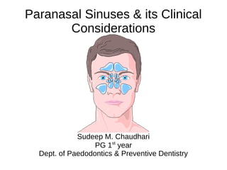
- 1. Paranasal Sinuses & its Clinical Considerations Sudeep M. Chaudhari PG 1st year Dept. of Paedodontics & Preventive Dentistry
- 2. Contents ● Introduction ● Types of paranasal sinuses 1)Maxillary sinus 2)Frontal sinus 3)Sphenoidal sinus 4)Ethmoidal sinus ● Functional importance ● Clinical considerations ● Conclusion ● References
- 3. Introduction ● The paranasal sinuses are air-filled spaces located within the bones of the skull and facial bones. The sinus is regarded by some as an accessory space to the nasal cavity, occurring only as a result of an inadequate process of ossification. In contrast, others report the functional contributions of the maxillary sinus in many aspects of olfactory and respiratory physiology. ● The sinuses are rudimentary or even absent at birth. They enlarge rapidly during the ages of six to seven years, i.e. time of eruption of permanent teeth and then after puberty. From birth to adult life the growth of the sinuses is due to enlargement of the bones; in old age it is due to resorption of the surrounding cancellous bone.
- 4. Types of paranasal sinuses - There are 4 pairs of paranasal sinuses 1) Frontal sinus 2) Maxillary sinus 3) Sphenoidal sinus 4) Ethmoidal sinus
- 5. 1) Maxillary Sinus ● Definition- The maxillary sinus is the pneumatic space that is lodged inside the body of the maxilla and that communicates with the environment by way of the middle nasal meatus and the nasal vestibule. ● Anatomy of the maxillary sinus was first described by the British surgeon and anatomist Nathaniel Highmore in 1651. ● It is also known as the “Antrum of Highmore”.
- 6. Develomental aspect ● Maxillary sinus is the first of the paranasal sinus to develop. ● 4th week intra-uterine life – dorsal portion of 1st Pharyngeal arch forms the Maxillary process, which extends forward and beneath the developing eye to give rise to the maxilla.
- 7. Horizontal shift of the palatal shelves & fusion with one another. Nasal septum separates the oral cavity from the two nasal chambers. Influences further expansion of the lateral nasal wall & 3 wall begin to fold. 3 conchae & meatuses arise
- 8. Superior & Inferior Meatus Remain as shallow depressions along the lateral nasal wall for first half of I.U. life 3 Meatus Middle Meatus Expands immediately into lateral nasal wall in an inferior direction occupying more of the further maxillary body
- 9. ● Development of sinus starts at 12 weeks as an evagination of the mucous membrane in the lateral wall of the middle meatus of the nose when the nasalepithelium invades the maxillary mesenchyme.
- 10. Age changes of Maxillary Sinus Tubular at birth Ovoid in childhood Pyramidal in adulthood
- 11. 0-3 years ● At birth filled with deciduous tooth germs ● Size : 7mm X 4mm X 4mm ● Volume 6-8ml ● 20th month – posterior development ● 3rd year : 1/2 adult size 3-4 years ● Increase in width with facial growth ● Position : 2nd deciduous molar & 1st permanent molar 5-9 years ● Size : 27mm X 18mm X 17mm ● Growth corresponds to permanent teeth eruption
- 12. 9-12 years ● Antral floor same level with nasal floor 12-15 years ● Floor of sinus 5- 12mm below nasal floor ● Size : 32mm X 33mm X 25mm ● Volume : 15-20ml ● Floor : 1st molar < 2nd molar < 2nd premolar Old age ● Resorption of ridge with continued sinus pneumatization which leaves a thin layer of corticle bone separating the sinus mucosa from oral mucosa
- 14. Anatomy of Maxillary Sinus ● Largest of the paranasal sinus ● Pyramidal shaped cavity within the body of the Maxilla ● The base of the pyramid forming the lateral nasal wall and apex at the root of the zygoma.
- 15. Roof of the antrum ● Formed by floor of the orbit and is transversed by the infraorbital nerves.It is flat and slopes slightly anteriorly and laterally.
- 16. Floor of the sinus ● Curved rather than flat formed by alveolar process of the maxilla and lies about 1cm below the level of the floor of the nose. ● Closely related to root apices of the maxillary premolar and molar.
- 17. Anterior wall ● Formed by the facial surface of the maxilla. ● Extends from pyriform aperture anteriorly to zygomaticomaxillary suture & inferior orbital rim superiorly to alveolar process inferiorly. ● Convexity towards sinus thinnest in canine fossa ● Important structures - Infraorbital foramen Anterior superior alveolar nerve (ASA) Middle superior alveolar nerve (MSA). Canine Fossa
- 18. Posterior wall ● Made of zygomatic and greater wing of sphenoid bone. ● A thin plate of bone separate the antral cavity from the infratemporal fossa. ● Thick laterally, thin medially. ● Important structures - ➢ Posterior superior alveolar nerve ➢ Maxillary artery ➢ Pterygopalatine ganglion ➢ Nerve of pterygoid canal
- 19. Medial wall ● Bounded by the nasal cavity ● The opening of the sinus is closer to the roof and thus at a higher level than the floor. ● Formed by lateral nasal wall ● Inferior - nasal conchae ● Posterior - palatine bone ● Superior -uncinate process of ethmoid,lacrimal bone ● Contains double layer of mucous membrane(pars membranacea) ● Important structures - ➢ Sinus ostium ➢ Hiatus semilunaris ➢ Ethmoidal bulla ➢ Uncinate process ➢ Infundibulum
- 20. Ostium ● Opening of the maxillary sinus is called osteum. ● Ostium of the maxillary sinus is situated high up in medial wall and opens into the middle meatus of the nose in the lower part of the hiatus semilunaris. ● Lies above the level of nasal floor. ● The ostium lies approximately 2/3rd up the medial wall of the sinus, making drainage of the sinus inherently difficult. ● Blockage of the ostium can easily occur when there is inflammation of the mucosal lining of the ostium. ● An accessory ostium is also present behind the main ostium in 30% cases.
- 21. Neurovascular supply ● Arterial supply : ➔ By facial artery branch of external carotid artery. ➔ By infra orbital & greater palatine arteries branch of maxillary artery which is branch of external carotid artery. ● Venous: ➔ To anterior facial vein & pterygoid plexus. ➔ Infection from the maxillary sinus may spread to involve cavernous sinus via any of its draining veins as the pterygoid plexus communicates with the cavernous sinus by emissary vein.
- 22. Neurovascular supply ● Nerve supply : ➔ Anterior superior alveolar nerve ➔ Middle superior alveolar nerve ➔ Posterior superior alveolar nerve ● Lymphatic Drain : ➔ The lymphatic drains in to submandibular lymph nodes.
- 23. Microscopic Features ● The epithelial layer of maxillary sinus lining is thinner than that of nasal cavity. ● Lined by Ciliated pseudostratified columnar epithelium derived from olfactory epithelium of middle nasal meatus ● Most numerous cells -Columnar ciliated cells ● Additional cells- Basal cells,Columnar non-ciliated cells,Goblet cells
- 24. Ciliated Cells ● The cilia is composed of typical 9+1 pairs of microtubules & provide mobile apparatus to the sinus epithelium which moves the debris, microorganisms and the mucous film lining the epithelial surface of the sinus into the nasal cavity through the ostium.
- 25. Goblet Cells ● It is mucous synthesizing and secreting cells. ● It resembles an inverted wine glass with a short stack like basal end containing the nucleus and a swollen apical end containing mucin. ● It is an apocrine gland, i.e it pours its secretion through rupture of its apical cell membrane that get regenerated. So it has all the criteria of the synthesizing and secreting cells.
- 26. 2) Frontal Sinus ● Situated in between inner and outer table of frontal bone ● Usually asymmetrical ● Pyramidal in shape with apex upwards and base is formed by the floor ● Boundaries ➔ Anterior wall-outer table of frontal bone ➔ Posterior wall- inner table of frontal bone separates the sinus from cranial cavity ➔ Floor- formed by thin bone separating sinus from orbit ➔ Medial wall- forms the septum between two frontal sinuses
- 27. ● Average measurements: ➔ Heightt.: 3.2cm ➔ Width: 2.4cm, ➔ Depth: 1.6cm ➔ Capacity- 5-10 ML ● NEUROVASCULAR SUPPLY : ➔ Blood supply - Supra orbital artery ➔ Venous return - Anastomotic veins in supra orbital notch, connecting supra orbital and superior ophthalmic veins. ➔ Lymphatic drainage – Submandibular nodes. ➔ Nerve supply - Supra orbital nerve(ophthalmic nerve)
- 28. 3) Sphenoidal Sinus ● There are two sphenoidal sinuses in the sphenoid bone divided unequally by a thin bony septum ● Relations- ● Laterally- cavernous sinus containing 3rd ,4th ,5th ,6th cranial nerves, internal carotid artery, optic nerve ● Superiorly- pituitary gland, optic chiasma, olfactory bulb, frontal lobe ● Inferiorly- nasopharynx and vidian nerve ● Posteriorly- brainstem, basilar artery
- 29. ● NEUROVASCULAR SUPPLY : ➔ Blood supply: Posterior ethmoidal and internal carotid arteries. ➔ Venous drainage: Pterygoid venous plexuses and cavernous sinus. ➔ Nerve supply: Posterior ethmoidal nerve and orbital branches of pterygopalatine ganglion. ➔ Lymphatic drainage: Retropharyngeal nodes
- 30. 4)Ethmoidal Sinus ● Thin walled air cavities in the lateral masses of ethmoid bone. ● Relations - ➔ Roof- anterior cranial fossa lateral to cribriform plate ➔ Lateral wall- orbit, optic nerve, nasolacrimal duct separated by thin bone called lamina papyracea ➔ Inferior- maxillary sinus ➔ Posteriorly- sphenoid sinus ➔ Medially- superior and middle turbinate
- 31. ● Divided into three groups – anterior, middle, posterior 1) ANTERIOR ETHMOIDAL SINUS : ➔ Has 1 to 11 air cells. ➔ NEUROVASCULAR SUPPLY : Anterior ethmoidal nerve and vessels. ➔ LYMPHATICS : Submandibular nodes. ➔ Some of the important anterior group cells includes a) Agger nasi cells b) Ethmoidal bulla c) Supraorbital cells d) Frontoethmoidal cells e) Haller cells
- 32. 2)MIDDLE ETHMOIDAL SINUS : ➢Has 1 to 7 air cells. ➢NEUROVASCULAR SUPPLY : Anterior ethmoidal nerve and vessels and orbital branches of pterygopalatine ganglion. ➢LYMPHATICS : Submandibular nodes. 3)POSTERIOR ETHMOIDAL SINUS : ➢Has 1 to 7 air cells. ➢NEUROVASCULAR SUPPLY : Posterior ethmoidal nerve and vessels and orbital branches of pterygopalatine ganglion. ➢LYMPHATICS : Retropharyngeal nodes. ➢Onodi cells – one of the most important air cell of this group.
- 33. Functional Importance ➔ Imparts resonance to the voice ➔ Increases the surface area and lightens the skull ➔ Moistens and warms inspired air ➔ Filters the debris from the inspired air ➔ Mucus production and storage ➔ Limit extent of facial injury from trauma ➔ Provides thermal insulation to important tissues ➔ Serves as accessory olfactory organs.
- 35. Developmental anomalies • Aplasia – complete absence • Agenesis – functionally underdevelopment • Hypoplasia – altered development ➢ Occurs either alone or in association with other anamolies - ➔ Choanal atresia ➔ Cleft palate ➔ High palate ➔ Mandibulofacial dysostosis
- 36. ● Supernumerary maxillary sinus - ➔ Two competely separated sinuses on the same side ➔ Results in two permanently separated ostia of the sinus ● Pituitary gigantism – all sinuses assume a much larger volume ● Congenital syphilis – small sinuses
- 37. Maxillary Sinusitis ● Acute Maxillary Sinusitis ➔ Sudden onset ➔ Duration of 4weeks or less ● Subacute Maxillary Sinusitis ➔ Duration of 4 – 12 weeks ● Chronic Maxillary Sinusitis ➔ Duration of atleast 12 weeks
- 38. Odontogenic origin ● Periapical infection from the teeth ● Oroantral fistula ● Dental material in antrum ● Traumatic injuries ● Implant ● Infected dental cyst ● Periodontitis Non-odontogenic origin ● Mechanical obstruction of ostium ● Bacterial contamination ● Immune deficiency
- 39. ● Severs pain, constant and localized ● Area: area of eyeball cheek frontal region. ● Exacerbated by stooping or lowering head. ● Referred to : Teeth, orbit,ear ● Pain increased by biting on affected side but unaffected by drinking hot cold or sweet fluids. ● Generalized Toxemia: Fever with chills & nausea Symptoms ● Nasal discharge : watery in start, later mucopurulent. ● Nasal mucosa: show reddening and inflammation with pus. ● In case of odontogenic sinusitis: discharge foul odor ● Tenderness to pressure on swelling over the sinus Signs
- 40. Periapical infection from the teeth ● The spread of pulpal disease beyond the confines of the dental supporting tissues into the maxillary sinus was termed Endo – antral syndrome (EAS) by Selden (1974). ● The anatomic proximity of the roots of the maxillary bicuspid and molar teeth to the floor of the sinus leads to potential infection of the sinus by direct extension of an apical abscess.
- 41. Oroantral fistula ● Oroantral communication: Abnormal connection between the oral and antral cavities. When oroantral communication is left open, epithelial tissue may develop in its track -"oroantral fistula".
- 42. Etiology ● Extraction of teeth ● Destruction of portion of floor of sinus by periapical lesion ● Injudicious use of instruments ● Forcing a tooth or a root during the attempted removal ● Extensive trauma to face ● Surgery of maxillary sinus ● Chronic infections ● Implant dentures
- 43. Symptoms of fresh oroantral communication: ● Escape of fluids ● Epistaxis ● Escape of air ● Enhanced column of air ● Excruciating pain Symptoms of established oroantral fistula: ● Pain. ● Persistent purulent unilateral nasal discharge. ● Post nasal drip. ● Popping out of antral polyp
- 44. Dental Material In The Antrum ● During obturation, the sinus may be invaded by either sealer or by solid materials such as gutta percha or silver cones ● The material produces an inflammatory reaction with an area of rarefaction in the periapical tissues. Such inflammation is likely to persist until the foreign object is removed. Overextended obturating material into the maxillary sinus
- 45. Displacement of A Root or Tooth into Maxillary Sinus ● It is basically a mishap incident results from a neglected act by the operator while applying wrong force. ● Occurs rarely but the 3rd molar and 2nd premolar are the most at risk of dislodgement. ● May occur with severe maxillofacial injuries. ● Removal of root tip can be done through the tooth socket or through the canine fossa by Caldwell Luc approach.
- 46. Conclusion Due to close proximity of maxillary sinus to orbit, alveolar ridge, diseases involving these structures may produce confusing symptoms. Hence a precise information about the surgical anatomy is essential to dentist. The close anatomical relationship of the maxillary sinus and the roots of maxillary molars, premolars and in some instances canines, can also lead to several endodontic complications. Clinicians must be particularly cautious when performing dental procedures involving the maxillary posterior teeth.
- 47. References ● Chaurasia BD. BD Chaurasia's human anatomy regional and applied, dissection and clinical volume 3: head, neck and brain. CBS publishers; 2004. ● Kumar GS. Orban's oral histology & embryology. Elsevier Health Sciences; 2014 Feb 10. ● Rajendran R. Shafer's textbook of oral pathology. Elsevier India; 2009. ● Ghom AG, Ghom SA, editors. Textbook of oral medicine. JP Medical Ltd; 2014 Sep 30. ● Neelima AM. Textbook of oral maxillofacial surgery. Jaypee brothers medical publisher ltd. 2008;1:549-65.
Editor's Notes
- The palatal shelves shifts horizontally & fuses with each other.