Nrm.2015.28
•
0 likes•129 views

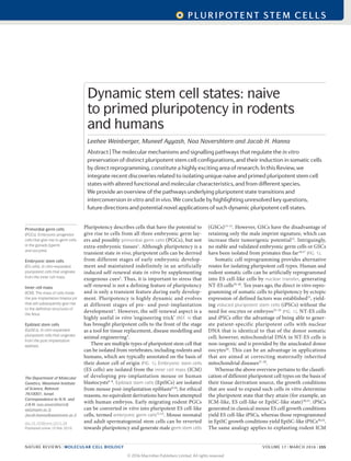
Pluripotency describes cells that have the potential to give rise to cells from the three germ layers but not extraembryonic tissues. There are multiple types of pluripotent stem cells that can be isolated from different stages of embryonic development in rodents and humans. Naive pluripotent stem cells, like mouse embryonic stem cells, resemble the pre-implantation embryo state while primed pluripotent stem cells, like mouse epiblast stem cells, resemble the post-implantation embryo state. Growth conditions influence whether pluripotent stem cells attain a naive or primed state in vitro.

Recommended
Recommended
More Related Content
What's hot
What's hot (20)
Similar to Nrm.2015.28
Similar to Nrm.2015.28 (20)
Recently uploaded
Recently uploaded (20)
Nrm.2015.28
- 1. Pluripotency describes cells that have the potential to give rise to cells from all three embryonic germ lay- ers and possibly primordial germ cells (PGCs), but not extra-embryonic tissues1 . Although pluripotency is a transient state in vivo, pluripotent cells can be derived from different stages of early embryonic develop- ment and maintained indefinitely in an artificially induced self-renewal state in vitro by supplementing exogenous cues2 . Thus, it is important to stress that self-renewal is not a defining feature of pluripotency and is only a transient feature during early develop- ment. Pluripotency is highly dynamic and evolves at different stages of pre- and post-implantation development3 . However, the self-renewal aspect is a highly useful in vitro ‘engineering trick’ (REF. 4) that has brought pluripotent cells to the front of the stage as a tool for tissue replacement, disease modelling and animal engineering5 . There are multiple types of pluripotent stem cell that can be isolated from vertebrates, including rodents and humans, which are typically annotated on the basis of their donor cell of origin (FIG. 1). Embryonic stem cells (ES cells) are isolated from the inner cell mass (ICM) of developing pre-implantation mouse or human blastocysts6–8 . Epiblast stem cells (EpiSCs) are isolated from mouse post-implantation epiblasts9,10 ; for ethical reasons, no equivalent derivations have been attempted with human embryos. Early migrating rodent PGCs can be converted in vitro into pluripotent ES cell-like cells, termed embryonic germ cells11,12 . Mouse neonatal and adult spermatogonial stem cells can be reverted towards pluripotency and generate male germ stem cells (GSCs)13–15 . However, GSCs have the disadvantage of retaining only the male imprint signature, which can increase their tumorigenic potential15 . Intriguingly, no stable and validated embryonic germ cells or GSCs have been isolated from primates thus far16,17 (FIG. 1). Somatic cell reprogramming provides alternative routes for isolating pluripotent cell types. Human and rodent somatic cells can be artificially reprogrammed into ES cell-like cells by nuclear transfer, generating NT-ES cells18–20 . Ten years ago, the direct in vitro repro- gramming of somatic cells to pluripotency by ectopic expression of defined factors was established21 , yield- ing induced pluripotent stem cells (iPSCs) without the need for oocytes or embryos22–25 (FIG. 1). NT-ES cells and iPSCs offer the advantage of being able to gener ate patient-specific pluripotent cells with nuclear DNA that is identical to that of the donor somatic cell; however, mitochondrial DNA in NT-ES cells is non-isogenic and is provided by the anucleated donor oocytes26 . This can be an advantage in applications that are aimed at correcting maternally inherited mitochondrial diseases27–29 . Whereas the above overview pertains to the classifi cation of different pluripotent cell types on the basis of their tissue derivation source, the growth conditions that are used to expand such cells in vitro determine the pluripotent state that they attain (for example, an ICM-like, ES cell-like or EpiSC-like state)30,31 . iPSCs generated in classical mouse ES cell growth conditions yield ES cell-like iPSCs, whereas those reprogrammed in EpiSC growth conditions yield EpiSC-like iPSCs30,32 . The same analogy applies to explanting rodent ICM The Department of Molecular Genetics, Weizmann Institute of Science, Rehovot 7610001, Israel. Correspondence to N.N. and J.H.H. noa.novershtern@ weizmann.ac.il; Jacob.hanna@weizmann.ac.il doi:10.1038/nrm.2015.28 Published online 10 Feb 2016 Primordial germ cells (PGCs). Embryonic progenitor cells that give rise to germ cells in the gonads (sperm and oocytes). Embryonic stem cells (ES cells). In vitro-expanded pluripotent cells that originate from the inner cell mass. Inner cell mass (ICM). The mass of cells inside the pre-implantation blastocyst that will subsequently give rise to the definitive structures of the fetus. Epiblast stem cells (EpiSCs). In vitro-expanded pluripotent cells that originate from the post-implantation epiblast. Dynamic stem cell states: naive to primed pluripotency in rodents and humans Leehee Weinberger, Muneef Ayyash, Noa Novershtern and Jacob H. Hanna Abstract | The molecular mechanisms and signalling pathways that regulate the in vitro preservation of distinct pluripotent stem cell configurations, and their induction in somatic cells by direct reprogramming, constitute a highly exciting area of research. In this Review, we integrate recent discoveries related to isolating unique naive and primed pluripotent stem cell states with altered functional and molecular characteristics, and from different species. We provide an overview of the pathways underlying pluripotent state transitions and interconversion in vitro and in vivo. We conclude by highlighting unresolved key questions, future directions and potential novel applications of such dynamic pluripotent cell states. REVIEWS NATURE REVIEWS | MOLECULAR CELL BIOLOGY VOLUME 17 | MARCH 2016 | 155 pluripotent stem cells © 2016 Macmillan Publishers Limited. All rights reserved
- 2. Nature Reviews | Molecular Cell Biology Blastocyst Post-implantation embryo Pre-implantation embryo PGCs ICM Postnatal or adult ES cell EpiSC Embryonic germ cell Somatic cells GSC iPSC In vitro isolated pluripotent cell type In vivo stage of development Yes Yes No Yes No Yes No Yes Yes Yes NT-ES cellYes Yes Male imprinting pattern only Non-isogenic mitochondria In vitro established Post- implantation epiblast Spermatogonial progenitors Oocyte Nucleus Somatic cell Nuclear transfer Cloned blastocyst Human Mouse Embryonic germ cells In vitro-expanded pluripotent cells that are derived from embryonic primordial germ cells (PGCs). Germ stem cells (GSCs). In vitro-expanded pluripotent stem cells that originate from neonatal or adult testis-derived spermatogonial stem cells. Nuclear transfer The cloning of a somatic cell-derived nucleus and its introduction into an anucleated host oocyte. Induced pluripotent stem cells (iPSCs). In vitro-generated pluripotent cells derived by the ectopic expression of defined exogenous factors in somatic cells. X inactivation Dosage compensation of the X chromosome in females, whereby one of the X chromosomes is epigenetically silenced. cells in growth conditions for ES cells or EpiSCs30,33 . In comparison to developmentally restricted mouse EpiSCs, ES cells are highly competent in generating high-contribution chimeric mice after microinjection into host blastocysts, retain a pre-X inactivation state in female cell lines and have reduced expression of line- age commitment factors30,31 . Such attributes influence the use of pluripotent cells in cell-differentiation assays and in animal transgenics. Thus, it is of importance to understand and define different pluripotent states and configurations across different species4 . In this Review, we provide an integrated perspective on recent breakthroughs in our understanding of the diversity and complexity of the regulation of the pluri- potent state in vitro. This includes advances in preserv- ing naive pluripotency from non-rodent species and alternative pluripotent states. We highlight unresolved issues, key questions and future directions in this exciting area of stem cell research. Murine pluripotent states Mouse ES cells were shown to reside in an ICM-like state30 , referred to as naive pluripotency31 , as they retain several molecular characteristics of the ICM. A novel type of pluripotent cell, EpiSCs, were later derived from post-implantation rodent epiblasts9,10 . In comparison to naive ES cells, EpiSCs retain an alternative pluri- potency configuration, which is referred to as primed pluripotency31 . There are marked molecular and func- tional differences between pluripotent cell types, which subsequently influence their characteristics, function and safety. Growth conditions for naive pluripotency. To fully understand the biology of mouse naive ES cells and their developmental context, it is of relevance to review the evolution of growth conditions that have been devised to isolate such cells over the past 30 years. ES cells were originally derived from the 129 mouse strain7,8 by using Figure 1 | Deriving different types of pluripotent stem cell in mouse and human. Various pluripotent cell types can be derived from different types of embryonic cell harvested at various stages of mouse or human development. Alternatively, somatic cells can be reprogrammed by somatic cell nuclear transfer (producing nuclear transfer embryonic stem (ES) cells (NT-ES cells)) or by the expression of exogenous transcription factors (generating induced pluripotent stem cells (iPSCs)). Pluripotent cells derived from post-implantation epiblast (epiblast stem cells (EpiSCs)) or from the germ cell lineage (embryonic germ cells and spermatogonial germ stem cells (GSCs)) have not yet been stably derived in humans or other primates. For therapeutic purposes, iPSCs and NT-ES cells have the advantage that their nuclear DNA is genetically identical to that of the somatic cells of the donor patient. However, NT-ES cells will retain mitochondrial DNA of the anucleated female oocyte into which the donor somatic cell nucleus is transferred. Spermatogonial GSCs are generated from spermatogonial stem cells that have already established an exclusive male (paternal) imprinting pattern; thus, the GSCs that are derived from them have the same imprinting pattern. If spermatogonial GSCs can be established from adult human males in the future, the fact that these cells lack the maternal imprint will probably limit their therapeutic potential. ICM, inner cell mass; PGCs, primordial germ cells. REVIEWS 156 | MARCH 2016 | VOLUME 17 www.nature.com/nrm REVIEWS © 2016 Macmillan Publishers Limited. All rights reserved
- 3. Nature Reviews | Molecular Cell Biology Potential cross-species differences Potential link BMP4 BMPR Activin A or TGFβ FGF2 FGFR LIF WNT LIFR or gp130 Frizzled Insulin or stress Insulin receptor β-catenin β-catenin β-catenin β-catenin MEK ERK MEK ERK P P P P NuRD NuRD PKC PKC p38 p38 JNK JNK BMP4 Activin A or TGFβ FGF2 LIF WNT Insulin or stress Cytoplasm Nucleus ESRRβNANOG STELLA KlF2,4,5 OCT4SOX2 OCT4 OTX2 ZIC2 SOX2 GSK3β GSK3β TCF3 TCF3 STAT3 STAT3 SMAD4 SMAD1,5,8 SRC SRC Primed Naive ? ? SMAD4 SMAD4 SMAD2,3 SMAD2,3 P P SMAD4 SMAD1,5,8 Naive pluripotency A pluripotent state that resembles the pre-implantation embryonic configuration(s). Primed pluripotency A pluripotent state that resembles the post-implantation embryonic configuration(s). mitotically inactive mouse embryonic fibroblasts (MEFs) as feeder cells and fetal bovine serum (FBS)34 . Leukaemia inhibitory factor (LIF), which activates the JAK–STAT3 (Janus kinase–signal transducer and acti- vator of transcription 3) pathway, was later identified as a key ingredient that enabled the proliferation of mouse ES cells in FBS/LIF conditions without MEFs35,36 (FIG. 2). Such naive mouse ES cells express hallmark pluripotency factors (such as octamer-binding protein 4 (OCT4; also known as POU5F1), homeobox protein NANOG and oestrogen-related receptor-β (ESRRβ; also known as ERR2)) and retain a pre-X inactivation state in female cell lines37 (FIG. 3). Functionally, ES cells can populate the host pre-implantation mouse ICM following micro injection into blastocysts and generate high-contribution chimeras with colonization of the germ line37 . Thefirstserum-andfeeder-freedefinedconditionsfor expanding mouse ES cells were developed by combining low doses of bone morphogenetic protein 4 (BMP4) with LIF38 . The addition of small-molecule inhibitors for Figure2|Signalling pathways and their influence on naive and primed pluripotent states. Differentsignalling pathwayscanpositivelyornegativelyregulatenaiveandprimedmurinepluripotentstemcells.Notethatthemajority of the signallingpathwaysshownhaveopposingeffectsonthenaiveandprimedpluripotentstatesinmice(forexample, the leukaemiainhibitoryfactor(LIF)–signaltransducerandactivatoroftranscription3(STAT3)andfibroblastgrowthfactor2 (FGF2)–ERKsignallingpathways).Itisimportanttohighlightthatotherpathwaysnotincludedinthisschemearelikelyto alsobeinvolvedinsuchregulationandwillprobablybefurthercharacterizedinthefuture.Suchpathwaysmayinclude HIPPO,RHO,NOTCHandnuclearfactor-κBsignalling.Pinkboxeshighlightsignallingpathwaysthatmayfunction differentlyintheregulationofmouseandhumanpluripotentcells.Morespecifically,itremainstobefullyunderstood whethersignallinginducedbylowdosesoftransforminggrowthfactor-β(TGFβ),activin–NODAL,nuclearβ-cateninorFGF2 (MEK–ERKindependent)influenceshumannaivepluripotencyinadifferentmannertothatpreviouslyobservedinrodent naiveembryonicstemcells. Dashedarrowsindicatepotentiallinksthatremaintobeestablished.BMP,bonemorphogenetic protein;ESRRβ,oestrogen-relatedreceptor-β;GSK3β,glycogensynthasekinase3β;JNK,Jun-likekinase;NuRD,nucleosome remodellinganddeacetylases;OCT4,octamer-bindingprotein4;PKC,proteinkinaseC;TCF3,transcriptionfactor3. AdaptedfromPosterhttp://www.nature.com/nrm/posters/pluripotency/index.html,NaturePublishingGroup. REVIEWS NATURE REVIEWS | MOLECULAR CELL BIOLOGY VOLUME 17 | MARCH 2016 | 157 pluripotent stem cells © 2016 Macmillan Publishers Limited. All rights reserved
- 4. Nature Reviews | Molecular Cell Biology Pluripotent cell property Naive pluripotent pluripotent cell cell Primed Mouse Human 2i, LIF FBS, LIF FGF2, Activin A or FGF2, TGFβ OCT4, KLF2, KLF4, 2i, LIF 2i, LIF, p38i, JNKi, PKCi, ROCKi, FGF2, TGFβ 2i, LIF, PKCi, MEF +KLF2, NANOG 2i, BRAFi, ROCKi, SRCi, LIF, Activin A, MEF (FGF2, JNKi) FGF2, TGFβ, 2i, BMPi, LIF, MEF FGF2, TGFβ or FGF2, Activin A or FGF2, MEF MEK–ERK dependence No Yes Long-term dependence on FGF2 signalling No Yes Long-term dependence on TGFβ or Activin A signalling No Yes Dominant OCT4 enhancer Distal Proximal H3K27me3 on developmental regulators Low High Global DNA hypomethylation Yes No Mild Strong X chromosome inactivation No Yes Dependence on DNMT1, DICER, METTL3, MBD3 No Yes Priming markers (OTX2, ZIC2) ↓ ↑ Pluripotency markers (NANOG, KLFs, ESRRβ) ↑ ↓ * Mild* Strong* Strong* Mild* * TFE3 nuclear localization High Low CD24/MHC class 1 Low/low High/mod Expressed adhesion molecules E-cadherin N-cadherin Promotion of pluripotency maintenance by NANOG or PRDM14 Yes No Metabolism OxPhos, Glycolytic Glycolytic Competence as initial starting cells for PGCLC induction High Low Capacity for colonization of host pre-implantation ICM and contribution to advanced embryonic chimeras High Low ‡ ‡ Hypomethylation of promoter and enhancer regions Yes No KIT Yes No Tolerance for absence of exogenous L-glutamine Yes No Mitochondrial membrane activity and depolarization High Low Competence as initial starting cell for TSC induction High Low MEK signalling increased the derivation efficiency of ES cells and their stability39 . This approach was extended by developing different defined conditions, all involving MEKinhibitors,thatcanbeusedtoisolatemouseEScells. A combination of three inhibitors, termed 3i conditions, was shown to stabilize pluripotent cells without LIF, indi- cating the existence of redundant pathways that can be used to isolate ES cells in vitro and can compensate for Figure 3 | Naive and primed pluripotent cell properties in mouse and human isolated pluripotent stem cells (PSCs). The first column on the left lists the properties that can be used to distinguish between murine embryonic stem cells expanded in 2i/leukaemia inhibitory factor (LIF) conditions(naivepluripotentcells)andmurineepiblaststemcellsexpanded in fibroblast growth factor 2 (FGF2)/Activin A conditions (primed pluripotent cells). These two reference states are used to annotate various other growth conditions that have been used for mouse or human pluripotentstemcells.Foreachgrowthcondition,weindicatewhethercells cultured in that condition have naive-like properties (shown in orange) or primed-like properties (shown in blue). Empty boxes indicate lack of characterization.Thislistofpropertiesislikelytoincreasewithtimeandcan be used to systematically annotate new pluripotent states isolated in uniqueconditionsandfromotherspecies;foranextendedtableincluding pluripotent cells derived from mouse, rat, human and rhesus macaque, see Supplementary information S1 (figure). BMPi, bone morphogenetic protein inhibitor; DNMT1, DNA methyltransferase 1; FBS, fetal bovine serum; H3K27me3, histone H3 Lys27 trimethylation; ICM, inner cell mass; JNKi, Jun-like kinase inhibitor; KLF, Krüppel-like factor; MBD3, methyl CpG-bindingdomainprotein3;MEF,mouseembryonicfibroblast;METTL3, methyltransferase-like protein 3; OCT4, octamer-binding protein 4; OxPhos, oxidative phosphorylation; PGCLC, primordial germ cell-like cell; PKCi, protein kinase C inhibitor; PRDM14, PR domain zinc-finger protein 14; ROCKi, RHO-associated protein kinase 1 inhibitor; TFE3, transcription factor E3; TGFβ, transforming growth factor-β; TSC, trophoblast stem cell. *Nooestrogen-relatedreceptor-β(ESRRβ).‡ Mousehostembryos.Adapted fromPosterhttp://www.nature.com/nrm/posters/pluripotency/index.html, Nature Publishing Group. REVIEWS 158 | MARCH 2016 | VOLUME 17 www.nature.com/nrm REVIEWS © 2016 Macmillan Publishers Limited. All rights reserved
- 5. 3i conditions Defined naive pluripotency growth conditions combining three inhibitors (i) for MEK, fibroblast growth factor (FGF) and glycogen synthase kinase 3 (GSK3) signalling. Ground state pluripotency Originally described as a state of pluripotency that is independent of exogenous activator signalling input or stimulation. 2i/LIF conditions Defined naive pluripotency growth conditions containing two inhibitors (i) for MEK and GSK3, together with LIF cytokine. Alternative 2i conditions Defined naive pluripotency growth conditions containing two inhibitors (i) for the glycogen synthase kinase 3 (GSK3) and SRC pathways. LIF/MEKi/aPKCi conditions Defined naive pluripotency growth conditions containing two inhibitors (i) for MEK and atypical protein kinase C (aPKC) signalling, together with the leukaemia inhibitory factor (LIF) cytokine. a lack of LIF–STAT3 signalling40 . Notably, this cell con- figuration was labelled as ground state pluripotency, as the cells cultured in 3i conditions were reported to grow inde- pendently of any exogenous signalling stimuli. However, the use of this term is challenged by the fact that growth in these conditions relies heavily on the inhibition of glyco- gen synthase kinase 3 (GSK3), which mimics stimulation of the WNT signalling pathway, and on exogenous insu- lin, which activates PI3K–AKT signalling40 . Furthermore, 2i/LIF conditions were adopted as an enhanced means to expandmouseEScellpopulations,andthereducedprolif- eration in 3i conditions compared with 2i/LIF conditions indicates a role for autocrine-secreted fibroblast growth factor 4 (FGF4) in promoting the growth of naive ES cells independently of MEK–ERK signalling30,41,42 (FIG. 2). Notably, complete and combined genetic ablation of Erk1 and Erk2 is detrimental to maintaining rodent naive ES cell survival and does not yield an identical phenotype to that resulting from MEK inhibition, which indicates that MEKinhibitionhasadditionalrolesinmaintainingEScell stability that are independent of ERK43 . Alternative 2i conditions, involving small-molecule inhibitors for GSK3 and SRC pathways, yield germline-competent ES cells44 (FIG. 2). Go6983, which is a small-molecule inhibitor of atypical protein kinase C (aPKCi), was identified as another stimulator for isolating mouse ES cells, together with LIF and/or inhibitors of MEK45 . Single-cell RNA sequencing (RNA-seq) analysis has shown that there is equivalent global heterogeneity between different growth conditions for naive pluri potency; however, differences exist in the identity of the genes that underlie heterogeneity in each condition46 . Enriched conditions have been important for deriving ES cells from mouse strains that were, until recently, con- sidered to be non-permissive for deriving naive ES cells. WhereasEScellsderivedfromthe129mousestraincanbe expanded in FBS/LIF conditions, for other mouse strains, such as non-obese diabetic (NOD) mice, supplementa- tion with 2i conditions or with GSK3 inhibitor (GSKi) is essential for both the derivation and maintenance of naive pluripotent cells30,42 . 3i and 2i/LIF conditions have been used to yield rat ES cells, although these conditions are suboptimal47,48 . LIF/MEKi/aPKCi conditions are a more robust method of supporting rat ES cells49 (FIGS 2,3). The above findings underscore the relevance of analy sing rodent ES cells expanded in different naive growth conditions and from different genetic backgrounds. Furthermore, they emphasize the importance of other signalling pathways that remain to be more extensively characterized in the context of pluripotency (FIG. 2). SRC functions as a downstream target of MEK–ERK and calcineurin–nuclear factor of activated T cells (NFAT) signallingtopromoteES cell differentiation50 , and its inhi- bition promotes naive pluripotency44,50 . Nuclear factor-κB (NF-κB) inhibition has been identified as a downstream effector that mediates naive pluripotency supported by aPKC inhibition45 ; however, other pathways, such as the methyl CpG-binding domain protein 3 (MBD3)– nucleosome remodelling and deacetylases (NuRD) repressor complex, can also be neutralized by aPKCi (J.H.H., unpublished observations). The HIPPO signalling pathway regulates epiblast versus trophoblast segregation in late mouse morulas and is highly active in pluripotent epiblast cells, leading to the exclusion of YES-associated protein (YAP) and transcriptional co-activator with PDZ-binding motif (TAZ) effectors from the nucleus51,52 . Depletion of YAP and TAZ in mouse naive ES cells expanded in 2i/LIF conditions further enhances their resistance to differen- tiation52 , whereas another study indicated that YAP and TAZ are essential regulators of the stability of naive ES cells expanded in FBS/LIF53 . Reanalysis of these findings in different conditions might resolve these seemingly opposing results (FIG. 3). It should be noted that signalling pathways are often pleiotropic and may simultaneously have positive and negative effects on naive pluripotency. For example, stabilization of nuclear β-catenin following GSK3 inhi- bition promotes naive pluripotency by neutralizing the repressive activity of transcription factor 3 (TCF3) on its target genes in the nucleus54 . Cytoplasmic β-catenin promotes naive pluripotency by increasing E-cadherin membrane stability55 (FIG. 2). However, nuclear β-catenin can induce mesodermal gene expression through its LEF co-effectors47 , although such differentiation-priming effects are outweighed by the naive pluripotency- promoting functions of β-catenin under optimized con- ditions. LIF has also been shown to promote primitive endoderm specification in naive pluripotency growth conditions56 . Such ‘non-purist’ effects should be kept in mind when dissecting the role of signalling pathways on pluripotency. The large number of conditions in which naive murine ES cells can be grown have been important in better understanding and revisiting the roles of several classical pluripotency regulators. Whereas NANOG was first purported to be absolutely essential for establish- ing naive pluripotency through iPSC reprogramming, cell fusion or EpiSC reversion57 , various conditions have since enabled the reprogramming of NANOG-null donor cells in vitro58–60 . However, NANOG-null ES cells cannot be derived from mouse ICM, which indicates that although NANOG is indispensable for establishing pluripotency in vivo, it is dispensable during in vitro induction61 . This example highlights that in vitro main- tenance of pluripotency cannot be considered to be ‘authentic’, as some in vitro conditions can potentiate the robustness of the naive pluripotency programme and compensate for deficiencies that are not sustainable in vivo. Similarly, Krüppel-like factor 2 (Klf2)-knockout embryos do not present lethality at the pre-implantation stage, and naive ES cells in 2i/LIF or FBS/LIF conditions can tolerate ablation of Klf2 (REF. 62). However, ES cells in 2i‑only conditions cannot sustain loss of KLF2 (REF. 62), as LIF is required to compensate for the lack of KLF2. Another emerging regulatory principle is that not all factors that are expressed in the ICM or in ES cells neces- sarily promote naive pluripotency; some of them, in fact, promote its dissolution. However, these factors are toler ated by ES cells in vitro, owing to the optimized and enriched growth conditions that are used. For example, binding of TCF3 to its naive pluripotency-promoting REVIEWS NATURE REVIEWS | MOLECULAR CELL BIOLOGY VOLUME 17 | MARCH 2016 | 159 pluripotent stem cells © 2016 Macmillan Publishers Limited. All rights reserved
- 6. FGF2/Activin A conditions Defined primed pluripotency growth conditions for mouse epiblast stem cells, composed of recombinant fibroblast growth factor 2 (FGF2) and Activin A cytokines. Seed enhancers A subgroup of enhancers that are dormant in naive cells but become more active in primed pluripotent and somatic cells. GSK3i/IWR1 conditions Defined primed pluripotency growth conditions for mouse epiblast stem cells, containing a glycogen synthase kinase 3 (GSK3) pathway inhibitor and the small-molecule tankyrase inhibitor, IWR1. FGF2/IWR1 conditions Defined primed pluripotency growth conditions for mouse epiblast stem cells, containing recombinant fibroblast growth factor 2 (FGF2) and the small-molecule tankyrase inhibitor, IWR1. target genes leads to their partial repression but is tolerated in FBS/LIF growth conditions. However, neutralization of TCF3 by adding GSK3i boosts naive pluripotency54 . In a similar manner, mouse ES cells toler ate expression of the MBD3–NuRD complex, despite the fact that it partially represses naive pluripotency targets63 . However, genetic ablation of Mbd3 leads to upregulated expression of master regulators of naive pluripotency and enables LIF-independent cell growth63 . Consistently, the derivation of Mbd3‑knockout ES cells from the ICM is not compromised in 2i/LIF conditions64 . In summary, both TCF3 and MBD3 are expressed in the ICM and in ES cells, where they are likely to set the stage for terminating naive pluripotency. Thus, the molecular characteristics of a pluripotent state in a certain growth condition represent the net outcome of conflicting stabilizing and destabilizing factors that simultaneously reside in that state65 . Collectively, when analysing the function of pluri- potency regulators, it is important to systematically compare different naive growth conditions, genetic backgrounds and in vivo contexts66 . Such integrated analysis is likely to unravel additional layers of underappreciated complexity and may resolve some conflicting results52,53 . Growth conditions for primed pluripotency. Primed EpiSCs were derived from post-implantation epiblasts of rodents in FGF2/Activin A conditions9,10 (FIG. 1). EpiSCs are capable of differentiating into cells of all three germ layers in vitro or in a teratoma assay, and thus they are pluripotent. However, they are inefficient in yielding chimeric animals once injected in pre-implantation epiblasts (FIG. 3), probably because they have altered molecular characteristics and correspond to a more advanced developmental stage in comparison to the host pre-implantation environment9,10 . EpiSCs can, however, form low-contribution chimeric embryos when injected into host post-implantation embryos ex vivo67 . EpiSCs maintain OCT4 and SOX2 expression, but they downregulate expression of most of the other pluripotency factors, including NANOG, ESRRβ, KLF2 and KLF4 (REF. 3). EpiSCs have not undergone differenti ation, but they upregulate lineage commitment factors such as homeobox protein OTX2, Brachyury and zinc- finger protein ZIC2 (REF. 68). Epigenetically, EpiSCs have distinct characteristics from naive ES cells: they inacti vate the X chromosome in females, upregulate global DNA methylation levels and acquire histone H3 Lys27 trimethylation (H3K27me3) at developmental regu- lators69,70 . The enhancer landscape is rewired between naive and primed pluripotent states68 , and develop- mental regulator gene-associated seed enhancers con- vert from a dormant to an active state in EpiSCs, thus pre-marking the lineage differentiation bias of primed PSCs71 . FIGURES 2,3 summarize the divergent signalling and molecular characteristics of murine primed and naive pluripotent cells. At the regulatory level, naive and primed pluripotent cells have been shown to have opposing dependence on epigenetic repressors66 (FIG. 4). Naive ES cells expanded in FBS/LIF or 2i/LIF conditions tolerate loss of epi- genetic repressors such as DNA methyltransferase 1 (DNMT1), DICER, Polycomb protein EED, MBD3 and methyltransferase-like protein 3 (METTL3)66 , which renders these cells ‘hyper-naive’ and resistant to differentiation66,72 (FIG. 4). Conversely, the mainten ance and viability of murine primed pluripotent cells depend on these regulators, and their ablation destabil izes the murine primed pluripotent state66 (FIG. 4). Defining precisely how the depletion of each of these repressors destabilizes the primed configuration is of future interest. Alternative growth conditions to expand murine EpiSCs have begun to emerge. The simultaneous use of a GSK3i (which induces β-catenin stabilization) together with a small-molecule inhibitor of tankyrase, IWR1 (which upregulates levels of axin 1 and axin 2, thus leading to the retention of β-catenin in the cyto plasm) — known as GSK3i/IWR1 conditions — main- tains novel primed EpiSCs without exogenous FGF2/Activin A supplementation73 (FIG. 3). Removal of IWR1 leads to increased nuclear shuttling of β-catenin and EpiSC differentiation73 . The mechanisms by which cytoplasmic β-catenin prevents EpiSC differentiation remain to be uncovered73 . It is tempting to speculate that the recently described ability of the cytoplasmic anaphase-promoting complex (APC)–axin–β-catenin destruction complex to function as a sequestration ‘sink’ for YAP and TAZ and prevent their nuclear shuttling52 is involved in the ability of GSK3i/IWR1 conditions to maintain EpiSCs. Notably, the latter alternative EpiSC state is different from EpiSCs expanded in classical FGF2/Activin A conditions, in that it retains higher levels of expression of naive markers73 and is thus relatively less primed (FIG. 3). Recent studies indicate that different primed condi- tions can endow EpiSCs with region-specific character- istics of post-implantation epiblasts. EpiSCs expanded in FGF2/Activin A conditions correspond transcrip- tionally and functionally to anterior late-gastrula primitive streak cells74 . Alternative FGF2/IWR1 conditions generate murine EpiSCs that correspond to posterior- proximal epiblasts75 . Furthermore, even in classical FGF2/Activin A conditions, distinct subpopulations of EpiSCs can co-exist, each representing different stages of post-implantation embryonic development76 . Finally, the length of time for which pluripotent cells are maintained under primed conditions greatly influences their characteristics and functionality77 . Counter-intuitively, whereas mouse PGCs are speci- fied from the post-implantation epiblast in vivo, EpiSCs that are maintained in vitro for more than 7 days in FGF2/Activin A conditions lose competence to gener- ate PGCs in response to BMP4 (REF. 77). Starting with naive cells and inducing brief priming for 2–4 days yields distinct primed cells that are highly competent for generating PGC-like cells, termed Epi-like cells77 . The latter are transcriptionally more similar to in vivo post-implantation epiblasts than to EpiSCs77 . Thus, the above paradigm indicates another aspect of the artifi- cial features that can be acquired by pluripotent cells REVIEWS 160 | MARCH 2016 | VOLUME 17 www.nature.com/nrm REVIEWS © 2016 Macmillan Publishers Limited. All rights reserved
- 7. Ablation of epigenetic repressors Ablation of epigenetic repressorsPrimed pluripotent cell Nature Reviews | Molecular Cell Biology Pluripotency factors Pluripotency factors Lineage-priming factors Lineage-priming factors Primed pluripotency Mouse EpiSC (FGF2/Activin A conditions) Naive pluripotency Mouse ES cell (FBS/LIF or 2i/LIF conditions) Ablation of epigenetic repressors Ablation of epigenetic repressors Differentiation and/or cell death Naive pluripotent cell once they are expanded indefinitely in vitro, in contrast to their in vivo counterparts that exist only transiently during development. Studies involving clonal lines and single-cell analy- sis will provide deeper understanding of the features of region-specific EpiSCs and of EpiSCs shortly after their in vitro induction from a naive state under different priming conditions74 . This may help us to understand how lineage priming is established at the single-cell level during these key early developmental transitions74,75 and may be relevant for optimizing other differentiation protocols and predicting the behaviour of PSCs. Conversion between naive and primed states. Similar to the reprogramming of somatic cells into a naive ES cell- like state by the combined overexpression of pluripotency factors together with LIF, primed EpiSCs can also be reverted to naive iPSCs. Overexpression of KLF4 or MYC in EpiSCs, under LIF-containing conditions, generates naive ES cells30,78 . FBS/LIF signalling alone can be suffi cient to induce such conversion in cells from permis- sive mouse genetic backgrounds (such as 129 strains)79 , but not in cells from ‘non-permissive’ strains such as NOD mice, for which supplementation with small mol ecules, such as 2i conditions, is necessary30 . Other fac- tors, such as NANOG, PR domain zinc-finger protein 14 (PRDM14) and ESRRβ, have been shown to synergisti- cally induce and boost the efficiency of this process80,81 . Explanting post-implantation embryonic day 5.5 (E5.5)– E7.5 epiblasts in naive conditions also reverts them to naive PSCs30,79 . The opposite conversion can be achieved for in vitro and in vivo isolated naive cells, as expanding murine naive PSCs or ICM cells under primed conditions leads them to gradually adopt an EpiSC state30,33,78 . Studies focusing on the molecular changes that accompany the in vitro conversion from naive to primed pluripotent states have unravelled key events in the mech- anisms of reprogramming69 . Naive ES cells expanded in 2i/LIF conditions retain global levels of hypomethyl ation in both promoters and gene bodies, highly similar to those measured in ICM cells82,83 . When naive ES cells are transferred into FBS/LIF naive conditions, there is an increase in global DNA methylation levels, although promoter and enhancer regulatory regions remain pro- tected from DNA methylation84 . Only after transfer into primed FGF2/Activin A EpiSC-inducing growth condi- tions does DNA methylation accumulate over enhancer and promoter regulatory elements84 . Transitioning naive FBS/LIF-cultured PSCs into 2i/LIFconditionsinitiallyleadstochangesinthepromoter occupancy of the genes encoding the OCT4, SOX2 and NANOG pluripotency factors85 . Changes in H3K27me3 deposition and the enhancer landscape follow later, prob- ably in response to the rewiring of transcription factor binding85 . Downregulation of DNA methylation follows next and has mainly been attributed to downregulation in expression of de novo DNA methyltransferase enzymes82 . It should be noted, however, that ablation of DNMT3A and DNMT3B in ES cells in FBS/LIF conditions does not lead to such rapid loss of DNA methylation86 , and other yet-to-be-identified events might be involved in this rapid 2i‑induced epigenetic response. MEK–ERK inhibition influences Polycomb interactions and leads to decreased promoter occupancy by Polycomb repres- sive complex 2 (PRC2) and decreased phosphorylation on the carboxy-terminal domain of RNA polymerase II (Pol II) on lineage-commitment genes87 , leading to loss of H3K27me3 and increased Pol II pausing at biva- lent developmental regulatory loci69 . Analysis of other defined naive pluripotency growth conditions (such as 2i/LIF/PKCi and alternative 2i conditions) and studies in other rodents will be important to discern the redundan- cies and specificities of different signalling pathways and their crosstalk with chromatin organization. Figure 4 | The opposing influence of epigenetic repressors on murine naive and primed pluripotent cells. Murine naive and primed pluripotent cells differ not only in their dependence on distinct signalling pathways and in their epigenetic profiles, but also in their lineage decision-making. Murine naive embryonic stem cells (ES cells) expanded in either 2i/leukaemia inhibitory factor (LIF) or fetal bovine serum (FBS)/LIF conditions tolerate complete loss of epigenetic repressors such as DNA methyltransferase 1 (DNMT1), methyl CpG-binding domain protein 3 (MBD3), DICER, DGCR8, methyltransferase-like protein 3 (METTL3), EED and EZH2. Furthermore, the loss of epigenetic repressors strengthens the equilibrium in favour of pluripotency- promoting factors and generates ‘hyper-naive’ pluripotent cells that are relatively more resistant to differentiation and can tolerate withdrawal of LIF cytokine. Murine primed epiblast stem cells (EpiSCs) generally respond in the opposite manner to the complete ablation of epigenetic repressors. Established murine primed EpiSCs naturally downregulate pluripotency factors and upregulate lineage-priming factors compared with naive pluripotent cells. Ablation of epigenetic repressors at the primed pluripotency stage tips the balance towards differentiation and/or compromises cell survival. It should be noted that the ablation of various epigenetic repressors (such as METTL3 or DGCR8) overall negatively influences the stability of primed pluripotency; however, the downstream events leading to the state collapse are not necessarily identical in each case and thus should be thoroughly dissected in vitro and in vivo. FGF2, fibroblast growth factor 2. Adapted with permission from REF. 66, AAAS. REVIEWS NATURE REVIEWS | MOLECULAR CELL BIOLOGY VOLUME 17 | MARCH 2016 | 161 pluripotent stem cells © 2016 Macmillan Publishers Limited. All rights reserved
- 8. Human conventional pluripotent cells The first human ES cells to be isolated from blastocysts6 were markedly different from murine ES cells in their characteristics and tissue culture requirements. FGF2 and transforming growth factor-β1 (TGFβ1)/Activin A signalling (but not LIF signalling) are the core signal- ling modules that maintain such conventional human ES cells derived from the ICM, or iPSCs obtained by direct in vitro reprogramming88 . A primed pluripotent state. Differences between con- ventional human and mouse ES cells had initially been attributed to unknown genetic differences between species, as human ES cells were also derived from the ICM and not from post-implantation stages. However, studies of stem cells derived from different mouse strains have discerned a scenario whereby ICM cells can adapt in vitro into a primed state if naive conditions are not devised to match the requirements of the particu- lar genetic background of the donor embryos used30 . Specifically, NOD mice are relatively ‘less permissive’ than 129 mice to yielding naive ES cells and iPSCs, as LIF alone is not sufficient to maintain naive pluripotency of cells from NOD mice, and 2i/LIF conditions are per- manently required to stabilize and maintain naive pluri- potency in vitro in NOD PSCs30 . Furthermore, ICM cells from both 129 and NOD mice expanded under primed conditions yield EpiSC-like cells that are indistinguish able from EpiSCs derived from E6.5 embryos30 or in vitro from already established ES cells30 . The relevance of the latter in vitro priming scenario to determining the identity of conventional human ES cells is supported by the fact that human conven- tional ES cells and iPSCs retain a large number of primed pluripotency features. These include low levels of expression of naive pluripotency markers (such as KLF17 and developmental pluripotency-associated protein 3 (DPPA3)), deposition of H3K27me3 over developmental genes, lack of exclusive nuclear local ization of transcription factor E3 (TFE3), loss of pluri potency upon inhibition of MEK–ERK signalling, lack of global hypomethylation as seen in ICM cells, and lack of a pre-X inactivation state in most conventional female PSC lines70,89,90 . Furthermore, human primed ES cells do not tolerate complete loss of DNMT1 (REF. 86), similar to what has been shown for mouse EpiSCs66 (FIG. 4). Complete knockout of DICER, MBD3 or METTL3 has not been achieved so far for human ES cells64,66 (FIG. 3). Less primed than murine EpiSCs. Despite the above, it is important to realize that human conventional or primed ES cells are not identical to murine EpiSCs and can be considered to be relatively less primed. For example, human ES cells do not upregulate the expression of FGF5 or N-cadherin (as seen in murine EpiSCs), and human ES cells express high levels of E-cadherin (as detected in mousenaiveEScells)70 .HumanEScellsexpresshighlevels of some naive pluripotency markers such as NANOG, PRDM14 and reduced expression protein 1 (REX1; also known as ZFP42) that are not expressed or are residu- ally expressed by mouse EpiSCs91 . Moreover, human primed ES cells are functionally dependent on NANOG and PRDM14, and ablation of these factors induces the differentiation of human ES cells91 . The distribution of DNA methylation in human ES cells corresponds to that of murine naive ES cells expanded in FBS/LIF con- ditions, rather than that of FGF2/Activin A-expanded mouse EpiSCs, as the promoters of human ES cells and mouse naive ES cells are protected from invasion by repressive DNA methylation84,92 . Furthermore, whereas murine EpiSCs demonstrate exclusive cytoplasmic local- ization of TFE3, and naive 2i/LIF-cultured ES cells show exclusive nuclear localization of TFE3 (REF. 93), human primed ES cells show an intermediate configuration, in which TFE3 is present in both the cytoplasm and the nucleus70 . Human naive pluripotent cells The metastability of naive and primed pluripotent states depending on the growth conditions used30 , and the stringent requirements for exogenous factors to promote the naive pluripotency of isolated naive PSCs from pre- viously ‘non-permissive’ rodent strains30,31 , have led to a consideration of whether unique and more stringent conditions can be used to isolate previously unidentified alternative naive-like pluripotent states in humans. Transgene-dependent generation. 2i/LIF conditions are notsufficienttomaintainnaivehumanEScellsoriPSCs94 . However, additional transgene expression can induce an artificial transgene-dependent state that may be of considerable interest. Continued exogenous OCT4 and KLF4,orKLF2andKLF4,transgeneexpressioncanmain- tain human ES cells and iPSCs in a unique pluripotent state in 2i/LIF conditions94 . Recently, these observations were extended by optimizing the overexpression of KLF2 and NANOG transgenes, allowing the expansion of humannaiveiPSCsin2i/LIFconditions95 .Overexpression of KLF2 and NANOG transgenes in primed ES cells also enabled their expansion in 2i/LIF/aPKCi conditions96 . These cells had extensive DNA hypomethylation and marked upregulation of naive pluripotency markers such as transcription factor CP2‑like protein 1 (TFCP2L1), KLF2 and KLF4. However, as KLF2 is not expressed in the human ICM97 , as 2i/LIF/aPKCi conditions are insuffi- cient to convert primed ES cells without exogenous trans- gene induction96 and as transgene-free cells remain to be validated under 2i/LIF/aPKCi conditions, it is unclear whether this state is indefinitely stable without retain- ing possibly leaky transgenes or MEFs. Furthermore, independent examination of the DNA methylation land- scape in these cells indicates an aberrant global loss of imprintingandexcessivehypomethylationofendogenous retroviral genes89,98 . Finally, although 2i/LIF/aPKCi condi- tions do not contain exogenous FGF or TGFβ1/Activin A cytokines, short-term inhibition of FGF receptor (FGFR) and TGF receptor (TGFR) signalling is not sufficient evi- dence to validate the independence of the cells from FGF, TGF and Activin A signalling96 (FIG. 2). Indeed, unlike in mice, the human pluripotent ICM differentiates following treatment of blastocysts with small-molecule inhibitors for TGF and activin–NODAL signalling97 . REVIEWS 162 | MARCH 2016 | VOLUME 17 www.nature.com/nrm REVIEWS © 2016 Macmillan Publishers Limited. All rights reserved
- 9. Although the field has shifted to studying trans- gene-independent conditions, as detailed below, it should be noted that transgene-dependent states may nevertheless be important, as it is possible that the robust naive pluripotency currently obtained in mouse ES cells is a rodent-specific phenomenon. Capturing human naive PSCs identical to those obtained from mice might still involve genetic modifications. Nevertheless, the following studies provide evidence that it is possible to generate alternative pluripotent states in humans and other species30,94 . Transgene-independent generation. Our team was the first to describe naive pluripotency growth con- ditions — designated NHSM (naive human stem cell medium) — that involve the complete ablation of MEK– ERK signalling and are compatible with the indefinite expansion of genetically unmodified human PSCs, in both MEF-containing and MEF-free conditions70 . These naive MEK-independent pluripotent cell lines could be derived from human pre-implantation embryos, through de novo iPSC generation, or from previously established primed ES cells and iPSCs70 . NHSM condi- tions contain 2i/LIF together with p38 inhibitor (p38i), Jun N-terminal kinase inhibitor (JNKi), aPKCi, RHO- associated protein kinase 1 inhibitor (ROCKi), and low doses of FGF2 and TGFβ1 (or Activin A); they render human PSCs more similar, but not identical, to murine naive PSCs70 (FIGS 2,3). In fact, human cells cultured in NHSM have features of so-called naive 2i/LIF-induced ground state pluripotency, which are not found even in naive mouse ES cells expanded in FBS/LIF conditions. These features include exclusive nuclear localization of TFE3 and demethylation of H3K27me3 marks over developmental genes69,70,93 . Transcriptionally, these cells have downregulated expression of lineage-commitment markers such as OTX2, ZIC2 and CD24, and moderately upregulated expression of pluripotency genes (more prominently when cultured on MEFs and when aPKCi is used)70,99 . Enhancer rewiring has been attained in these human naive PSCs, as seen with mouse cells71 . The cells had downregulation of expression of DNMT3B100 and a small global decrease in levels of DNA methyl ation, while maintaining imprinting integrity and chromosomal stability70 . Although chimeric analysis with human PSCs and the use of human embryos as hosts are ethically and legally forbidden, these human naive PSCs showed, for the first time, authentic inte- gration into blastocysts following microinjection into host mouse morulas and were able to contribute at low- grade levels in mouse embryos up to E10.5–E17.5 that underwent advanced organogenesis70 (FIG. 3). Important publications describing alternative condi- tions that yield human MEK-independent naive pluri- potent cells have emerged since then, each describing the production of cells with different enhanced molecu lar properties (FIG. 3). A combination of 2i/LIF, ROCKi, BMP receptor inhibitor (BMPRi), and high doses of FGF2 and TGFβ1 could maintain human PSCs only in the presence of MEFs101 . These PSCs had transcrip- tional upregulation of pluripotency markers such as STELLA (also known as DPPA3) and KLF5. Another study described culture conditions95 that used most of the same components found in NHSM70 (2i/LIF, ROCKi and Activin A (instead of TGFβ1) — with or without FGF2 and JNKi) but were supplemented with inhibitors for the Ser/Thr protein kinase BRAF and SRC path- ways (termed 5i/LA-MEF conditions). In comparison to previous studies, cells in 5i/LA-MEF conditions had a more marked upregulation of naive pluripotency mark- ers. However, the cells did not downregulate expression of DNMT3B, maintained an inactive X chromosome state in female cell lines and had an unusual pre-ICM transcriptional signature95 . Intriguingly, the process of converting primed cells back to a naive state in 5i/LA-MEF conditions is inefficient, taking 2 weeks to isolate initial clones that retain a slow growth rate95 . Furthermore, these conditions exclusively yield chromosomally abnormal cell lines95 . Thus, it remains to be determined whether such chromosomal abnor- malities are in fact inherent to 5i/LA-MEF-expanded cells and determine the properties described for this state95 , and whether they are being selected for dur- ing this inefficient conversion process. Finally, DNA methylation profiling and the epigenetic imprinting integrity of these cells are important aspects that remain to be evaluated. It is clear from the above summaries that none of the many published conditions generates human naive PSCs that are identical to mouse ES cells or human ICM97,102,103 . However, these studies implicate new sig- nalling pathways as being involved in human naive pluripotency and suggest avenues of research for the further optimization and characterization of such novel PSCs (FIG. 2). Mechanistically, it will be interest- ing to test whether there is a connection between RAF and aPKC inhibition and the influence of modulating WNT signalling by applying GSK3i/IWR1 conditions73 to human naive PSCs. Furthermore, RHO signalling has been shown to promote the nuclear localization of YAP and TAZ in primed human ES cells and to sustain their pluripotency104 . Thus, it remains to be defined whether ROCKi influences naive pluripotency characteristics70 through the modulation of YAP and TAZ. The role of MEK-independent FGF2 and TGFβ1/Activin A signalling, either in an autocrine manner or exogenously provided at low doses, remains to be understood in human naive PSCs. These cells show upregulation of the activin-like ligand growth- differentiation factor 3 (GDF3)96 , and human (but not mouse) ICM cells abundantly express activin recep- tors97 . Thus, it is tempting to speculate that primed human ES cells are relatively less primed than murine EpiSCs, owing to differences in their response to activin-like ligands, which might promote some naive features in human cells70,95 but not in mouse cells. To conclude, systematic analysis of the response of pluri- potent cells of different states from different species to a variety of TGF-ligand family members is of importance (FIG. 2). The possibility of generating human PSCs that are entirely independent of FGF and/or TGF signalling cannot be excluded. REVIEWS NATURE REVIEWS | MOLECULAR CELL BIOLOGY VOLUME 17 | MARCH 2016 | 163 pluripotent stem cells © 2016 Macmillan Publishers Limited. All rights reserved
- 10. Nature Reviews | Molecular Cell Biology Primed pluripotent states Naive pluripotent states Gene Human Mouse KLF2 No Yes KLF17 Yes No ERAS No Yes XIST Low No ESRRB No Yes DNMT3L High Low Human a c b Mouse Pre-implantation blastocyst (E6–E7) Pre-implantation blastocyst (E3.5–E4.5) Early blastocyst Post-implantation embryo (E9–E12) Extra-embryonic endoderm Trophectoderm Primitive endoderm Trophectoderm Epiblast ICM Ectoplacental cone Post-implantation embryo (E6–E7.5) Extra-embryonic ectoderm Extra-embryonic endoderm Morula Morula Zygote Early blastocyst Naive pluripotent cells Features of naive pluripotency Features of primed pluripotency Primed EpiSCs Naive ES cells Naive ES cells Naive ES cells Prim ed conventionalES cells More naive Less naive Yes No More primedLess primed Rodent pluripotency Stabilized by MEK inhibition Prim ed EpiSCs No extra-embryonic ectoderm or ectoplacental cone Flat disc-shaped epiblast Primed pluripotent cells Cylinder-shaped epiblast ES cell (2i/LIF/PKCi) (2i/LIF/TGFRi) (2i/LIF/FGFRi) (2i/LIF/NOTCHi) ?? ES cell (2i/LIF) ES cell (FBS/LIF) EpiSC (GSK3i/IWR1) Somatic lineage differentiation EpiSC (FGF2/Activin A) Differences between mouse and human epiblasts Recent studies focusing on single-cell RNA-seq of human pre-implantation embryos are starting to pro- vide answers to some of the questions highlighted above. Although there are no morphological differ- ences between human and mouse blastocysts, they have marked molecular differences at the cellular level97 (FIG. 5a,b). Human ICM epiblast cells do not express genes such as KLF2 and ESRRB that are thought to be important pluripotency factors in mice. Instead, KLF17 may have a human-specific role in the ICM97 . The GTPase ERAS, which is an ES cell-specific form of REVIEWS 164 | MARCH 2016 | VOLUME 17 www.nature.com/nrm REVIEWS © 2016 Macmillan Publishers Limited. All rights reserved
- 11. RAS, has become a pseudogene in humans105 , whereas ERAS-null mouse ES cells propagate slowly in FBS/LIF conditions105 . Non-human primate (marmoset) pluri- potent epiblasts have a similar transcriptional signature to that of human epiblasts, which is very different to that of mouse epiblasts106 . The marmoset and human naive ICM signatures include a lack of transcription of KLF2, nuclear receptor subfamily 0 group B member 1 (NR0B1) and BMP4, with high levels of expression of NODAL and its downstream signalling mediators106 . At the post-implantation stage, there are major dif- ferences between rodent and human embryos (FIG. 5a,b). Rodents are unusual in that their post-implantation epiblast assumes an egg-like cylinder shape, whereas in humans, the post-implantation epiblast assumes a flat disc shape, similar to that of most other mammals. Although it is not possible to carry out single-cell analy sis on early human post-implantation epiblasts, the study of non-human primates may provide some rele vant insights. Collectively, these differences between species may directly influence the distinct pluripotent characteristics that are observed in PSCs from different species in vitro and their distinct growth requirements. Furthermore, they are of relevance for understanding the developmental context of human pluripotent cells isolated in vitro. Classification of pluripotent states The characterization of different conditions to isolate human naive PSCs with distinct characteristics, and the limitations to conducting chimeric analysis in humans, have stimulated discussions regarding the classification of pluripotent states. It is often claimed that the ability to derive ES cells from human ICM cells in a newly devised growth condition constitutes the ‘gold standard’ for proving naivety. However, it should be kept in mind that the pluripotent state identity is eventually dictated by the derivation growth conditions, and not by whether the source of the cells was the pre- or post-implantation epiblast30,33 or PGCs82 . Use of the OCT4 distal versus proximal enhancer element as a binary distinguishing marker can also be misinterpreted95 . Both distal and proximal enhancer elements of OCT4 are active in naive and primed pluripotent states, both in humans and in mice107,108 . The difference emerges from their relative activity levels (high versus low) and dominance. Relying on a single attribute marker or functional test to define the pluripotent state is limiting and must be accompanied by systematic analysis of the ever-increasing number of characteristics that con- tinue to be identified for different pluripotent states (FIGS 3,4). Nevertheless, in our opinion, a molecular and functional characteristic that can be considered to be a major divider between naive and primed pluri- potent states is the response of cells to the challenge of blocking MEK signalling (FIG. 5c). Human conven- tional ES cells and mouse EpiSCs rapidly differentiate following MEK inhibition, whereas naive pluripotent cells tolerate MEK inhibition and consolidate their naivety following this challenge94 . The significance of this characteristic is supported by the ability of MEK or ERK inhibition to expand murine epiblast in ICMs, and by the fact that it signifies consolidation of naive pluripotency in vivo109 . Within the naive and primed ground states of pluri- potency, it is clear that if one considers the many naive and primed pluripotency features that were originally described for mouse naive 2i/LIF-cultured ES cells and primed FGF2/Activin A-cultured EpiSCs, different pluripotency growth conditions can simultaneously endow a mixture of primed and naive properties in the same cell type (FIG. 3). As such, pluripotent states can be classified as ‘more naive’ or ‘more primed’ by vir- tue of having more of such properties (FIG. 5c). Human primed ES cells have several features of naive pluri potency (such as protection of promoter regions from hypermethylation and dependence on NANOG), and recent comparative analysis with single-cell RNA-seq of human blastocysts suggested that some conventional human ES cell lines may be transcriptionally relatively less primed than previously thought97 . Murine naive ES cells expanded in FBS/LIF conditions can give rise to ‘all-ES cell’ chimeric embryos and tolerate abla- tion of Dnmt1 and Mettl3; however, they are globally hypermethylated and acquire H3K27me3 marks over developmental genes, as seen in EpiSCs (FIGS 3,5c). Thus, FBS/LIF conditions seem to endow mouse ES cells with relatively less-naive properties than 2i/LIF conditions. Figure 5 | A model to classify ‘relative naivety’ within the spectrum of naive to primed pluripotency. a | There are important similarities and differences in early pre- and post-implantation in vivo development in mice and humans. Although mouse and human embryos are morphologically similar until the blastocyst stage, there are important transcriptional differences, as summarized in part b. Furthermore, at the post-implantation stage, morphological differences in embryo shape become apparent between mice and humans, including differences in epiblast shape and in extra-embryonic structures. In mice, naive embryonic stem cells (ES cells) can be derived from the inner cell mass (ICM) of pre-implantation blastocysts or from post-implantation epiblasts when naive growth conditions are applied. Primed epiblast stem cells (EpiSCs) can be derived from post-implantation epiblasts or from the ICM when primed culture conditions are used. Thus, the growth conditions rather than the cell source determine the pluripotent state configuration that is acquired in vitro. A similar scenario applies for the derivation of both naive and primed pluripotent cells from human ICM, depending on the growth conditions used. Pluripotent cells cannot be derived from human post-implantation embryos, owing to ethical issues; therefore, a molecular analysis of these cells is lacking. c | A model to explain ‘relative naivety’ within the spectrum of naive to primed pluripotency. One major molecular and functional criterion that, in our opinion, can be used to separate naive and primed pluripotent cells is their ability to maintain and stabilize their pluripotent state upon blockade of MEK activity (dashed black line). Within the naive pluripotent state, it is difficult to describe the pluripotent state of cells in absolute terms, as naive cells can also have, to some extent, features of primed pluripotency. Similarly, within the spectrum of primed pluripotency, primed pluripotent cells cultured in different conditions have different features and varying degrees of naivety (FIG. 3). Finally, it is possible that supplementation of 2i/leukaemia inhibitory factor (LIF) conditions with small molecules such as atypical protein kinase C inhibitor (aPKCi), fibroblast growth factor receptor inhibitor (FGFRi) or NOTCH inhibitor (NOTCHi) can be used to consolidate naive pluripotency features, particularly for other rodents such as rats, for which the stability of naive cells in 2i/LIF feeder-free conditions should be further improved. Full annotation of the different human pluripotent states will enable charting of an equivalent landscape for human and other primate pluripotent stem cells. DNMT3L, DNA methyltransferase 3‑like; ERAS, ES cell-expressed RAS; ESRRB, oestrogen-related receptor-β; FBS, fetal bovine serum; GSK3i, glycogen synthase kinase 3 inhibitor; KLF, Krüppel-like factor; TGFRi, transforming growth factor receptor inhibitor; XIST, X-inactive specific transcript. Parts a and b are adapted from Poster http://www.nature.com/nrm/posters/pluripotency/index.html, Nature Publishing Group. Part c is adapted with permission from REF. 4, Elsevier. ◀ REVIEWS NATURE REVIEWS | MOLECULAR CELL BIOLOGY VOLUME 17 | MARCH 2016 | 165 pluripotent stem cells © 2016 Macmillan Publishers Limited. All rights reserved
- 12. Another functional test that can be used to assess the stringency and extent of naivety in different pri- mate naive PSCs is whether the cells can tolerate com- plete ablation of epigenetic repressors such as METTL3, DNMT1, DGCR8 and MBD3 (REFS 4,66) (FIGS 3,4). Furthermore, such tests might be useful for optimizing conditions that can close the gap between mouse and human naive pluripotent cells (BOX 1; FIG. 4). It will also be informative to annotate different naive and primed states from other species that have been isolated thus far according to such criteria (FIG. 3). Implications and future directions The breakthrough discovery that it is possible for somatic cells to be reprogrammed to pluripotency21 has provided the foundation for a deeper investigation Box 1 | A ‘dark side’ of naive pluripotency? With the development of naive pluripotency culture conditions and efforts to endow cells with more features of naivety, it has become relevant to ask how much naivety is needed, and whether there is a ‘dark side’ to permanently maintaining pluripotent stem cells (PSCs) under certain naive conditions. Rodent embryonic stem cells (ES cells) expanded in 2i/leukaemia inhibitory factor (LIF) conditions have an increased tendency to acquire genomic abnormalities41 , and it remains unclear whether these alterations occur as a by-product of non-specific activity of the small-molecule inhibitors used95,116 or as a direct result of intrinsic molecular features of naive pluripotency (for example, increased activity of endogenous retroviral elements or a reduction in epigenetic repressive marks). One can envision a scenario in which such features can be tolerated in vivo because this configuration exists for only 1–2 days, whereas prolonged in vitro expansion of this state might increase the frequency of unwanted damaging events. This concern may also relate to safeguarding the integrity of DNA methylation and imprinting in naive PSCs expanded in vitro over an extended period of time. Studies focusing on the loss of DNA methylation following the transfer of mouse PSCs into 2i/LIF conditions have quantified methylation levels 10–24 days after transfer and have documented a rapid global loss of DNA methylation accompanied by the relative resistance of retrotransposons and imprinting regions to such demethylation83 . However, it is unclear whether this methylation state represents a final plateau that naive cells achieve, or whether further culture in 2i/LIF conditions would lead to a decrease in the relative resistance of such regions to demethylation. Indeed, methylation over imprinted genes and retrotransposons is partially, yet significantly, reduced in 2i/LIF conditions83 . If such effects are frequent, researchers will have to re-evaluate how to optimize the application of naive conditions. One scenario might involve decreasing inhibitor levels to avoid excessive hypomethylation or other unwanted effects. An alternative scenario is to maintain cells in primed pluripotency culture conditions and transfer them into naive culture conditions only for a short time before the initiation of differentiation. of pluripotent states, and for the understanding that pluripotent configurations can be rewired. The ability to rewire cells for pluripotency has a direct influence on current hurdles and limitations related to the quality and characteristics of human iPSCs (BOXES 1,2). One of the most fascinating questions related to the naive-to-primed pluripotency continuum is why these divergent pluripotent configurations exist. This is often accompanied by the pragmatic consideration of which cells — naive or primed — are better to work with. In our opinion, as this phenomenon is deeply rooted in early embryonic development in vivo, it is likely that both naive and primed configurations constitute essential and integral components of the developmental process, to optimize and maximize the benefits of multipotency and lineage specification simultaneously. We hypothe- size that naive pluripotency emerged as an epigenetic erasure state that renders pluripotent cells free of lineage and epigenetic restriction, while simultaneously making these cells relatively less responsive to signalling path- ways that might interfere with the establishment of such a lineage-neutral state. The induction of specification by morphogens may not be efficiently enforced during or immediately after establishing naive pluripotency, with- out a short ‘delay period’. As such, the naive pluripotency network is gradually resolved and becomes more recep- tive to inductive cues at the post-implantation stage, and PSCs are differentially patterned and primed accord- ing to their spatial localization before overt somatic differentiation occurs. At the functional level, it remains to be established whether using human naive PSCs as a starting material, with or without a brief period of priming, would resolve the problems that are currently associated with proto- cols for the in vitro differentiation of human PSCs. Will human naive PSCs yield increased consistency in differ- entiation between independent iPSC lines110 ? Can naive PSC conditions yield better quality cells in differenti- ation protocols when used as a starting material? Can human naive PSCs be used in differentiation protocols that have not been successful with human conventional PSCs? Encouraging support for this possibility has recently been provided by results showing the enhanced ability of human PSCs expanded in NHSM conditions (even in the absence of aPKCi) to undergo in vitro dif- ferentiation into PGCs, which is a protocol that was Box 2 | Potential implications of induced pluripotent stem cell (iPSC) reprogramming Recent studies have elucidated how certain epigenetic regulators have opposing effects on the maintenance of naive and primed murine PSCs66 (FIG. 4). These findings may be relevant when comparing the induction of pluripotency mechanisms in humans versus mice, as human iPSCs, but not mouse iPSCs, are typically reprogrammed in conventional (primed) pluripotency conditions117 . Consequently, some of the differences observed between human and mouse iPSC regulators may be related not to species differences but rather to the fact that distinct pluripotent states are being induced. Therefore, it will be imperative to expand screens of human iPSC reprogramming regulators to include different pluripotency conditions, as these may yield different outcomes. Another consideration related to the effect of pluripotent state characteristics on reprogramming is whether naive conditions might improve the quality of the iPSCs obtained and facilitate the loss of residual epigenetic memory and heterogeneity118,119 . Similarly, subtle epigenetic differences in DNA methylation between nuclear transfer embryonic stem cells and iPSCs generated from the same human donor cells120 might also be neutralized when deriving iPSCs in naive conditions that mimic more closely the epigenetic features of the inner cell mass. REVIEWS 166 | MARCH 2016 | VOLUME 17 www.nature.com/nrm REVIEWS © 2016 Macmillan Publishers Limited. All rights reserved
- 13. inefficient for primed human PSCs111 . The molecular rationale for evaluating the potential benefits highlighted above is that naive pluripotency is more associated with the removal of epigenetic repressive marks in regulatory regions, compared with primed pluripotency70,96 . This might enable more efficient activation of lineage speci fiers during differentiation of naive pluripotent cells. Furthermore, lineage biases in human primed PSCs are heavily associated with the localized accumulation of repressive marks such as DNA methylation112 . The recent advances in generating human naive PSCs will continue to boost attempts to generate naive-like PSCs from other species and to test same-species and inter-species embryo chimerism assays113 . Cynomolgus monkey naive ES cells derived in NHSM conditions gave rise to the first chimera-competent ES cells from a non-human primate114 . Developmentally advanced mouse embryos (E10.5–E17.5) with low levels of chimerism were obtained following injection of naive human70 or monkey iPSCs115 . These observations raise various questions related to defining the frequency, lineage preference and developmental quality of such integrated primate iPSC-derived cells. Systematic efforts will be key to determining whether humanized animal models70 might become relevant to disease modelling, the study of human development or the generation of transplantable human organs113 . Continued breakthroughs in single-cell technolo- gies and their application to different pluripotent cell types and embryonic samples will facilitate identification of the properties that are relevant for adequate function- ality of PSCs. This will help to set standards for optimal starting materials for stem cell-based therapeutics and research (BOX 1). It is expected that while this previously under-appreciated complexity of pluripotency is being investigated to enable scientists to better control cell fate, proposed criteria and standards will need to be debated and revised. Note added in proof A recent study has reported and confirmed the aber- rant and inevitable global loss of DNA imprinting in human naive pluripotent cells generated in 5i/LA or 2i/LIF/aPKCi/NANOG/KLF2 conditions121 . 1. Hanna, J. H., Saha, K. & Jaenisch, R. Pluripotency and cellular reprogramming: facts, hypotheses, unresolved issues. Cell 143, 508–525 (2010). 2. Nichols, J. & Smith, A. Pluripotency in the embryo and in culture. Cold Spring Harb. Perspect. Biol. 4, a008128 (2012). 3. Hackett, J. A. & Surani, M. A. Regulatory principles of pluripotency: from the ground state up. Cell Stem Cell 15, 416–430 (2014). 4. Manor, Y. S., Massarwa, R. & Hanna, J. H. Establishing the human naive pluripotent state. Curr. Opin. Genet. Dev. 34, 35–45 (2015). 5. Inoue, H., Nagata, N., Kurokawa, H. & Yamanaka, S. iPS cells: a game changer for future medicine. EMBO J. 33, 409–417 (2014). 6. Thomson, J. A. et al. Embryonic stem cell lines derived from human blastocysts. Science 282, 1145–1147 (1998). The first study to derive primed ES cells from human blastocysts. 7. Evans, M. J. & Kaufman, M. H. Establishment in culture of pluripotential cells from mouse embryos. Nature 292, 154–156 (1981). 8. Martin, G. R. Isolation of a pluripotent cell line from early mouse embryos cultured in medium conditioned by teratocarcinoma stem cells. Proc. Natl Acad. Sci. USA 78, 7634–7638 (1981). 9. Tesar, P. J. et al. New cell lines from mouse epiblast share defining features with human embryonic stem cells. Nature 448, 196–199 (2007). 10. Brons, I. G. et al. Derivation of pluripotent epiblast stem cells from mammalian embryos. Nature 448, 191–195 (2007). References 9 and 10 describe the derivation of primed EpiSC lines from rodent post-implantation epiblasts. 11. Leitch, H. G. et al. Embryonic germ cells from mice and rats exhibit properties consistent with a generic pluripotent ground state. Development 137, 2279–2287 (2010). 12. Matsui, Y., Zsebo, K. & Hogan, B. L. Derivation of pluripotential embryonic stem cells from murine primordial germ cells in culture. Cell 70, 841–847 (1992). The first study to describe the generation of pluripotent ES cell-like cells from mouse embryonic PGCs. 13. Kanatsu-Shinohara, M. et al. Generation of pluripotent stem cells from neonatal mouse testis. Cell 119, 1001–1012 (2004). The first study to describe the generation of pluripotent ES cell-like cells from mouse spermatogonial stem cells. 14. Tanaka, T., Kanatsu-Shinohara, M., Hirose, M., Ogura, A. & Shinohara, T. Pluripotent cell derivation from male germline cells by suppression of Dmrt1 and Trp53. J. Reprod. Dev. 61, 473–484 (2015). 15. Ko, K. et al. Induction of pluripotency in adult unipotent germline stem cells. Cell Stem Cell 5, 87–96 (2009). 16. Ko, K. et al. Human adult germline stem cells in question. Nature 465, E1; discussion E3 (2010). 17. Shamblott, M. J. et al. Derivation of pluripotent stem cells from cultured human primordial germ cells. Proc. Natl Acad. Sci. USA 95, 13726–13731 (1998). 18. Yamada, M. et al. Human oocytes reprogram adult somatic nuclei of a type 1 diabetic to diploid pluripotent stem cells. Nature 510, 533–536 (2014). 19. Tachibana, M. et al. Human embryonic stem cells derived by somatic cell nuclear transfer. Cell 153, 1228–1238 (2013). The first study to generate validated human NT-ES cells from somatic fibroblasts. 20. Wakayama, T. et al. Differentiation of embryonic stem cell lines generated from adult somatic cells by nuclear transfer. Science 292, 740–743 (2001). This study shows the feasibility of somatic cell nuclear transfer in mice. 21. Takahashi, K. & Yamanaka, S. Induction of pluripotent stem cells from mouse embryonic and adult fibroblast cultures by defined factors. Cell 126, 663–676 (2006). This is the first study to show direct in vitro reprogramming of somatic cells into iPSCs using defined transcription factors. 22. Takahashi, K. et al. Induction of pluripotent stem cells from adult human fibroblasts by defined factors. Cell 131, 861–872 (2007). 23. Wernig, M. et al. In vitro reprogramming of fibroblasts into a pluripotent ES-cell-like state. Nature 448, 318–324 (2007). 24. Yu, J. et al. Human induced pluripotent stem cells free of vector and transgene sequences. Science 324, 797–801 (2009). 25. Yu, J. et al. Induced pluripotent stem cell lines derived from human somatic cells. Science 318, 1917–1920 (2007). 26. Deuse, T. et al. SCNT-derived ESCs with mismatched mitochondria trigger an immune response in allogeneic hosts. Cell Stem Cell 16, 33–38 (2015). 27. Ma, H. et al. Metabolic rescue in pluripotent cells from patients with mtDNA disease. Nature 524, 234–238 (2015). 28. Tachibana, M. et al. Mitochondrial gene replacement in primate offspring and embryonic stem cells. Nature 461, 367–372 (2009). 29. Paull, D. et al. Nuclear genome transfer in human oocytes eliminates mitochondrial DNA variants. Nature 493, 632–637 (2013). 30. Hanna, J. et al. Metastable pluripotent states in NOD- mouse-derived ESCs. Cell Stem Cell 4, 513–524 (2009). The first study to define distinct requirements in different mouse strains for in vitro and ex vivo conversions between naive and primed pluripotent cells. 31. Nichols, J. & Smith, A. Naive and primed pluripotent states. Cell Stem Cell 4, 487–492 (2009). 32. Han, D. W. et al. Direct reprogramming of fibroblasts into epiblast stem cells. Nat. Cell Biol. 13, 66–71 (2011). 33. Najm, F. J. et al. Isolation of epiblast stem cells from preimplantation mouse embryos. Cell Stem Cell 8, 318–325 (2011). 34. Martin, G. R. & Evans, M. J. Differentiation of clonal lines of teratocarcinoma cells: formation of embryoid bodies in vitro. Proc. Natl Acad. Sci. USA 72, 1441–1445 (1975). 35. Smith, A. G. et al. Inhibition of pluripotential embryonic stem cell differentiation by purified polypeptides. Nature 336, 688–690 (1988). 36. Williams, R. L. et al. Myeloid leukaemia inhibitory factor maintains the developmental potential of embryonic stem cells. Nature 336, 684–687 (1988). 37. Orkin, S. H. & Hochedlinger, K. Chromatin connections to pluripotency and cellular reprogramming. Cell 145, 835–850 (2011). 38. Ying, Q. L., Nichols, J., Chambers, I. & Smith, A. BMP induction of Id proteins suppresses differentiation and sustains embryonic stem cell self renewal in collaboration with STAT3. Cell 115, 281–292 (2003). 39. Burdon, T., Stracey, C., Chambers, I., Nichols, J. & Smith, A. Suppression of SHP‑2 and ERK signalling promotes self-renewal of mouse embryonic stem cells. Dev. Biol. 210, 30–43 (1999). 40. Ying, Q. L. et al. The ground state of embryonic stem cell self-renewal. Nature 453, 519–523 (2008). A study describing defined 3i naive conditions capable of generating LIF- and STAT3‑independent mouse ES cells. 41. Buehr, M. et al. Capture of authentic embryonic stem cells from rat blastocysts. Cell 135, 1287–1298 (2008). 42. Nichols, J. et al. Validated germline-competent embryonic stem cell lines from nonobese diabetic mice. Nat. Med. 15, 814–818 (2009). 43. Chen, H. et al. Erk signaling is indispensable for genomic stability and self-renewal of mouse embryonic stem cells. Proc. Natl Acad. Sci. USA 112, E5936–E5943 (2015). 44. Shimizu, T. et al. Dual inhibition of Src and GSK3 maintains mouse embryonic stem cells, whose differentiation is mechanically regulated by Src signaling. Stem Cells 30, 1394–1404 (2012). REVIEWS NATURE REVIEWS | MOLECULAR CELL BIOLOGY VOLUME 17 | MARCH 2016 | 167 pluripotent stem cells © 2016 Macmillan Publishers Limited. All rights reserved
- 14. 45. Dutta, D. et al. Self-renewal versus lineage commitment of embryonic stem cells: protein kinase C signaling shifts the balance. Stem Cells 29, 618–628 (2011). The first study using aPKCi to boost the generation of naive murine iPSCs and ES cells. 46. Kolodziejczyk, A. A. et al. Single cell RNA- sequencing of pluripotent states unlocks modular transcriptional variation. Cell Stem Cell 17, 471–485 (2015). 47. Chen, Y., Blair, K. & Smith, A. Robust self-renewal of rat embryonic stem cells requires fine-tuning of glycogen synthase kinase‑3 inhibition. Stem Cell Rep. 1, 209–217 (2013). 48. Meek, S. et al. Tuning of β-catenin activity is required to stabilize self-renewal of rat embryonic stem cells. Stem Cells 31, 2104–2115 (2013). 49. Rajendran, G. et al. Inhibition of protein kinase C signaling maintains rat embryonic stem cell pluripotency. J. Biol. Chem. 288, 24351–24362 (2013). 50. Li, X. et al. Calcineurin-NFAT signaling critically regulates early lineage specification in mouse embryonic stem cells and embryos. Cell Stem Cell 8, 46–58 (2011). The first study to show that Src inhibition promotes murine naive pluripotency. 51. Nishioka, N. et al. The Hippo signaling pathway components Lats and Yap pattern Tead4 activity to distinguish mouse trophectoderm from inner cell mass. Dev. Cell 16, 398–410 (2009). This study identifies the role of the HIPPO signalling pathway in epiblast versus trophoblast specification in pre-implantation mouse embryos. 52. Azzolin, L. et al. YAP/TAZ incorporation in the β-catenin destruction complex orchestrates the Wnt response. Cell 158, 157–170 (2014). 53. Lian, I. et al. The role of YAP transcription coactivator in regulating stem cell self-renewal and differentiation. Genes Dev. 24, 1106–1118 (2010). 54. Wray, J. et al. Inhibition of glycogen synthase kinase‑3 alleviates Tcf3 repression of the pluripotency network and increases embryonic stem cell resistance to differentiation. Nat. Cell Biol. 13, 838–845 (2011). 55. Faunes, F. et al. A membrane-associated β-catenin/ Oct4 complex correlates with ground-state pluripotency in mouse embryonic stem cells. Development 140, 1171–1183 (2013). 56. Morgani, S. M. & Brickman, J. M. LIF supports primitive endoderm expansion during pre- implantation development. Development 142, 3488–3499 (2015). 57. Silva, J. et al. Nanog is the gateway to the pluripotent ground state. Cell 138, 722–737 (2009). 58. Maza, I. et al. Transient acquisition of pluripotency during somatic cell transdifferentiation with iPSC reprogramming factors. Nat. Biotechnol. 33, 769–774 (2015). 59. Carter, A. C., Davis-Dusenbery, B. N., Koszka, K., Ichida, J. K. & Eggan, K. Nanog-independent reprogramming to iPSCs with canonical factors. Stem Cell Rep. 2, 119–126 (2014). 60. Schwartz, B. A. et al. Nanog is dispensable for the generation of induced pluripotent stem cells. Curr. Biol. 24, 347–350 (2014). 61. Festuccia, N. et al. Esrrb is a direct Nanog target gene that can substitute for Nanog function in pluripotent cells. Cell Stem Cell 11, 477–490 (2012). 62. Yeo, J. C. et al. Klf2 is an essential factor that sustains ground state pluripotency. Cell Stem Cell 14, 864–872 (2014). 63. Reynolds, N. et al. NuRD suppresses pluripotency gene expression to promote transcriptional heterogeneity and lineage commitment. Cell Stem Cell 10, 583–594 (2012). This study establishes MBD3–NuRD as a repressor of naive pluripotency-promoting genes in mouse ES cells. 64. Rais, Y. et al. Deterministic direct reprogramming of somatic cells to pluripotency. Nature 502, 65–70 (2013). 65. Loh, K. M. & Lim, B. A precarious balance: pluripotency factors as lineage specifiers. Cell Stem Cell 8, 363–369 (2011). 66. Geula, S. et al. m6 A mRNA methylation facilitates resolution of naive pluripotency toward differentiation. Science 347, 1002–1006 (2015). The first study to show an opposing dependence on epigenetic repressors between mouse naive and primed PSCs. 67. Huang, Y. et al. In vivo differentiation potential of epiblast stem cells revealed by chimeric embryo formation. Cell Rep. 2, 1571–1578 (2012). 68. Buecker, C. et al. Reorganization of enhancer patterns in transition from naive to primed pluripotency. Cell Stem Cell 14, 838–853 (2014). 69. Marks, H. et al. The transcriptional and epigenomic foundations of ground state pluripotency. Cell 149, 590–604 (2012). 70. Gafni, O. et al. Derivation of novel human ground state naive pluripotent stem cells. Nature 504, 282–286 (2013). The first study to generate genetically unmodified and indefinitely stable human MEK-independent naive PSCs, which were also capable of generating advanced mouse–human chimeric embryos. 71. Factor, D. C. et al. Epigenomic comparison reveals activation of “seed” enhancers during transition from naive to primed pluripotency. Cell Stem Cell 14, 854–863 (2014). 72. Kumar, R. M. et al. Deconstructing transcriptional heterogeneity in pluripotent stem cells. Nature 516, 56–61 (2014). 73. Kim, H. et al. Modulation of β-catenin function maintains mouse epiblast stem cell and human embryonic stem cell self-renewal. Nat. Commun. 4, 2403 (2013). 74. Kojima, Y. et al. The transcriptional and functional properties of mouse epiblast stem cells resemble the anterior primitive streak. Cell Stem Cell 14, 107–120 (2014). 75. Wu, J. et al. An alternative pluripotent state confers interspecies chimaeric competency. Nature 521, 316–321 (2015). References 74 and 75 are the first two studies describing region-specific features of mouse EpiSCs expanded in vitro. 76. Han, D. W. et al. Epiblast stem cell subpopulations represent mouse embryos of distinct pregastrulation stages. Cell 143, 617–627 (2010). 77. Hayashi, K., Ohta, H., Kurimoto, K., Aramaki, S. & Saitou, M. Reconstitution of the mouse germ cell specification pathway in culture by pluripotent stem cells. Cell 146, 519–532 (2011). 78. Guo, G. et al. Klf4 reverts developmentally programmed restriction of ground state pluripotency. Development 136, 1063–1069 (2009). Together with reference 30, one of two studies that are the first to show conversion between murine naive and primed pluripotent cells. 79. Bao, S. et al. Epigenetic reversion of post-implantation epiblast to pluripotent embryonic stem cells. Nature 461, 1292–1295 (2009). 80. Gillich, A. et al. Epiblast stem cell-based system reveals reprogramming synergy of germline factors. Cell Stem Cell 10, 425–439 (2012). 81. Greber, B. et al. Conserved and divergent roles of FGF signaling in mouse epiblast stem cells and human embryonic stem cells. Cell Stem Cell 6, 215–226 (2010). 82. Leitch, H. G. et al. Naive pluripotency is associated with global DNA hypomethylation. Nat. Struct. Mol. Biol. 20, 311–316 (2013). 83. Ficz, G. et al. FGF signaling inhibition in ESCs drives rapid genome-wide demethylation to the epigenetic ground state of pluripotency. Cell Stem Cell 13, 351–359 (2013). 84. Hackett, J. A. et al. Synergistic mechanisms of DNA demethylation during transition to ground-state pluripotency. Stem Cell Rep. 1, 518–531 (2013). 85. Galonska, C., Ziller, M. J., Karnik, R. & Meissner, A. Ground state conditions induce rapid reorganization of core pluripotency factor binding before global epigenetic reprogramming. Cell Stem Cell 17, 462–470 (2015). 86. Liao, J. et al. Targeted disruption of DNMT1, DNMT3A and DNMT3B in human embryonic stem cells. Nat. Genet. 47, 469–478 (2015). 87. Tee, W. W., Shen, S. S., Oksuz, O., Narendra, V. & Reinberg, D. Erk1/2 activity promotes chromatin features and RNAPII phosphorylation at developmental promoters in mouse ESCs. Cell 156, 678–690 (2014). 88. Bertero, A. et al. Activin/Nodal signaling and NANOG orchestrate human embryonic stem cell fate decisions by controlling the H3K4me3 chromatin mark. Genes Dev. 29, 702–717 (2015). 89. Smith, Z. D. et al. DNA methylation dynamics of the human preimplantation embryo. Nature 511, 611–615 (2014). 90. Mekhoubad, S. et al. Erosion of dosage compensation impacts human iPSC disease modeling. Cell Stem Cell 10, 595–609 (2012). 91. Chia, N. Y. et al. A genome-wide RNAi screen reveals determinants of human embryonic stem cell identity. Nature 468, 316–320 (2010). 92. Shipony, Z. et al. Dynamic and static maintenance of epigenetic memory in pluripotent and somatic cells. Nature 513, 115–119 (2014). 93. Betschinger, J. et al. Exit from pluripotency is gated by intracellular redistribution of the bHLH transcription factor Tfe3. Cell 153, 335–347 (2013). 94. Hanna, J. et al. Human embryonic stem cells with biological and epigenetic characteristics similar to those of mouse ESCs. Proc. Natl Acad. Sci. USA 107, 9222–9227 (2010). This study provides the first evidence for alternative transgene-dependent human PSCs that can be expanded in conditions containing 2i and LIF. 95. Theunissen, T. W. et al. Systematic identification of culture conditions for induction and maintenance of naive human pluripotency. Cell Stem Cell 15, 471–487 (2014). 96. Takashima, Y. et al. Resetting transcription factor control circuitry toward ground-state pluripotency in human. Cell 158, 1254–1269 (2014). 97. Blakeley, P. et al. Defining the three cell lineages of the human blastocyst by single-cell RNA-seq. Development 142, 3151–3165 (2015). 98. Gkountela, S. et al. DNA demethylation dynamics in the human prenatal germline. Cell 161, 1425–1436 (2015). 99. Shakiba, N. et al. CD24 tracks divergent pluripotent states in mouse and human cells. Nat. Commun. 6, 7329 (2015). 100. Barakat, T. S. et al. Stable X chromosome reactivation in female human induced pluripotent stem cells. Stem Cell Rep. 4, 199–208 (2015). 101. Chan, Y. S. et al. Induction of a human pluripotent state with distinct regulatory circuitry that resembles preimplantation epiblast. Cell Stem Cell 13, 663–675 (2013). 102. Duggal, G. et al. Alternative routes to induce naive pluripotency in human embryonic stem cells. Stem Cells 33, 2686–2698 (2015). 103. Chen, H. et al. Reinforcement of STAT3 activity reprogrammes human embryonic stem cells to naive- like pluripotency. Nat. Commun. 6, 7095 (2015). 104. Ohgushi, M., Minaguchi, M. & Sasai, Y. Rho-signaling- directed YAP/TAZ activity underlies the long-term survival and expansion of human embryonic stem cells. Cell Stem Cell 17, 448–461 (2015). A study connecting RHO and HIPPO signalling pathways in the maintenance of human primed pluripotency. 105. Kameda, T. & Thomson, J. A. Human ERas gene has an upstream premature polyadenylation signal that results in a truncated, noncoding transcript. Stem Cells 23, 1535–1540 (2005). 106. Boroviak, T. et al. Lineage-specific profiling delineates the emergence and progression of naive pluripotency in mammalian embryogenesis. Dev. Cell 35, 366–382 (2015). 107. Buecker, C. & Wysocka, J. Enhancers as information integration hubs in development: lessons from genomics. Trends Genet. 28, 276–284 (2012). 108. Karwacki-Neisius, V. et al. Reduced Oct4 expression directs a robust pluripotent state with distinct signaling activity and increased enhancer occupancy by Oct4 and Nanog. Cell Stem Cell 12, 531–545 (2013). 109. Boroviak, T., Loos, R., Bertone, P., Smith, A. & Nichols, J. The ability of inner-cell-mass cells to self- renew as embryonic stem cells is acquired following epiblast specification. Nat. Cell Biol. 16, 516–528 (2014). 110. Osafune, K. et al. Marked differences in differentiation propensity among human embryonic stem cell lines. Nat. Biotechnol. 26, 313–315 (2008). 111. Irie, N. et al. SOX17 is a critical specifier of human primordial germ cell fate. Cell 160, 253–268 (2015). The first study to generate human PGC-like cells in vitro and demonstrate altered function of human MEK-independent naive PSCs. 112. Bock, C. et al. Reference maps of human ES and iPS cell variation enable high-throughput characterization of pluripotent cell lines. Cell 144, 439–452 (2011). 113. Kobayashi, T. et al. Generation of rat pancreas in mouse by interspecific blastocyst injection of pluripotent stem cells. Cell 142, 787–799 (2010). The first study to generate cross-species chimerism between mice and rats by microinjecting naive PSCs from one species into host blastocysts from the other. REVIEWS 168 | MARCH 2016 | VOLUME 17 www.nature.com/nrm REVIEWS © 2016 Macmillan Publishers Limited. All rights reserved
- 15. 114. Chen, Y. et al. Generation of cynomolgus monkey chimeric fetuses using embryonic stem cells. Cell Stem Cell 17, 116–124 (2015). A study demonstrating the first chimeric monkey fetuses to be generated, using naive monkey ES cells established in NHSM conditions supplemented with vitamin C. 115. Fang, R. et al. Generation of naive induced pluripotent stem cells from rhesus monkey fibroblasts. Cell Stem Cell 15, 488–496 (2014). 116. Bock, A. S., Leigh, N. D. & Bryda, E. C. Effect of Gsk3 inhibitor CHIR99021 on aneuploidy levels in rat embryonic stem cells. In Vitro Cell Dev. Biol. Anim. 50, 572–579 (2014). 117. Qin, H. et al. Systematic identification of barriers to human iPSC generation. Cell 158, 449–461 (2014). 118. Kim, K. et al. Donor cell type can influence the epigenome and differentiation potential of human induced pluripotent stem cells. Nat. Biotechnol. 29, 1117–1119 (2011). 119. Choi, J. et al. A comparison of genetically matched cell lines reveals the equivalence of human iPSCs and ESCs. Nat. Biotechnol. 33, 1173–1181 (2015). 120. Ma, H. et al. Abnormalities in human pluripotent cells due to reprogramming mechanisms. Nature 511, 177–183 (2014). 121. Pastor, W. A. et al. Naive human pluripotent cells feature a methylation landscape devoid of blastocyst or germline memory. Cell Stem Cell http://dx.doi.org/ 10.1016/j.stem.2016.01.019 (2016). Acknowledgements J.H.H. is supported by a generous gift from Ilana and Pascal Mantoux, the New York Stem Cell Foundation (NYSCF), the Flight Attendant Medical Research Institute (FAMRI), the Kimmel Innovator Research Award, the European Research Council (ERC) Starting Grant (StG‑2011‑281906) and ERC Proof of Concept Grant (PoC‑2015‑692945), Moross Cancer Institute, Israel Science Foundation – Natural Science Foundation of China programme, Morasha Biomed programme, ICORE programme, the ICRF Foundation, MINERVA fund, Helen and Martin Kimmel Institute for Stem Cell research, the Benoziyo Endowment fund, David and Fela Shapell Family Foundation INCPM Fund for Preclinical Studies, and an HFSPO research grant. J.H.H. is a NYSCF Robertson Investigator. We thank W. Greenleaf and members of the Hanna laboratory for discussions. We apologize to those whose work could not be covered or directly cited owing to space limitations. Competing interests statement The authors declare competing interests: see Web version for details. FURTHER INFORMATION Addgene plasmid repository: https://www.addgene.org/ CRISPR/CAS9 genome wide screen resource: http:// genome-engineering.org/gecko/ ENCODE project: http://www.nature.com/encode/ Epigenome Roadmap project: http://www.nature.com/ collections/vbqgtr Mouse ES cell ChIP compendium: http://bioinformatics.cscr. cam.ac.uk/ES_Cell_ChIP-seq_compendium.html Mouse ES single-cell RNA-seq resource — ESpresso: http:// www.ebi.ac.uk/teichmann-srv/espresso/ POSTER Stem cell states: naive to primed pluripotency: http:// www.nature.com/nrm/posters/pluripotency/index.html SUPPLEMENTARY INFORMATION See online article: S1 (figure) ALL LINKS ARE ACTIVE IN THE ONLINE PDF REVIEWS NATURE REVIEWS | MOLECULAR CELL BIOLOGY VOLUME 17 | MARCH 2016 | 169 pluripotent stem cells © 2016 Macmillan Publishers Limited. All rights reserved