Intensifying Screen.pptx
•Download as PPTX, PDF•
1 like•392 views

Radiographic intensifying screens are crucial components in radiography that enhance image quality and reduce patient radiation exposure. They consist of three layers - a phosphor layer that converts x-ray energy into visible light, a reflective layer that directs this light towards the film, and a supportive base layer. Together, these screens work with radiographic films to improve diagnostic images while minimizing the needed radiation dose. Regular maintenance of intensifying screens is important to ensure consistent, high-quality images over time.

Recommended
More Related Content
What's hot
What's hot (20)
Similar to Intensifying Screen.pptx
Similar to Intensifying Screen.pptx (20)
More from Dr. Dheeraj Kumar
More from Dr. Dheeraj Kumar (20)
Recently uploaded
Recently uploaded (20)
Intensifying Screen.pptx
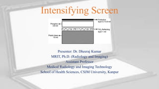
- 1. Intensifying Screen Presenter: Dr. Dheeraj Kumar MRIT, Ph.D. (Radiology and Imaging) Assistant Professor Medical Radiology and Imaging Technology School of Health Sciences, CSJM University, Kanpur
- 2. Introduction • Radiographic Intensifying Screens are crucial components in radiography, enhancing image quality while reducing patient exposure to X-rays. • Intensifying screen layers measure in mil, so 1 mil = 0.0254 millimetres. • These screens work in conjunction with radiographic films to convert X-ray energy into visible light, which is then captured by the film to create diagnostic images. • Their significance lies in their ability to improve image clarity and reduce the amount of radiation needed for diagnostic purposes. 14/08/2023 Intensifying Screen By- Dr. Dheeraj Kumar 2
- 3. Basic Components of Intensifying Screens • Radiographic Intensifying Screens consist of three primary layers: the phosphor layer, the reflective layer, and the base or support layer. • These layers work together to optimize the conversion of X-ray energy into visible light and direct it towards the film for image formation. 14/08/2023 Intensifying Screen By- Dr. Dheeraj Kumar 3
- 4. Phosphor Layer • The phosphor layer is the central element of the intensifying screen. It is made up of phosphor crystals that play a crucial role in the conversion of X-ray energy to visible light. • When X-rays interact with the phosphor crystals, they cause the crystals to fluoresce, emitting light photons. • The phosphor layer can be composed of different types of phosphors, with calcium tungstate and rare earth phosphors being common options due to their efficient light emission characteristics. 14/08/2023 Intensifying Screen By- Dr. Dheeraj Kumar 4
- 5. Reflective Layer • The reflective layer is positioned behind the phosphor layer. Its purpose is to reflect the emitted light photons forward, towards the radiographic film. • This layer helps optimize the efficiency of light collection and minimizes light scattering, ensuring that a higher proportion of emitted light contributes to the formation of the radiographic image. • The reflective layer is typically made of a highly reflective material, such as titanium dioxide or barium sulphate. These materials have high scattering coefficients for visible light, allowing the emitted light to be efficiently directed toward the film. 14/08/2023 Intensifying Screen By- Dr. Dheeraj Kumar 5
- 6. Base or Support Layer • The base or support layer is located behind the reflective layer. This layer provides structural support to the entire screen assembly. • Additionally, it prevents backscatter of X-rays that might penetrate the phosphor and reflective layers, ensuring that these X- rays do not contribute to image degradation or fogging of the film. • The base layer is usually made of a durable and flexible material, such as polyester or polycarbonate. These materials provide the necessary support while being able to withstand the handling and mechanical stress that screens may experience during use. 14/08/2023 Intensifying Screen By- Dr. Dheeraj Kumar 6
- 7. Functions of Intensifying Screens • Radiographic Intensifying Screens serve multiple functions in radiography. • They absorb X-rays that pass through the patient's body and convert this X-ray energy into visible light. This light then exposes the radiographic film, leading to image formation. • This process reduces patient exposure to X-rays while shortening exposure times, which is particularly important for minimizing patient motion Artifacts during the exposure. 14/08/2023 Intensifying Screen By- Dr. Dheeraj Kumar 7
- 8. Construction of Intensifying Screens • The construction of intensifying screens involves a sandwich-like structure of the phosphor layer, reflective layer, and base layer. • These layers are often bonded together using adhesive or lamination techniques. • Proper alignment during construction is crucial to ensure that the emitted light is directed towards the film accurately and consistently. • Quality control measures are essential to maintain optimal screen performance. 14/08/2023 Intensifying Screen By- Dr. Dheeraj Kumar 8
- 9. Types of Radiographic Intensifying Screens • Radiographic Intensifying Screens come in various types, including single-sided and double-sided screens. • Single-sided screens have a phosphor layer on one side, while double-sided screens have phosphor layers on both sides for increased efficiency. • High-speed screens are designed for fast exposures, reducing patient dose. Specialized screens are developed for specific applications, such as mammography and fluoroscopy, optimizing image quality for these procedures. 14/08/2023 Intensifying Screen By- Dr. Dheeraj Kumar 9
- 10. Screen-Film Combination • Radiographic Intensifying Screens work in combination with radiographic films. • The screens emit visible light when exposed to X-rays, and this emitted light exposes the film. • It's important to match the speed of the film with the speed of the screen to achieve optimal image quality. • A mismatch in speeds can result in suboptimal images or unnecessary radiation exposure to the patient. 14/08/2023 Intensifying Screen By- Dr. Dheeraj Kumar 10
- 11. Screen Cleaning and Maintenance • Regular maintenance of Radiographic Intensifying Screens is crucial to ensure consistent image quality. • Screens can accumulate dirt, dust, and artifacts over time, which can affect their performance. • Proper cleaning using non-abrasive cleaners and techniques is essential. Care should be taken to avoid scratching the phosphor layer or damaging the screen's structural integrity. 14/08/2023 Intensifying Screen By- Dr. Dheeraj Kumar 11
- 12. Advantages and Disadvantages • Radiographic Intensifying Screens offer several advantages. • They reduce patient dose by allowing lower X-ray exposure levels, leading to increased patient safety. • They also shorten exposure times, minimizing the risk of motion artifacts. • However, the use of screens can come with some disadvantages, including a reduction in spatial resolution due to the conversion of X-rays to visible light and the potential for artifacts in the image. 14/08/2023 Intensifying Screen By- Dr. Dheeraj Kumar 12
- 13. Comparison with Digital Radiography • In the era of digital imaging, the role of Radiographic Intensifying Screens has evolved. • Digital radiography systems directly capture X-rays and convert them to digital images, eliminating the need for traditional film. • However, Intensifying Screens are still employed in some digital systems to enhance image quality and reduce radiation exposure. • This demonstrates the continued relevance of these screens in modern radiography. 14/08/2023 Intensifying Screen By- Dr. Dheeraj Kumar 13
- 14. Practical Considerations • Proper handling, storage, and disposal of Radiographic Intensifying Screens are important considerations. • Screens should be stored in a clean and dry environment to prevent damage. When replacing old or damaged screens, proper disposal methods must be followed to ensure environmental safety. • Quality assurance programs that include regular testing and maintenance of screens are essential to maintain consistent image quality. 14/08/2023 Intensifying Screen By- Dr. Dheeraj Kumar 14
- 15. Conclusion • Radiographic Intensifying Screens play a vital role in traditional radiography by converting X-ray energy into visible light, resulting in high- quality diagnostic images. • They are integral components of radiographic equipment that enhance image clarity while minimizing patient exposure to X-rays. • As technology advances, these screens continue to be relevant in optimizing diagnostic imaging procedures. 14/08/2023 Intensifying Screen By- Dr. Dheeraj Kumar 15
- 16. References • Bushong, S. C. (2020). Radiologic Science for Technologists: Physics, Biology, and Protection. Elsevier. • Carlton, R. R., & Adler, A. M. (2013). Principles of Radiographic Imaging: An Art and a Science. Cengage Learning. • Huda, W., & Mahesh, M. (2017). Review of Radiologic Physics. Wolters Kluwer Health • Kim, J. H., & Kim, S. Y. (2015). Image quality and radiation dose comparison between a dual-side read CR system and a conventional CR system using an abdominal phantom. Radiation Protection Dosimetry, 165(1- 4), 188-193. • Smith-Bindman, R., Miglioretti, D. L., Larson, E. B., & Lee, C. I. (2012). Radiographic imaging for back pain: systematic review. JAMA, 307(11), 1148-1155. 14/08/2023 Intensifying Screen By- Dr. Dheeraj Kumar 16
- 17. Questions and Discussion • Open the floor for questions, comments, and discussions related to Radiographic Intensifying Screens. • This is a great opportunity for your audience to engage with the content, seek clarifications, and share their insights or experiences. 14/08/2023 Intensifying Screen By- Dr. Dheeraj Kumar 17
- 18. Thank You 14/08/2023 Intensifying Screen By- Dr. Dheeraj Kumar 18