Downloaded 75 times
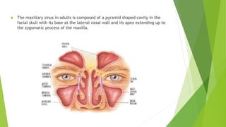
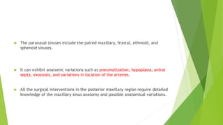
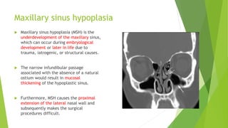
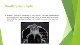
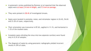

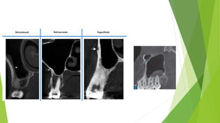


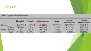


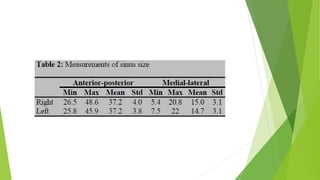


The document discusses various anatomical variations of the maxillary sinus that can be detected using CBCT imaging, including hypoplasia, septa, pneumatization, and variations in arterial locations. It summarizes a study that used CBCT to evaluate 198 scans from Iranian patients, finding a high prevalence of septa (over 50% of sinuses) and identifying locations of the posterior superior alveolar artery in most sinuses. The discussion emphasizes how recognizing these variations is important for surgical planning, as variations can increase surgical risks and affect procedures like sinus lifts or extractions. CBCT is considered useful for pre-operative evaluation to identify variations and adjust treatment plans accordingly.


![谷歌留痕技术 [ 𝙩𝙤𝙥 𝟮𝟯𝟯. 𝙘 𝙤𝙢 ]](https://cdn.slidesharecdn.com/ss_thumbnails/top233-260130174328-3833018c-thumbnail.jpg?width=640&height=640&fit=bounds)


