Downloaded 560 times


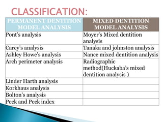
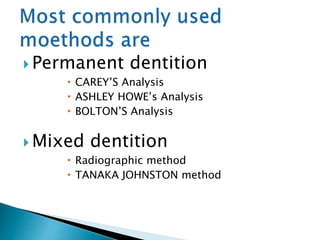

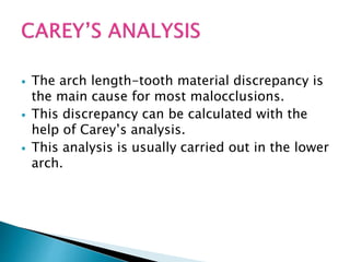
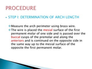


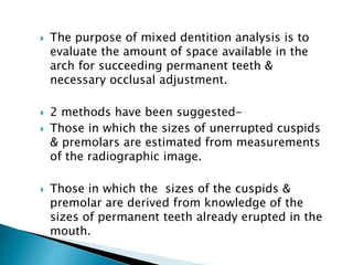


Study models are essential records in orthodontics that provide a 3D representation of the teeth and occlusion. A study model has two parts - the anatomic portion showing the teeth and soft tissues, and the artistic portion which is the stone base. Models need to be trimmed accurately to reproduce the dental anatomy and occlusion. Various analyses like Carey's, Ashley Howe's and Bolton's can be done on study models to assess discrepancies and plan treatment. Mixed dentition analysis using Moyer's or Tanaka-Johnston method helps predict the size of unerupted teeth.


