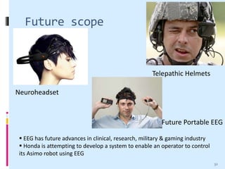
ELECTROENCEPHALOGRAM
(EEG)
Presented by
Vajarala Ashikh
1
Introduction
 Electroencephalography is a technique that
records the electrical activity of the brain
 During an EEG test, small electrodes like cup or
disc type are placed on the scalp
 They pick up the brain's electrical signals and
send them to a machine called
electroencephalogram
 It records the signals as wavy lines on to a
computer screen or paper in order of microvolt
2
History of EEG
 In 1875, Sir Richard Caton presented his findings
about electrical phenomena of the exposed
cerebral hemispheres of rabbits and monkeys
 In 1890, Sir Adolf Beck published an
investigation of spontaneous electrical activity of
the brain of rabbits and dogs
 In 1924, Sir Hans Berger recorded the first
human EEG
3
Why EEG is used
 An EEG is mainly used when there is a need to
diagnose and manage epilepsy
 It can also be used to investigate other
conditions such as encephalitis, dementia, head
injuries, brain tumors, hemorrhage
 An EEG can identify areas of the brain that are
not working properly
 EEGs are also used to determine the level of
brain function in people who are in a coma
4
Parts of Brain
 Cerebrum
 Frontal Lobe
 Parietal Lobe
 Temporal Lobe
 Occipital Lobe
 Cerebellum
 Brain stem
Parts of brain
5
Source of EEG activity
 Neurons are electrically
charged by membrane
transport proteins that
pump ions across their
membranes
 When the wave of ions
reaches the electrodes on
the scalp, they can push or
pull electrons on the metal
of the electrodes
 Push or pull difference
measured as voltage across
time is referred as EEGElectrode on scalp
6
Types of electrode placement
EEG electrodes placed
separately on scalp
EEG electrodes mounted as
special band on head
7
International 10-20 System
8
Closely spaced electrodes
9
Types of EEG
• Around 20 electrodes are stuck to the scalp using a special
paste and EEG signals are recorded
Routine EEG
• The EEG tracing will be recorded along with the heart rate,
airflow, respiration, oxygen saturation and limb movement
Sleep EEG
• It involves recording the brain activity throughout the day and
night
• A small portable EEG recorder is clipped on to the clothing
Ambulatory EEG
10
Types of EEG electrodes
11
Ear clip electrode
Disk electrodes
Intracortical electrodes
Electrode
Montage
Selector
Hi-pass Low-pass Notch Sensitivity
Amplifiers
Filters
Electrode
test/calibrate
Jackbox
Analog to digital
converter
Oscilloscope Computer
Chart
drive
Ink-writing
oscillograph
Writer unit
EEG
Subject
Electrodes
Schematic diagram of an EEG machine
12
Montages
Sequential montage
• Each channel represents the difference between two
adjacent electrodes
Referential montage
• Each channel represents the difference between a
certain electrode and a designated reference electrode
Average reference montage
• The outputs of all of the amplifiers are summed and averaged
Laplacian montage
• Each channel represents the difference between an electrode
and a weighted average of the surrounding electrodes
13
Wave patterns
 Delta waves
 Frequency range 0.5-4 Hz
 Slow-sleep wave for adults
 Theta waves
 Frequency range 4-7 Hz
 Drowsiness in older children and adults
14
 Alpha waves
 Frequency range from 7-14 Hz
 Closing of the eyes, relaxation and attenuation with
eye opening or mental exertion
 Mu rhythm
 Frequency range from 8-13 Hz
 Shows rest-state motor neurons
15
 Beta waves
 Frequency range 15 – 30 Hz
 Active, busy, or anxious thinking, active concentration
 Gamma waves
 Frequency range approximately 30–100 Hz
 Perception that combines two different senses, such as
sound and sight
 Short-term memory matching of recognized objects,
sounds, or tactile sensations
16
EEG displaying epilepsy
EEG waveforms detecting epileptic spikes
17
EEG Results
Normal EEG Abnormal EEG
 A brainwave pattern called
alpha rhythm should be
seen when sitting quietly
with eyes closed
 EEG results are
often normal because
recording a person's brain
activity during the times it
is abnormal is difficult
 People with epilepsy may
have abnormal brain
activity detected
 People who do not have
epilepsy may also have an
abnormal EEG result
indicating any other
disorder
18
EEG Artifacts
• Mains voltage of 110/230 volts, exceeds the EEG's 50
to 100 microvolts by 126dB
• Amplifier notch filters are designed to suppress a
certain amount of mains interference
Mains
Interference
• Eye-induced artifacts - eye blinks, eye movements
• ECG and EMG induced artifacts
• Glossokinetic artifacts
Biological
Artifacts
• Movement by the patient, or even settling of the
electrodes
• Presence of an IV drip that can cause rhythmic, fast,
low-voltage bursts, which may be confused for spikes
Environmental
Artifacts
19
Mains interference artifacts
Eye blink artifacts
20
Artifact correction
 Independent component analysis techniques
have been used to correct or remove EEG
contaminants
 This would result in clean EEG by nullifying
(zeroing) the weight of unwanted components
 Surface Laplacian has been shown to be
effective in eliminating muscle artefact
21
Risks and Precautions
 Slight redness may occur in the locations where
the electrodes were placed
 In rare cases, the cleaning liquid or paste may
cause temporary skin irritation
 The person is instructed not to take food that
contains caffeine
 Not to have oiled hair on the day of test
22
Advantages
 Hardware costs are significantly lower than those of
most other techniques
 EEG has very high temporal resolution, on the order
of milliseconds rather than seconds
 Extremely non-invasive
 EEG is silent, which allows for better study of the
responses to auditory stimuli
 EEG does not involve exposure to high-intensity
(>1 Tesla) magnetic fields
23
Disadvantages
 Low spatial resolution on the scalp
 EEG determines neural activity that occurs
below the upper layers of the brain poorly
 Often takes a long time to connect a subject to
EEG
 Signal-to-noise ratio is poor
24
Uses of EEG
 Clinical Use
 Distinguish epileptic seizures from non-epileptic
seizures, syncope (fainting) and sub-cortical
movement disorders
 To serve as an adjunct test of brain death
 To determine whether to use anti-epileptic
medications
 Research Use
 Cognitive science, cognitive psychology, neuro -
linguistics and psycho physiological research
25
Some more uses
 Investigate epilepsy and locate seizure origin
 Monitor cognitive engagement (alpha rhythm)
 Monitor human and animal brain development
 Test epilepsy drug effects
 Test afferent pathways (by evoked potentials)
 Investigate sleep disorder and physiology
 Control anesthesia depth
26
Modern clinical EEG system
 It is a 36 channel cEEG system
 The system incorporates digital
video with the traditional EEG
 It is unaffected by electrical,
radio and magnetic interference
27
Research on infant attention
 The goal of the research was to examine the
role of the brain in development of infant
attention
 Baby with EEG recording net that
measures 128 channels of EEG
activity
 Pictures and movies with sounds
were shown to check baby’s
response
infantlab.psych.sc.edu
Baby with EEG
28
Recent Development
InteraXon EEG Headset
29
Software for EEG
 EEG recording can be analyzed using various
programs
 EEGLAB
 Fieldtrip
 NBT
 Tucker-Davis Technologies
 Brainvision Analyzer
30
Companies
 Some of these companies have built commercial
EEG devices
 NeuroSky
 OCZ Technology
 Square Enix
 Mattel
 Emotiv
31
Future scope
Telepathic Helmets
Neuroheadset
 EEG has future advances in clinical, research, military & gaming industry
 Honda is attempting to develop a system to enable an operator to control
its Asimo robot using EEG
Future Portable EEG
32
References
 Handbook of Biomedical Instrumentation-R.S Khandpur, 2nd edition
 Haas, L F (2003). "Hans Berger (1873-1941), Richard Caton (1842-
1926), and electroencephalography". Journal of Neurology,
Neurosurgery & Psychiatry
 www.nhschoices.com
 infantlab.psych.sc.edu
 www.ece.mcmaster.ca
 guile3d.com
 viasyshealthcare.com
 24megabytes.com
 tuckerdavistechnologies.com
 thefuturist.com
 E. Niedermeyer, F. H. Lopes da Silva. 1993. Electroencephalography:
Basic principles, clinical applications and related fields, 3rd edition,
Lippincott,Williams &Wilkins, Philadelphia
33
Thank you
34

Electroencephalogram(EEG)

  • 1.
  • 2.
    Introduction  Electroencephalography isa technique that records the electrical activity of the brain  During an EEG test, small electrodes like cup or disc type are placed on the scalp  They pick up the brain's electrical signals and send them to a machine called electroencephalogram  It records the signals as wavy lines on to a computer screen or paper in order of microvolt 2
  • 3.
    History of EEG In 1875, Sir Richard Caton presented his findings about electrical phenomena of the exposed cerebral hemispheres of rabbits and monkeys  In 1890, Sir Adolf Beck published an investigation of spontaneous electrical activity of the brain of rabbits and dogs  In 1924, Sir Hans Berger recorded the first human EEG 3
  • 4.
    Why EEG isused  An EEG is mainly used when there is a need to diagnose and manage epilepsy  It can also be used to investigate other conditions such as encephalitis, dementia, head injuries, brain tumors, hemorrhage  An EEG can identify areas of the brain that are not working properly  EEGs are also used to determine the level of brain function in people who are in a coma 4
  • 5.
    Parts of Brain Cerebrum  Frontal Lobe  Parietal Lobe  Temporal Lobe  Occipital Lobe  Cerebellum  Brain stem Parts of brain 5
  • 6.
    Source of EEGactivity  Neurons are electrically charged by membrane transport proteins that pump ions across their membranes  When the wave of ions reaches the electrodes on the scalp, they can push or pull electrons on the metal of the electrodes  Push or pull difference measured as voltage across time is referred as EEGElectrode on scalp 6
  • 7.
    Types of electrodeplacement EEG electrodes placed separately on scalp EEG electrodes mounted as special band on head 7
  • 8.
  • 9.
  • 10.
    Types of EEG •Around 20 electrodes are stuck to the scalp using a special paste and EEG signals are recorded Routine EEG • The EEG tracing will be recorded along with the heart rate, airflow, respiration, oxygen saturation and limb movement Sleep EEG • It involves recording the brain activity throughout the day and night • A small portable EEG recorder is clipped on to the clothing Ambulatory EEG 10
  • 11.
    Types of EEGelectrodes 11 Ear clip electrode Disk electrodes Intracortical electrodes
  • 12.
    Electrode Montage Selector Hi-pass Low-pass NotchSensitivity Amplifiers Filters Electrode test/calibrate Jackbox Analog to digital converter Oscilloscope Computer Chart drive Ink-writing oscillograph Writer unit EEG Subject Electrodes Schematic diagram of an EEG machine 12
  • 13.
    Montages Sequential montage • Eachchannel represents the difference between two adjacent electrodes Referential montage • Each channel represents the difference between a certain electrode and a designated reference electrode Average reference montage • The outputs of all of the amplifiers are summed and averaged Laplacian montage • Each channel represents the difference between an electrode and a weighted average of the surrounding electrodes 13
  • 14.
    Wave patterns  Deltawaves  Frequency range 0.5-4 Hz  Slow-sleep wave for adults  Theta waves  Frequency range 4-7 Hz  Drowsiness in older children and adults 14
  • 15.
     Alpha waves Frequency range from 7-14 Hz  Closing of the eyes, relaxation and attenuation with eye opening or mental exertion  Mu rhythm  Frequency range from 8-13 Hz  Shows rest-state motor neurons 15
  • 16.
     Beta waves Frequency range 15 – 30 Hz  Active, busy, or anxious thinking, active concentration  Gamma waves  Frequency range approximately 30–100 Hz  Perception that combines two different senses, such as sound and sight  Short-term memory matching of recognized objects, sounds, or tactile sensations 16
  • 17.
    EEG displaying epilepsy EEGwaveforms detecting epileptic spikes 17
  • 18.
    EEG Results Normal EEGAbnormal EEG  A brainwave pattern called alpha rhythm should be seen when sitting quietly with eyes closed  EEG results are often normal because recording a person's brain activity during the times it is abnormal is difficult  People with epilepsy may have abnormal brain activity detected  People who do not have epilepsy may also have an abnormal EEG result indicating any other disorder 18
  • 19.
    EEG Artifacts • Mainsvoltage of 110/230 volts, exceeds the EEG's 50 to 100 microvolts by 126dB • Amplifier notch filters are designed to suppress a certain amount of mains interference Mains Interference • Eye-induced artifacts - eye blinks, eye movements • ECG and EMG induced artifacts • Glossokinetic artifacts Biological Artifacts • Movement by the patient, or even settling of the electrodes • Presence of an IV drip that can cause rhythmic, fast, low-voltage bursts, which may be confused for spikes Environmental Artifacts 19
  • 20.
  • 21.
    Artifact correction  Independentcomponent analysis techniques have been used to correct or remove EEG contaminants  This would result in clean EEG by nullifying (zeroing) the weight of unwanted components  Surface Laplacian has been shown to be effective in eliminating muscle artefact 21
  • 22.
    Risks and Precautions Slight redness may occur in the locations where the electrodes were placed  In rare cases, the cleaning liquid or paste may cause temporary skin irritation  The person is instructed not to take food that contains caffeine  Not to have oiled hair on the day of test 22
  • 23.
    Advantages  Hardware costsare significantly lower than those of most other techniques  EEG has very high temporal resolution, on the order of milliseconds rather than seconds  Extremely non-invasive  EEG is silent, which allows for better study of the responses to auditory stimuli  EEG does not involve exposure to high-intensity (>1 Tesla) magnetic fields 23
  • 24.
    Disadvantages  Low spatialresolution on the scalp  EEG determines neural activity that occurs below the upper layers of the brain poorly  Often takes a long time to connect a subject to EEG  Signal-to-noise ratio is poor 24
  • 25.
    Uses of EEG Clinical Use  Distinguish epileptic seizures from non-epileptic seizures, syncope (fainting) and sub-cortical movement disorders  To serve as an adjunct test of brain death  To determine whether to use anti-epileptic medications  Research Use  Cognitive science, cognitive psychology, neuro - linguistics and psycho physiological research 25
  • 26.
    Some more uses Investigate epilepsy and locate seizure origin  Monitor cognitive engagement (alpha rhythm)  Monitor human and animal brain development  Test epilepsy drug effects  Test afferent pathways (by evoked potentials)  Investigate sleep disorder and physiology  Control anesthesia depth 26
  • 27.
    Modern clinical EEGsystem  It is a 36 channel cEEG system  The system incorporates digital video with the traditional EEG  It is unaffected by electrical, radio and magnetic interference 27
  • 28.
    Research on infantattention  The goal of the research was to examine the role of the brain in development of infant attention  Baby with EEG recording net that measures 128 channels of EEG activity  Pictures and movies with sounds were shown to check baby’s response infantlab.psych.sc.edu Baby with EEG 28
  • 29.
  • 30.
    Software for EEG EEG recording can be analyzed using various programs  EEGLAB  Fieldtrip  NBT  Tucker-Davis Technologies  Brainvision Analyzer 30
  • 31.
    Companies  Some ofthese companies have built commercial EEG devices  NeuroSky  OCZ Technology  Square Enix  Mattel  Emotiv 31
  • 32.
    Future scope Telepathic Helmets Neuroheadset EEG has future advances in clinical, research, military & gaming industry  Honda is attempting to develop a system to enable an operator to control its Asimo robot using EEG Future Portable EEG 32
  • 33.
    References  Handbook ofBiomedical Instrumentation-R.S Khandpur, 2nd edition  Haas, L F (2003). "Hans Berger (1873-1941), Richard Caton (1842- 1926), and electroencephalography". Journal of Neurology, Neurosurgery & Psychiatry  www.nhschoices.com  infantlab.psych.sc.edu  www.ece.mcmaster.ca  guile3d.com  viasyshealthcare.com  24megabytes.com  tuckerdavistechnologies.com  thefuturist.com  E. Niedermeyer, F. H. Lopes da Silva. 1993. Electroencephalography: Basic principles, clinical applications and related fields, 3rd edition, Lippincott,Williams &Wilkins, Philadelphia 33
  • 34.