Download to read offline
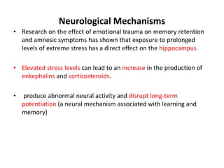
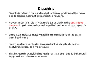

This document discusses the pathophysiology of retrograde amnesia and its distinction from anterograde amnesia, highlighting the complexities of post-traumatic amnesia (PTA) and the neurological mechanisms involved. It emphasizes the impact of emotional trauma on memory, the role of diaschisis in memory impairment, and the effects of increased acetylcholine levels following brain injury. Additionally, it reviews brain imaging studies that reveal significant reductions in blood flow in key brain areas during episodes of PTA.