Submit Search
Upload
如何初步判讀腹部X光
•
4 likes
•
7,780 views
T
TsungAnChen
Follow
在這個PPT裡面,簡單介紹了判讀腹部X光的幾個面向以及常見X光變化,希望對大家有所幫助。
Read less
Read more
Health & Medicine
Report
Share
Report
Share
1 of 39
Download now
Download to read offline
Recommended
Update of atlanta classification of acute pancreatitis published
Dr Priyanka Vishwakarma
Acute Abdomen-Radiology
Parvathy Nair
Abdominal Xrays for undergraduates
Roshan Valentine
Biliary atresia
Biliary atresia
Phòng Khám An Nhi
Pancreatic Trauma
Pancreatic Trauma
Cody Starnes
Ct Imaging of Abdomen Dr. Muhammad Bin Zulfiqar
Dr. Muhammad Bin Zulfiqar
Imaging in acute abdominal pain
SCGH ED CME
Lirads new 2018
Lirads new 2018
Dr.Anmol Uberoi
Appendicectomy
Appendicectomy
srinivas reddy
Presentation1.pptx, radiological imaging of intusussception.
Abdellah Nazeer
Ultrasound of acute & chronic cholecystitis
Samir Haffar
Interpretation of X-Ray and other imaging
drmainuddin
Radiological anatomy & Techniques of the Ventricular system
Mohamed M.A. Zaitoun
How read chest ct 3
How read chest ct 3
ANAS ALSOHLE
CT Urography
CT Urography
Dr. Yash Kumar Achantani
Bowel Preparation for Colonoscopy or Surgery
MarketingTeamBiz
Head ct
Head ct
Kebede Gofer
Presentation1.pptx, radiological imaging of obstructive jaundice.
Abdellah Nazeer
Genito urinary tuberculosis
Annie Agarwal
Pigtail catheter
Pigtail catheter
DR.MARIMUTHU SARAVANAMUTHU
CT urography
CT urography
SabitaMandal1
Post Operative (Gastro-Jejunostomy) Efferent Loop Obstruction due to Recurren...
Hriday Ranjan Roy
Abdominal CT scan made easy
Masrur Akbar Khan
Radiology of Tuberculosis
Mohit Goyal
Abdoiminal examination
Chiranjeevi JIPMER Puducherry
Acute abdomen
Acute abdomen
Walid Agmy
Ct urinary system in radiology
Mahesh Kumar
Introduction to abdominal x ray interpretation
George Abraham
受傷的醫者
受傷的醫者
TsungAnChen
全民身體活動指引整理
全民身體活動指引整理
TsungAnChen
More Related Content
What's hot
Appendicectomy
Appendicectomy
srinivas reddy
Presentation1.pptx, radiological imaging of intusussception.
Abdellah Nazeer
Ultrasound of acute & chronic cholecystitis
Samir Haffar
Interpretation of X-Ray and other imaging
drmainuddin
Radiological anatomy & Techniques of the Ventricular system
Mohamed M.A. Zaitoun
How read chest ct 3
How read chest ct 3
ANAS ALSOHLE
CT Urography
CT Urography
Dr. Yash Kumar Achantani
Bowel Preparation for Colonoscopy or Surgery
MarketingTeamBiz
Head ct
Head ct
Kebede Gofer
Presentation1.pptx, radiological imaging of obstructive jaundice.
Abdellah Nazeer
Genito urinary tuberculosis
Annie Agarwal
Pigtail catheter
Pigtail catheter
DR.MARIMUTHU SARAVANAMUTHU
CT urography
CT urography
SabitaMandal1
Post Operative (Gastro-Jejunostomy) Efferent Loop Obstruction due to Recurren...
Hriday Ranjan Roy
Abdominal CT scan made easy
Masrur Akbar Khan
Radiology of Tuberculosis
Mohit Goyal
Abdoiminal examination
Chiranjeevi JIPMER Puducherry
Acute abdomen
Acute abdomen
Walid Agmy
Ct urinary system in radiology
Mahesh Kumar
Introduction to abdominal x ray interpretation
George Abraham
What's hot
(20)
Appendicectomy
Appendicectomy
Presentation1.pptx, radiological imaging of intusussception.
Ultrasound of acute & chronic cholecystitis
Interpretation of X-Ray and other imaging
Radiological anatomy & Techniques of the Ventricular system
How read chest ct 3
How read chest ct 3
CT Urography
CT Urography
Bowel Preparation for Colonoscopy or Surgery
Head ct
Head ct
Presentation1.pptx, radiological imaging of obstructive jaundice.
Genito urinary tuberculosis
Pigtail catheter
Pigtail catheter
CT urography
CT urography
Post Operative (Gastro-Jejunostomy) Efferent Loop Obstruction due to Recurren...
Abdominal CT scan made easy
Radiology of Tuberculosis
Abdoiminal examination
Acute abdomen
Acute abdomen
Ct urinary system in radiology
Introduction to abdominal x ray interpretation
More from TsungAnChen
受傷的醫者
受傷的醫者
TsungAnChen
全民身體活動指引整理
全民身體活動指引整理
TsungAnChen
奴工島
奴工島
TsungAnChen
在宅醫療,從cure到care
在宅醫療,從cure到care
TsungAnChen
初步了解腫瘤指標
初步了解腫瘤指標
TsungAnChen
原子習慣
原子習慣
TsungAnChen
More from TsungAnChen
(6)
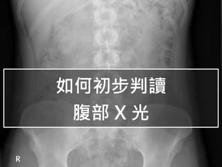
受傷的醫者
受傷的醫者
全民身體活動指引整理
全民身體活動指引整理
奴工島
奴工島
在宅醫療,從cure到care
在宅醫療,從cure到care
初步了解腫瘤指標
初步了解腫瘤指標
原子習慣
原子習慣
如何初步判讀腹部X光
1.
如何初步判讀 腹部 X 光
2.
常見腹部 X 光種類 Abdominal
plain film 站著的PA view,上緣包含橫膈膜 KUB 躺著的AP view,照到kidney、ureter、bladder Left decubitus 左側躺,肝臟在上
3.
腹部 X 光觀察重點 A
Air B Bone C Calcification D Soft tissue Density
4.
Air – 正常腹部可看到的
air 胃 左上腹,有時會看 到air-fluid level
5.
Air – 正常腹部可看到的
air 小腸 可以看到plicae circulares
6.
Air – 正常腹部可看到的
air 大腸 可以看到Haustra, 裡面可能有stool
7.
Air – 異常的
bowel gas Perforation 小腸阻塞 大腸阻塞 Volvulus 腸壁發炎
8.
Air – 異常的
bowel gas Perforation 在CXR看到橫膈膜 下有free air
9.
Air – 異常的
bowel gas Perforation 在Left decubitus下 看到肝臟上方有air
10.
Air – 異常的
bowel gas 小腸阻塞 小腸>3cm,有stack of coin sign。可能是 obstruction or ileus
11.
Air – 異常的
bowel gas 小腸阻塞 若附近局部發炎造成 ileus,也可能看到 sentinel loop
12.
Air – 異常的
bowel gas 大腸阻塞 大腸>6cm或直腸>9cm 阻塞近端脹,但遠端無腸氣 可能是obstruction或 pseudo-obstruction
13.
Air – 異常的
bowel gas Volvulus 乙狀結腸volvulus 會有coffee bean sign
14.
Air – 異常的
bowel gas Volvulus 直腸 volvulus
15.
Air – 異常的
bowel gas 腸壁發炎 腸壁變厚,形成 thumbprinting sign
16.
Air – 異常的
bowel gas 腸壁發炎 Lead pipe colon 長期ulcerative colitis 造成haustrum消失
17.
Air – 異常的
bowel gas 腸壁發炎 Toxic megacolon 可以看到很多腸壁發炎腫脹 形成pseudo-polyps
18.
Bone – 腹部可看到的
bone Rib L spine 沒有rib相接 sacrum Iliac bone Femur head
19.
Bone – 腹部可看到的異常 骨折
髖關節腔狹窄 脊椎退化 蝕骨性變化 成骨性變化
20.
Bone – 腹部可看到的異常 退化性骨刺 髖關節腔狹窄 骨折
21.
Bone – 腹部可看到的異常 蝕骨性變化 有些骨轉移會有蝕骨性變化, 如本片的左側femur
head
22.
Bone – 腹部可看到的異常 成骨性變化 有些骨轉移會有成骨性變化, 本片為攝護腺髖骨轉移
23.
Calcification – 腹部的異常鈣化點 腎結石
膽結石 淋巴結鈣化 糞石 腎上腺鈣化 血管鈣化 慢性胰臟炎
24.
Calcification – 腹部的異常鈣化點 腎結石 90%腎結石在腹部X光中可 見。尿酸結石則看不到。
25.
Calcification – 腹部的異常鈣化點 腎結石 較小的結石會 卡在輸尿管中
26.
Calcification – 腹部的異常鈣化點 膽結石 在右上腹,只有10-15% 膽結石可以被看見 淋巴結鈣化 位置有時候需要與腎 結石做鑑別
27.
Calcification – 腹部的異常鈣化點 糞石 在Right
iliac fossa中,與 闌尾炎有高度相關性
28.
Calcification – 腹部的異常鈣化點 血管鈣化 在腹部X光中看到血管鈣化, 代表嚴重的動脈硬化
29.
Calcification – 腹部的異常鈣化點 慢性胰臟炎 在腹部X光中看到胰臟的鈣 化,暗示有慢性胰臟炎
30.
Calcification – 腹部的異常鈣化點 腎上腺鈣化 在腎臟上方,呈現三角形的鈣化 多與之前出血或TB感染有關
31.
Density – 腹部正常軟組織 Liver spleen Kidney bladder Psoas
muscle
32.
Density – 腹部異常軟組織 肝臟腫大
脾臟腫大 腎臟擴大 腹水 骨盆腔腫塊
33.
Density – 腹部異常軟組織 肝臟腫大 可以看到腸子被 推開
34.
Density – 腹部異常軟組織 脾臟腫大 可以看到腸子被 推開
35.
Density – 腹部異常軟組織 腎臟腫大 腎臟在後腹腔,不 會影響腸子空間
36.
Density – 腹部異常軟組織 腹水 KUB,腸氣飄在中央,後 面是不透光的腹水
37.
Density – 腹部異常軟組織 骨盆腔腫塊 可以看到腸子 被向上推開
38.
Summary Air Perforation、小腸阻塞、大腸阻塞、volvulus、腸壁發炎 Bone 骨折、髖關節腔狹窄、脊椎退化、蝕骨性變化、成骨性變化 Calcification 腎結石、膽結石、淋巴結鈣化、糞石、血管鈣化 慢性胰臟炎、腎上腺鈣化 Soft tissue Density 肝臟腫大、脾臟腫大、腎臟擴大、腹水、骨盆腔腫塊
39.
參考資料 Radiology Masterclass https://www.radiologymasterclass.co.uk/ Radiopaedia https://radiopaedia.org/?lang=us
Download now