
Recommended
More Related Content
What's hot
What's hot (20)
Similar to Effect of OLFM1 and LPHN2 on Glioblastoma Metastasis
Similar to Effect of OLFM1 and LPHN2 on Glioblastoma Metastasis (20)
Recently uploaded
Recently uploaded (20)
Effect of OLFM1 and LPHN2 on Glioblastoma Metastasis
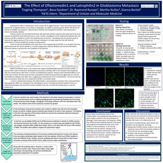
- 1. The Effect of Oflactomedin1 and Latrophilin2 in Glioblastoma Metastasis Tingting Thompson1, Beca Gardner2, Dr. Raymond Runyan2, Martha Nuñez2, Gianna Bortoli2 1KEYS Intern, 2Department of Cellular and Molecular Medicine Introduction Figure 4.1 Nucleus stain with Hoechst Figure 4.2 LPHN2 stain with LPHN2 primary antibody Figure 4.3 Overlap stain of nucleus and LPHN2 protein epithelial cell primary epithelial cancer cell metastatic cell endothelial cell basement membrane Methods Results Figure 5.1 OLFM1 Experimental: T-ARB with 1˚+2˚ Figure 5.2 OLFM1 Control: T-ARB with 2˚ only Since the staining signal continued to show the same strength in both the experimental and control samples, we failed to find the correct method of primary antibody attachment. Introduction Varying conditions: 6 wells 1. Fixation methods 2. Antigen retrieval buffers (ARB) Experimental vs. control: 1˚+ 2˚ v 2˚ only Figure 3.2 Staining: Round Two Tray with six varying well conditions to expose OLFM1 antigen. 1˚ means primary antibody, ø had no retrieval buffer, top labels were the different fixation methods: methanol vs. tannic Test 1 failure for OLFM1 primary antibody caused need to further explore how to expose the OLFM1 antigen for the primary antibody to attach to. Factors tested: 6 wells 1. Coverslip vs. Non-Coverslip 2. Cell density- 25,000 v 75,000 3. Primary antibodies Testing Figure 3.1 Staining: Round One Trays contain six varying wells that were designed to compare different factors for imaging Glioblastoma (GB) is a lethal brain cancer known for its aggressiveness. There are several factors that account for its aggressiveness, one of them is metastasis, the ability to spread quickly. Within the GB cancer cell line 62 (GB62), two proteins– olfactomedin1 (OLFM1) and latrophilin2 (LPHN2)– were observed to enhance metastatic abilities. Previous work in the lab determined that cells with both proteins present were observed to be more invasive compared to those with only one or none. Further investigation of the relationship between the two proteins and GB is needed to confirm the hypothesized connection. Identifying how the proteins work to intensify the aggressiveness of the cancer through metastasis is a major objective that will allow for explorations on how to disable the proteins causing cancer. There are several possibilities that could occur from suppressing the OLFM1 or its receptor that may significantly limit the cancer spread (1). If cancer progression could be delayed, less harmful alternative treatment options may become more available to the patients. LPHN2 and OLFM1 assist in the invasive late stage of Epithelial-Mesenchymal transition (EMT)– a process often used in embryonic development and wound mending. EMT is the transition from epithelial cells into mesenchymal cells which is similar to Glioblastoma Invasion. Previous work identified GB62 as one of the most invasive cell lines and one that responded to OLFM1 by becoming more aggressive (1). Cancers have different subtypes that often vary in aggressive characteristics such as invasiveness– cell line 62 was the most promising to study due to its response to OLFM1 in an earlier study. Is there a relationship between the olfactomedin-1 and latrophilin-2 proteins that could potentially have significant effects on metastasis–the spreading of cancer? A Duolink kit will test whether there is a relationship between two identified proteins related to the cell, not what the relationship is, only the existence of. Along with the antibody stains, Hoechst, a nucleus stain, was used to confirm cells. Photos were taken using a light microscope. During testing, the primary antibody was evaluated to determine if it worked. (1) Lencinas, Alejandro et al. “Olfactomedin-1 Activity Identifies a Cell Invasion Checkpoint during Epithelial-Mesenchymal Transition in the Chick Embryonic Heart.” Disease Models & Mechanisms 6.3 (2013): 632–642. PMC. Web. 26 June 2017. 1.1 Carrier, T., and John Allen. "What Is Tumor Progression?" WiseGEEK. Conjecture Corporation, 11 June 2017. Web. 12 July 2017. 1.2 Chiblak, Sara. "Pancreatic Cancer: Current Concepts in Invasion and Metastasis." Research Gate. N.p., Dec. 2011. Web. 1.3 "Mitral Valve Disease and the Cavalier King Charles Spaniel." CavalierHealth.org. N.p., n.d. Web. 12 July 2017. 2.1 Rastogi, Varun. "Diagnostic Procedures for Autoimmune Vesiculobullous Diseases: A Review." JOMFp. N.p., 13 Feb. 2015. Web. References I would first like to thank everyone in my lab for putting up with me for a wonderful six weeks! Next, I am extremely thankful to KEYS and KEYS staff for helping me succeed this summer and giving me the chance to get hands on lab experience. I will cherish the time I have spent with everyone this summer and all the new people I have met. I would also like to give thanks to my friends– Hannah Nguyen and Olivia Pietz– and family– Carol Thompson, Elizabeth Hurd, and Grace Thompson– for helping me adjust during the program. Acknowledgements Due to many circumstances such as cell contamination and troubleshooting problems, we were unable to perform the actual Duolink kit. Although, future steps were concluded for LPHN2. The LPHN2 primary antibody stained in the green and properly surrounded the blue nuclei since LPHN2 is in the membrane. It can also be concluded that the laminin coating on the coverslips successfully maintained a large cell count. Without the protein- protein relationship assessment from Duolink, support for the hypothesis is yet to be recorded. Conclusion Other methods will need to be tested in order to create the best environment for determining the relationship of the proteins. In the end, it is also possible that there is no relationship at all. To continue troubleshooting, other factors and methods may be explored, including: Adding extra OLFM1 protein variants if we are losing too many of the extracellular protein or if the cells are not secreting enough Finding a new primary antibody if the problem is the antibody itself Alternative technique to Duolink would be Immunoprecipitation Figure 1.2 An example of EMT in pancreatic cancer– a cancer that does involve epithelial cells. Figure 1.3 The process of EMT. Along side are characteristics and abilities of the two cell types. Cancerous Uncontrolled cell division Damaged cells Well differentiated cells Uniform cell growth Cancer Progression Discussion Successes: o Coverslip for imaging o 75,000 cells per well o LPHN2 primary antibody OLFM1 stain did not work, a possible factor was inability for the primary antibody to find the antigen on the protein. Fluorescent Label Labeled Secondary Antibody Primary Antibody Antigen Epithelial cells Metastatic cells Endothelial cells basement membrane Primary epithelial cancer cell Carcinoma in situ primary site Normal epithelium EMT Intravasation/ Extravasation Secondary tumour distant metastasis Figure 1.1 Explanation of cancer as regular cells who have uncontrollable and abnormal growth.