Alambagh Call Girl 9548273370 , Call Girls Service Lucknow
1
1. Adam Boulger 27040016
Discuss the characteristics and functionof T-lymphocyte cells
T-Lymphocytesare anessential partof the immune system, involvedinthe detectionand
destructionof pathogenicorganisms.Thisessaywillfocusonthe developmentof αβT cellsinside
the thymusand finishingwithabrief discussionuponthe importantrolesandfunctionof T-cells.
Originationof T cellsbeginsinthe bone marrow
T cellsoriginate fromhaematopoieticstemcells(HSC)inside the bone marrow.The firststage of
differentiationbeginswith HSC’sbeingconvertedintolymphoidprogenitorcells(LPC) (Schwarz&
Bhandoola,2006). These LPC’smigrate towardsthe thymusformaturation.However,recent
researchhas providedevidence thatTcell developmentandmaturationcanalsooccur inthe tonsils.
The focus of thisessaywill be onIntrathymic differentiationandmaturation. (McClory,Hughes,
Freud & Briercheck,2012)
Intrathymic T-Lymphocyte DifferentiationandMaturation
Once the LPC’s have migratedtowardsthe thymusviathe bloodfromthe bone marrow,the cells
enterthe thymusthroughthe corticomedullaryjunctionandare traffickedintothe thymuswhere
(Lind,Prockop,Porrit& Petrie,2001) the cellsproliferate innumbersbycellulardivisiontogenerate
a large populationof immature thymocytes(LPC’s).
Thymopoiesisisthe conversionof LPC’s intomature T Lymphocytes andsummarisedin figure 1.The
stagesof maturationare identifiedandclassifiedbythe expressionof differentcell surface
molecularmarkers,anexample isCD44,whichcharacterise the cells.
However, research hasshownthatmanynon-Tcell lineage cellsexpressCD44protein (Ponta,
Sherman& Herrlich,2003) and thatnon-T cellsare consistentlyCD25- and therefore this
complicatesnarrowingdownthe specificlineageforTcells.Recentresearchhasusedthe CD117+
cell surface protein(C-Kit) toidentifyTcell lineages.CD117positve cellsare the mostlikelytofully
mature intoCD4+ T helpercellsorCD8+ cytotoxicT cells. (Ceredig&Rolink,2002)
2. Adam Boulger 27040016
The main stagesinvolvedinthe developmentof mature T lymphocytesare displayed inFigure 1.
Thisoccurs withinthe thymusmicroenvironment (Liu,&etal.,2012) whichprovidesaplethoraof
the requireddifferentiationfactorstosupport T cell lineagerestriction(Ciofani M& Zuniga-Plfucker,
2007). The LPC’sundergothe start of the T-Lineage specification,thisisactivatedbymultiple
transcriptionfactors.Anexample isthe BcI11btranscriptionfactorwhichisessential forthe
developmentof T cellsthroughcommitmenttothe Tcell lineage andmaintenance of development
(Yu,& etal.,2015). This formsDN1 cellswhichare heterogenous,thereforeable togive rise to
alpha-beta,gamma-deltaandmanyotherimmune cells(Ciofani M& Zuniga-Plfucker,2007).This is
shownin Figure 1, where the gamma-deltacell isshowninred. The differentiationandmaturation
pathwayforγδ cellsisnotfully known. Due tothis,the focus in thisessaywill be onthe
developmentof mature αβT cell receptorcells(Xiong&Raulet.2007)
Transcriptionfactorsact uponthe DN1 CD44+CD25-CD117+ cell andthis causesfurthercommitment
to the T cell lineage andtherefore expressionof CD25 to formDN2 CD44+CD25+CD117+, therefore
positivelyexpressingthischaracteristic phenotype.
Table 1:An overview of the characteristic phenotypessurface proteins discussed inthe above paragraphs and their
functions. (Miettinen& Lasota, 2005. National Institute Health, N.D. NationalInstitute Health, Jacques Thèze Interleukin2.
(2012)
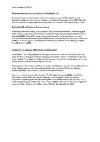
Figure 1:Overviewof Intrathymic T-CellProductionandDevelopment. The diagramshows the involvement of specific transcription
factors at eachdifferentiation stage. The expressionof cell surface proteins are also includedto enable cell type identification.
Abbreviations: LPC= Lymphoidprogenitor cells, DN = double negative, ISP= intermediate single positive, DP= double positive and CD =
cluster of differentiation. Data derived from the following references (Shah, & Zuniga-Pflucker, 2014. Avram & Califano, 2014.
Kratchmarox, Magnun& Reiner, 2018, Yu, et al.,2015)
3. Adam Boulger 27040016
As showninFigure 1, the difference intranscriptionfactorsbetweenDN1andDN2 is the absence
HES1. HES1 is a transcriptional repressor (De Obaldia,etal,.2013) and therefore the lackof HES1
allowsthe expressionof gene IL2RA.(Gene Cards, Geneticdatabase.N.D). Initiation of gene re-
arrangementsbeginsatDN2due toRAG1 and RAG2 activationproteins. Gene segments,suchasthe
TCR alphagene,whichare involvedinproducingthe proteins controllingthe developmentof the T-
cell receptor.These proteinscause the gene segmentstobe re-arrangedindifferentcombinations
to increase the variabilityof eachsegment,increasingthe chance of a unique receptor(NIH,
National Institute forHealth,N.D. Xiong&Raulet,2007).
Full T cell lineage commitmentoccursduringthisstage of transitiontoDN3 as the cellsare making
more specialisedchanges,suchasthe gene re-arrangementsstatedabove (Ciofani&Zuniga-
Pflucker,N.D).Asshowninfigure 1, a processthatoccurs at DN3 isthe re-arrangementof the β TCR
chainlocus,which triggersbeta-selection. Cellswhichdonotsuccessfullyperformthisenter
apoptosis. Thischange beginsthe transformationof cellsfrom DN3to DN4 and due to the induction
of the β-chainpairingupwiththe pre-Tα surrogate chain,formingPre-TCRβ-Dependentcomplex on
the surface of the DN4 cell whichisjustthe pre-TCRreceptorcomplex. (DivyaK,Shah,2014).
Thymocytesdifferentiatethroughavarietyof double negative (DN) stagesandthiscanbe tracked
by the expressionof cell surface proteins(Table 2)
Table 1: A summaryof the cell-surface markers used to identify each stage in figure 1, with the expression marked + and
absence - at each stage of DN differentiation.
The nextstage is the formationof the ISPcell,whichisjustan intermediate stage.Thisistriggered
by the transcriptionfactorTCF-1,whichupregulatesthe expressionof genesessentialinsuccessful
T-cell formation.Thiscausesthe CD8surface proteintobe expressedandbecome ISPCD8+onthe
surface of the cell (Weber,etal.,2011). Duringthisstage,re-arrangementof the TCRα chain occurs
and thisstimulates the formationof the DPCD8+CD4+ cells.Thisisthe DP stage,where rapid
proliferationoccurs(ErikJ. Peterson, JonathanS.(2019).The DP cells now undergopositive and
negative selection.
4. Adam Boulger 27040016
Positive SelectionofDP cells
Positive selectionoccursinthe cortex of the thymusas the immature thymocytesare trafficked
towardsthe medulla.Asshownin Figure 2the DPCD8+CD4+ cellswithasuccessfullyre-arranged
and expressedmature αβTcell receptor(TCR’s) are able to bindtoself-majorhistocompatibility
complex (MSC) proteinswithasufficientaffinity,suchasthe epithelialcell infigure 2.The MSC’scan
be dividedintoHLA-I(classI) andHLA-II(class2) moleculesandlessthan5% DP cellsare able to bind
to these testmolecules, withthe remainingcellsreceivinganapopticsignal.DPcellswhichrecognise
HLA-Idifferentiate andmature intocytotoxiccellsandthose thatrecognise HLA-2formT helper
cells.(Klein,Hinterberger&Wirnsberger,2009. Anderson&Takahama,2012)
Figure 2: Positive Selection of DPCD8+CD4+ cells. Cells which have successfullyformed a functional T-cell
receptor advance as the receptor is complementaryto the MHC molecules with a high affinity. Figure from
Kuby Immunology, Sixth Edition 2007.
5. Adam Boulger 27040016
Negative SelectionofDP cells
Afterpositive selection,the cellsenterthe medullaandthe secondcheck-pointoccurs.Thisis
negative selection,andthisiswhere the positivelyselectedcellsare exposedtoa varioussetof
diverse self-antigenspresentedonthe surface of epithelial cellsinfigure 3. SPcellswithhigh-affinity
receptorsforself-MHC’sare immediatelyidentifiedandselectedforapoptosis.If thisstage didnot
exist,itwouldcause the immune cellstoact againstthat relative antigen.Destroying/damaging
humancells(Anderson&Takahama,2012). Followingthe selection,the cellsare now classedas
mature naïve cellsand theyexitthe thymusandenterthe periphery.
Issueswithbothnegative andpositive selectioncancause a varietyof issues.Thisisbecause thymic
T cellscan escape thymicselectionandcause issuessuchasrheumatoidarthritis.Researchhas
shownthat byattenuatingTCRsignal intensityindevelopingTcellscancause systemicautoimmune
disease asT cellsreactwithself-antigens.
Cell-Cell interactions involving T Lymphocyte cells triggerthe secretion of molecules which
are vital for survival
T-Lymphocyte cell-cell interactions refer to the direct interaction between cells to allow
normal immune functions and survival. The elimination of cells infected by viruses is often
Figure 3: Negative selection of DPCD8+CD4+ cells. The cells are screened against a set of self-antigens to reduce the risk of
autoimmune disease. Figure 3 from Kuby Immunology, sixth edition 2007.
6. Adam Boulger 27040016
completed by cytotoxic T cells as most cells do not possess immuno-machinery. However, in
the case of a viral infection. Cytotoxic T cells identify the antigens produced by viral DNA.
This triggers secretion of cytotoxic molecules. The function of perforin-containing lytic
granules, cytotoxic t cells can destroy infected or cancerous cells (Krzewski & Brycescon,
2014) by perforating the cell surface. This perforin is also an issue as many autoimmune
diseases utilise this molecule. An example can be hemophagocytic lymphohistocytosis
(HLH), where hyperactivity and genetic issues cause fatal metabolic abnormalities. Research
has shown that mutations in the perforin gene could be the cause in familial HLH (Stepp, et
al., 1999).
Some forms of bacteria can survive and multiply inside a macrophage, an example of this is
M. tuberculosis. The cell-cell interaction between T helper cells and macrophages can detect
and destroy these bacteria as the T helper cell can bind to the antigen presented on the
macrophage surface and communicate to the macrophage via cytokines. This initiates the
destruction of the bacterium. This process can be seen below in Figure 4.
Figure 4: The process by which a macrophage ingests a pathogen and the T helper cell identifies the antigens presented by
the macrophage. Signalling further cells, such as the cytotoxic t cell to attack further infected cells. Figure 4 from Benjamin
Cummings, Pearson Education, 2004.
This release of cytokines, explained in Figure 4 also activates further immune pathways,
involving B lymphocytes’ which secrete antibodies by plasma cells. These antibodies travel
and interact with other infected cells, eventually causing the destruction. The origin of this is
due to the helper t cells.
In conclusion, the immune systemis incredibly complicated, often with multiple factors that
can produce the same desired effect. In my opinion, future research should focus on
utilising the immune system, especially in the elderly as reduced thymic activity leads to the
reliance on previously produced cells. As life expectancy is increasing (in correlation with the
advances in science and technology), the demand for medications to counter immune based
diseases will increase. Especially antibiotic resistance increases, this could become an issue.
7. Adam Boulger 27040016
References
Anderson, G., & Takahama, Y. (2012, June). Thymic epithelial cells: Working class heroes for
T cell development and repertoire selection. Retrieved from
https://www.ncbi.nlm.nih.gov/pubmed/22591984
Avram, D., & Califano, D. (2014, September 01). The Multifaceted Roles of Bcl11b in Thymic
and Peripheral T Cells: Impact on Immune Diseases. Retrieved from
http://www.jimmunol.org/content/193/5/2059
CD44 gene - Genetics Home Reference - NIH. (n.d.). Retrieved from
https://ghr.nlm.nih.gov/gene/CD44
Ceredig, R., & Rolink, T. (2002, November 01). A positive look at double-negative
thymocytes. Retrieved from https://www.nature.com/articles/nri937
Ciofani M, Zuniga-Plfucker. (2007, June) The Thymus as an inductive sit for T lymphopoiesis.
Retrieved from https://www.ncbi.nlm.nih.gov/pubmed/17506693?
Ciofani, M., & Zúñiga-Pflücker, J. C. (n.d.). The thymus as an inductive site for T
lymphopoiesis. Retrieved from https://www.ncbi.nlm.nih.gov/pubmed/17506693
D. (2014, June 12). T-cell development in thymus. Retrieved from
https://www.immunology.org/public-information/bitesized-immunology/immune-
development/t-cell-development-in-thymus
De Obaldia, M. E., Bell, J. J., Wang, X., Harly, C., Yashiro-Ohtani, Y., DeLong, J. H., Zlotoff, D.
A., Sultana, D. A., Pear, W. S., … Bhandoola, A. (2013). T cell development requires
constraint of the myeloid regulator C/EBP-α by the Notch target and transcriptional
repressor Hes1. Nature immunology. Retrieved from
https://www.ncbi.nlm.nih.gov/pmc/articles/PMC4038953/
Erik J. Peterson, Jonathan S. (2019). Retrieved from
https://www.sciencedirect.com/topics/medicine-and-dentistry/t-cell-proliferation
Gene Cards, Genetic database. Retrieved from https://www.genecards.org/cgi-
bin/carddisp.pl?gene=IL2RA
Helen E. Porritt, Lynn L. Rumfelt, Sahba Tabrizifard, Thomas M. Schmitt. (2004, June)
Heterogeneity among DN1 Prothymocytes Reveals Multiple Progenitors with Different
Capacities to Generate T Cell and Non-T Cell Lineages. Retrieved from
https://core.ac.uk/download/pdf/82672910.pdf
Jacques Thèze Interleukin 2. (2012). Interleukin 2. Retrieved from
https://www.sciencedirect.com/topics/neuroscience/interleukin-2
8. Adam Boulger 27040016
Klein, L., Hinterberger, M., Wirnsberger, G., & Kyewski, B. (2009, December). Antigen
presentation in the thymus for positive selection and central tolerance induction. Retrieved
from https://www.ncbi.nlm.nih.gov/pubmed/19935803
Kratchmarov, R., Magun, A. M., & Reiner, S. L. (2018). TCF1 expression marks self-renewing
human CD8+ T cells. https://www.ncbi.nlm.nih.gov/pmc/articles/PMC6058237/
Krzewski, K., & Bryceson, Y. T. (2014, June) Molecular Mechanisms Regulating Cytotoxic
Lymphocyte Development and Function, and Their Associations to Human Diseases.
Retrived from http://www.ncbi.nlm.nih.gov/pmc/articles/PMC4052198/
Lind, E. F., Prockop, S. E., Porritt, H. E., & Petrie, H. T. (2001, July 16). Mapping Precursor
Movement through the Postnatal Thymus Reveals Specific Microenvironments Supporting
Defined Stages of Early Lymphoid Development. Retrieved from
http://jem.rupress.org/content/194/2/127?ijkey=9dd411353f59518524042ca3bb3a0939f45
c320c&keytype2=tf_ipsecsha
Liu, Y., Chen, X. H., Si, Y. J., Li, Z. J., Gao, L., Gao, L., Zhang, C., … Zhang, X. (2012).
Reconstruction of hematopoietic inductive microenvironment after transplantation of
VCAM-1-modified human umbilical cord blood stromal cells. Retrieved from
https://www.ncbi.nlm.nih.gov/pmc/articles/PMC3285638/
McClory, S., Hughes, T., Freud, A. G., Briercheck, E. L., Martin, C., Trimboli, A. J., Yu, J.,
Zhang, X., Leone, G., Nuovo, G., … Caligiuri, M. A. (2012). Evidence for a stepwise program of
extrathymic T cell development within the human tonsil. The Journal of clinical
investigation, Received from https://www.ncbi.nlm.nih.gov/pmc/articles/PMC3314444/
Miettinen, M., & Lasota, J. (2005, September). KIT (CD117): A review on expression in
normal and neoplastic tissues, and mutations and their clinicopathologic correlation.
Retrieved from https://www.ncbi.nlm.nih.gov/pubmed/16082245
Notarangelo, L. D. (2014, October) Immunodeficiency and Immune Dysregulation Associated
with Proximal Defects of T Cell Receptor Signaling. Received from
https://www.ncbi.nlm.nih.gov/pmc/articles/PMC4254644/
Ponta, H., Sherman, L., & Herrlich, P. A. (2003, January). CD44: From adhesion molecules to
signalling regulators. Retrieved from https://www.ncbi.nlm.nih.gov/pubmed/12511867
Pullen, C. (2014, December 01). T Cells & Autoimmune Diseases. Retrieved from
https://www.the-rheumatologist.org/article/t-cells-autoimmune-diseases/
RAG1 gene - Genetics Home Reference - NIH. (n.d.). Retrieved from
https://ghr.nlm.nih.gov/gene/RAG1
RAG2 gene - Genetics Home Reference - NIH. (n.d.). Retrieved from
https://ghr.nlm.nih.gov/gene/RAG2
9. Adam Boulger 27040016
Schwarz, B. A., & Bhandoola, A. (2006, February). Trafficking from the bone marrow to the
thymus: A prerequisite for thymopoiesis. Retrieved from
https://www.ncbi.nlm.nih.gov/pubmed/16448533
Shah, D. K., & Zúñiga-Pflücker, J. C. (2014, May 01). An Overview of the Intrathymic
Intricacies of T Cell Development. Retrieved from
http://www.jimmunol.org/content/192/9/4017
Stepp, S. E. Dufourcq-Lagelouse, R., Le, F., Bhawan, S., Certain, S., Mathew, P. A., Kumar, V.
(1999, December 03). Perforin gene defects in familial hemophagocytic lymphohistiocytosis.
Retrieved from https://www.ncbi.nlm.nih.gov/pubmed/10583959
Weber, B. N., Chi, A. W., Chavez, A., Yashiro-Ohtani, Y., Yang, Q., Shestova, O., & Bhandoola,
A. (2011, August 03). A critical role for TCF-1 in T-lineage specification and differentiation.
Retrieved from https://www.nature.com/articles/nature10279
Xiong, N., & Raulet, D. H. (2007, February). Development and selection of gammadelta T
cells. Retrieved from https://www.ncbi.nlm.nih.gov/pubmed/17291276
Yu, Y., Wang, C., Clare, S., Wang, J., Lee, S. C., Brandt, C., . . . Liu, P. (2015, June 01). The
transcription factor Bcl11b is specifically expressed in group 2 innate lymphoid cells and is
essential for their development. Retrieved from
https://www.ncbi.nlm.nih.gov/pubmed/25964371